. 2017 Jan 17:5:4.
doi: 10.1186/s40425-016-0204-3. eCollection 2017.

Radiotherapy and MVA-MUC1-IL-2 vaccine act synergistically for inducing specific immunity to MUC-1 tumor antigen

Affiliations

Radiotherapy and MVA-MUC1-IL-2 vaccine act synergistically for inducing specific immunity to MUC-1 tumor antigen

Gilda G Hillman et al. J Immunother Cancer. .

Abstract

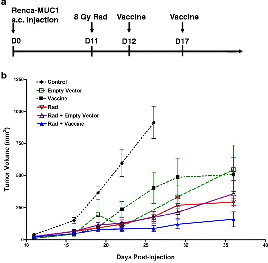
Background: We previously demonstrated that tumor irradiation potentiates cancer vaccines using genetic modification of tumor cells in murine tumor models. To investigate whether tumor irradiation augments the immune response to MUC1 tumor antigen, we have tested the efficacy of tumor irradiation combined with an MVA-MUC1-IL2 cancer vaccine (Transgene TG4010) for murine renal adenocarcinoma (Renca) cells transfected with MUC1.

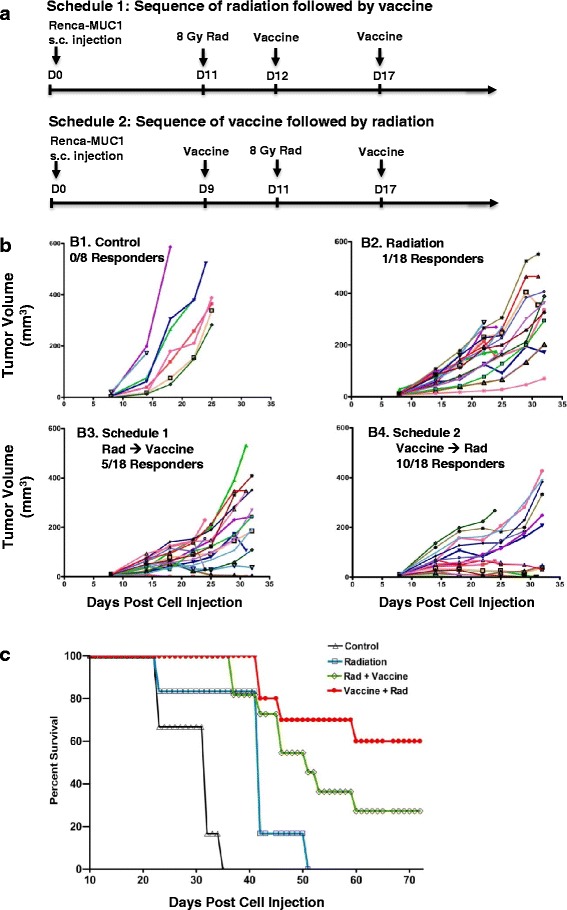
Methods: Established subcutaneous Renca-MUC1 tumors were treated with 8 Gy radiation on day 11 and peritumoral injections of MVA-MUC1-IL2 vector on day 12 and 17, or using a reverse sequence of vaccine followed by radiation. Growth delays were monitored by tumor measurements and histological responses were evaluated by immunohistochemistry. Specific immunity was assessed by challenge with Renca-MUC1 cells. Generation of tumor-specific T cells was detected by IFN-γ production from splenocytes stimulated in vitro with tumor lysates using ELISPOT assays.

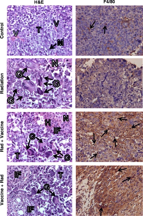
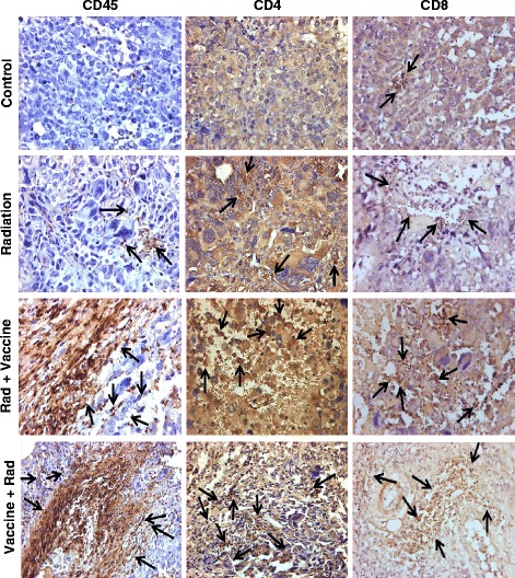
Results: Tumor growth delays observed by tumor irradiation combined with MVA-MUC1-IL-2 vaccine were significantly more prolonged than those observed by vaccine, radiation, or radiation with MVA empty vector. The sequence of cancer vaccine followed by radiation two days later resulted in 55-58% complete responders and 60% mouse long-term survival. This sequence was more effective than that of radiation followed by vaccine leading to 24-30% complete responders and 30% mouse survival. Responding mice were immune to challenge with Renca-MUC1 cells, indicating the induction of specific tumor immunity. Histology studies of regressing tumors at 1 week after therapy, revealed extensive tumor destruction and a heavy infiltration of CD45+ leukocytes including F4/80+ macrophages, CD8+ cytotoxic T cells and CD4+ helper T cells. The generation of tumor-specific T cells by combined therapy was confirmed by IFN-γ secretion in tumor-stimulated splenocytes. An abscopal effect was measured by rejection of an untreated tumor on the contralateral flank to the tumor treated with radiation and vaccine.

Conclusions: These findings suggest that cancer vaccine given prior to local tumor irradiation augments an immune response targeted at tumor antigens that results in specific anti-tumor immunity. These findings support further exploration of the combination of radiotherapy with cancer vaccines for the treatment of cancer.

Keywords: IL-2; MUC1; MVA vector; Radiation; Renal Cell Carcinoma.


Figures

Fig. 1
Fig. 1
Growth curves of Renca-MUC1 tumors treated with tumor irradiation and vaccine. Established Renca-MUC1 tumors were untreated (Control) or treated with MVA-empty (Empty Vector), MVA-MUC1-IL-2 vector (Vaccine), with or without radiation (Rad). Radiation was administered at 8 Gy photons on day 11. Two peritumoral injections of vectors were administered on days 11 and 17, at 107 PFU in 25 μl S08 buffer. a. Schedule of treatment. b. Tumor growth curves. Each symbol represents the mean tumor volume of 7-9 mice per group ± SEM at different time points post cell injection. Data were compiled from two experiments
Fig. 2
Fig. 2
Sequence of tumor irradiation and vaccine for the treatment of Renca-MUC1 tumors. Established Renca-MUC1 tumors were treated with 8 Gy radiation (Rad) and MVA-MUC1-IL-2 vector (Vaccine) administered peritumorally at 107 PFU in 25 μl S08 buffer. a. Schedule 1: Sequence of radiation followed by vaccine. Tumors were irradiated on day 11, followed by vaccine on day 12 and 17. Schedule 2: Sequence of vaccine followed by radiation. Tumors were first injected with vaccine on day 9, then were irradiated two days later on day 11, followed by a second treatment of vaccine on day 17. b. Tumor growth curves. B1. Control (Control) untreated mice. B2. Radiation treatment alone. B3. Schedule 1 sequence of radiation followed by vaccine. B4. Schedule 2 sequence of vaccine followed by radiation. Each symbol represents the tumor volume of individual mice with 8 mice per group for control and 18 mice per treatment group B2, B3, and B4 at different time points post cell injection. c. Survival curves. In a separate experiment identical to that shown in A, established Renca-MUC1 tumors were treated with 8 Gy radiation (Rad) and MVA-MUC1-IL-2 vector (Vaccine) administered peritumorally at 107 PFU using either Schedule 1 of radiation followed by two vaccine treatments or Schedule 2 of vaccine followed by radiation and a second vaccine treatment. Mice were followed for survival (n = 8 mice for control or radiation alone and n = 12 mice for combined radiation and vaccine)
Fig. 3
Fig. 3
Histology and macrophage infiltration in Renca-MUC1 tumors treated with radiation and vaccine. Established Renca-MUC1 tumors were treated with 8 Gy radiation (Rad) and MVA-MUC1-IL-2 vector (Vaccine) administered peritumorally at 107 PFU using either Schedule 1 of radiation followed by two vaccine treatments (Rad + Vaccine) or Schedule 2 of vaccine followed by radiation and a second vaccine treatment (Vaccine + Rad) as described in Fig. 2. Tumors sections, obtained at one week after the last vaccine treatment, were stained for H&E or by IHC for F4/80+ macrophages. The main findings were labeled with T for tumor, V for vessels, N for necrosis, M for mitosis, G for giant cells, H for hemorrhages and IF for inflammatory cells. In H&E stained tumor sections, control tumors presented as sheets of pleomorphic epithelial cells with frequent mitosis and minimal immune cells infiltrates. Radiation-treated tumors showed areas of necrosis, numerous giant cells with abnormal mitosis, eccentric nuclei or large vacuoles. Focal infiltration of immune cells was seen. Tumors treated with radiation and vaccine showed extensive tumor destruction with large areas of necrosis and hemorrhages, a few remaining giant tumor cells and overall decreased cellularity. Staining for F4/80+ macrophages showed few macrophages within control and radiation treated tumors but a heavy infiltration of macrophages in radiation and vaccine treated tumors (arrows). The extent of tumor destruction and immune infiltration was greater with Vaccine + Rad sequence than with Rad + vaccine sequence. All magnifications X40
Fig. 4
Fig. 4
Leukocyte infiltration in Renca-MUC1 tumors treated with radiation and vaccine. Tumor sections obtained from the experiment described in Fig. 3 were also stained by IHC for CD45+ leukocytes, CD4+ TH cells, and CD8+ CTL and cells positive for these markers are shown (arrows). Control tumors showed minimal immune cell infiltration. Radiation showed focal infiltration of CD45+, CD4+, and CD8+ cells. Following radiation and vaccine, a massive infiltration of CD45+, CD4+, and CD8+ cells was observed in areas of tumor destruction. The extent of tumor destruction and immune infiltration was greater with Vaccine + Rad sequence than with Rad + vaccine sequence. All magnifications X40

References

    1. Levy A, Chargari C, Cheminant M, Simon N, Bourgier C, Deutsch E. Radiation therapy and immunotherapy: implications for a combined cancer treatment. Crit Rev Oncol Hematol. 2013;85:278–287. doi: 10.1016/j.critrevonc.2012.09.001. - DOI - PubMed
    1. Vatner RE, Cooper BT, Vanpouille-Box C, Demaria S, Formenti SC. Combinations of immunotherapy and radiation in cancer therapy. Front Oncol. 2014;4:325. doi: 10.3389/fonc.2014.00325. - DOI - PMC - PubMed
    1. Formenti SC, Demaria S. Combining radiotherapy and cancer immunotherapy: a paradigm shift. J Natl Cancer Inst. 2013;105:256–265. doi: 10.1093/jnci/djs629. - DOI - PMC - PubMed
    1. Garnett-Benson C, Hodge JW, Gameiro SR. Combination regimens of radiation therapy and therapeutic cancer vaccines: mechanisms and opportunities. Semin Radiat Oncol. 2015;25:46–53. doi: 10.1016/j.semradonc.2014.07.002. - DOI - PMC - PubMed
    1. Vanneman M, Dranoff G. Combining immunotherapy and targeted therapies in cancer treatment. Nat Rev Cancer. 2012;12:237–251. doi: 10.1038/nrc3237. - DOI - PMC - PubMed

MeSH terms

LinkOut - more resources