

A Technical Note for Extracting an Incarcerated Femoral Kuntscher Nail
- PMID: 28116256
- PMCID: PMC5245924
- DOI: 10.13107/jocr.2250-0685.476
A Technical Note for Extracting an Incarcerated Femoral Kuntscher Nail
Abstract
Introduction: The use of the Kuntscher nail has been the most important advancement in trauma surgery. One of the problems is the difficulty to remove it. A new extraction technique is described in the present case report.
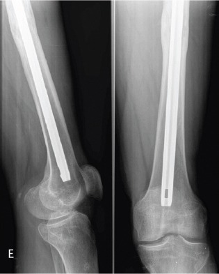
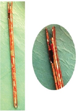
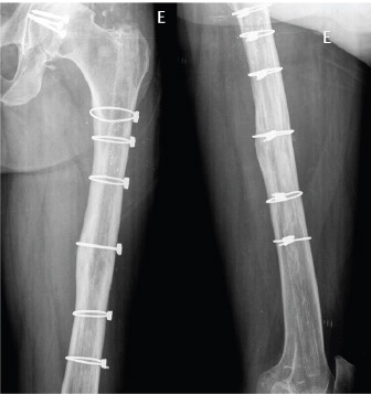
Case report: A 46-year-old man was referred for hip osteoarthritis. He had an acetabulum fracture and a femoral shaft fracture treated 30 years ago with a reamed Kuntscher femoral nail. Lateral hip approach was performed and after attempting to remove the nail with the specific tools being unsuccessful we decided to be more aggressive. Firstly, we performed a simple unicortical osteotomy on the lateral side from the proximal part to below the callus in order to decompress the femoral canal without success. Secondly, a trench in the greater trochanter around the proximal hole was performed to hit the nail from below which was still insufficient and furthermore, the hole broke when hitting the nail so we needed to drill a new hole distally. Finally, the Kuntscher nail was removed. Several cerclages closed the osteotomy and a bone graft was used to close the trench. The patient had a good evolution at one year of follow-up.
Conclusion: With this case report, we present a new salvage technique to remove an incarcerated Kuntscher nail when all the described methods have failed.
Keywords: Femoral nail; Incarcerated nail; Nail removal.
Conflict of interest statement
Conflict of Interest: Nil
Figures


References
-
- Georgiadis GM, Heck BE, Ebraheim NA. Technique for removal of intramedullary nails when there is failure of the proximal extraction device:a report of three cases. J Orthop Trauma. 1997;11(2):130–2. - PubMed
-
- Seligson D, Howard PA, Martin R. Difficulty in removal of certain intramedullary nails. Clin Orthop Relat Res. 1997;340:202–6. - PubMed
-
- Randall RL, Hall RJ, Slabaugh PB. Closed removal of a segmental intramedullary rod:a technical report. J Orthop Trauma. 1996;10(5):363–5. - PubMed
-
- Kuntscher G., M. R Hardware Removal of Intramedullary Nails:A surgical Technique. Georg Thieme Verlag. 1945:63–94.
-
- Seibert FJ, et al. Technique tip for the removal of a “non-removable” tibial nail (Expert Tibial Nail) Injury Extra. 2010;41(4):47–49.
Publication types
LinkOut - more resources
Full Text Sources