. 2017 Feb;10(1):47-65.
doi: 10.1007/s12265-016-9718-9. Epub 2017 Jan 23.

Implantation of a Poly-L-Lactide GCSF-Functionalized Scaffold in a Model of Chronic Myocardial Infarction

Affiliations

Implantation of a Poly-L-Lactide GCSF-Functionalized Scaffold in a Model of Chronic Myocardial Infarction

Cristiano Spadaccio et al. J Cardiovasc Transl Res. 2017 Feb.

Abstract

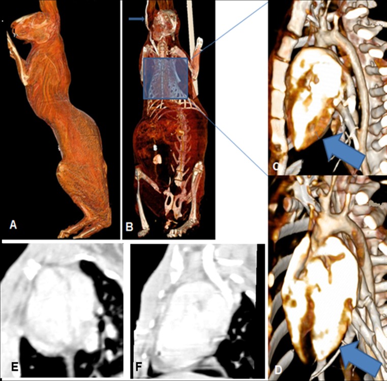
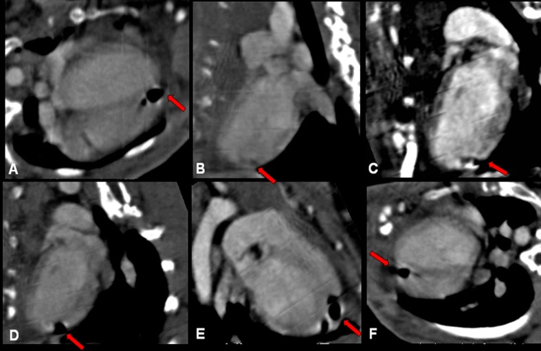
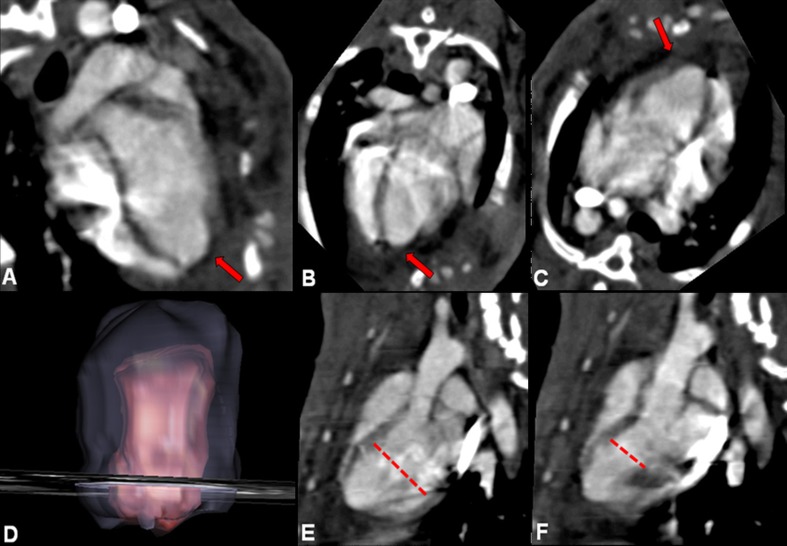
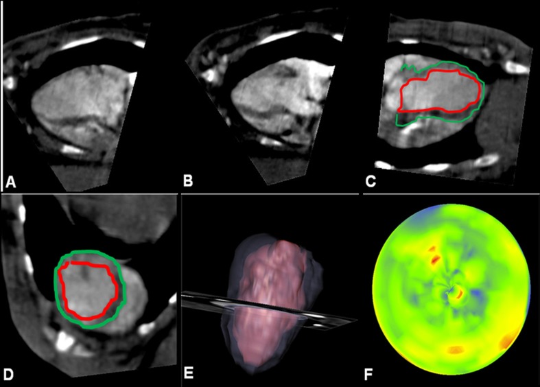
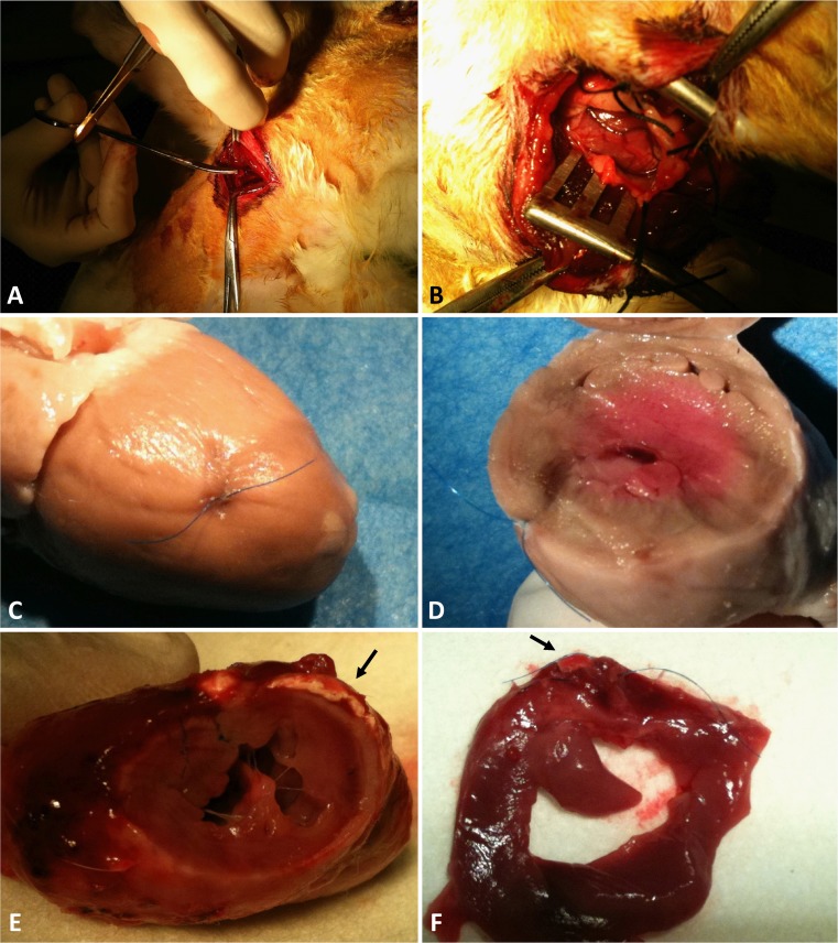
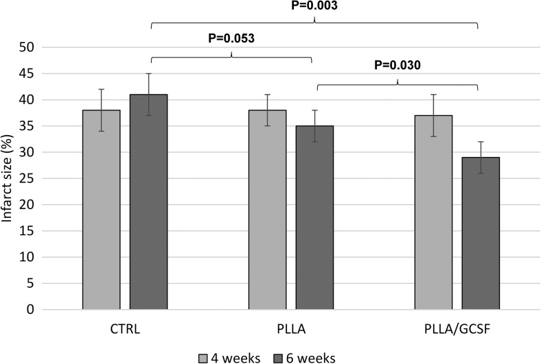
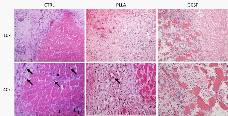
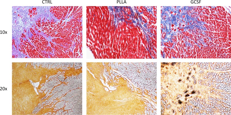
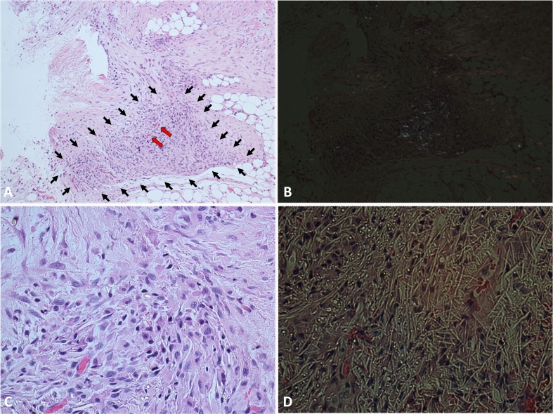
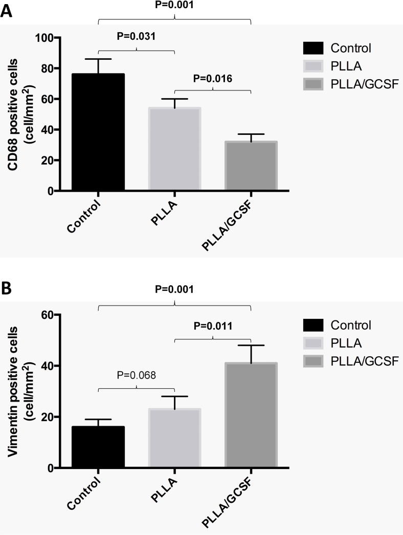
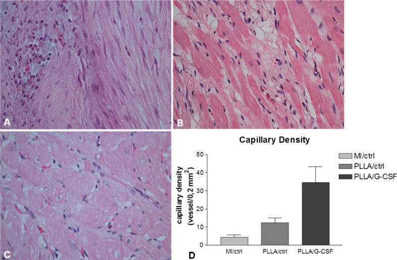
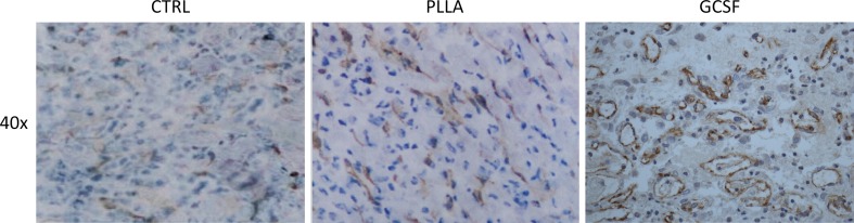
A previously developed poly-L-lactide scaffold releasing granulocyte colony-stimulating factor (PLLA/GCSF) was tested in a rabbit chronic model of myocardial infarction (MI) as a ventricular patch. Control groups were constituted by healthy, chronic MI and nonfunctionalized PLLA scaffold. PLLA-based electrospun scaffold efficiently integrated into a chronic infarcted myocardium. Functionalization of the biopolymer with GCSF led to increased fibroblast-like vimentin-positive cellular colonization and reduced inflammatory cell infiltration within the micrometric fiber mesh in comparison to nonfunctionalized scaffold; PLLA/GCSF polymer induced an angiogenetic process with a statistically significant increase in the number of neovessels compared to the nonfunctionalized scaffold; PLLA/GCSF implanted at the infarcted zone induced a reorganization of the ECM architecture leading to connective tissue deposition and scar remodeling. These findings were coupled with a reduction in end-systolic and end-diastolic volumes, indicating a preventive effect of the scaffold on ventricular dilation, and an improvement in cardiac performance.

Keywords: 3D scaffold; Cardiac graft; Electrospinning; GCSF; Myocardial infarction; PLLA; Tissue engineering.


Conflict of interest statement

Conflict of Interest

The authors declare that they have no conflict of interest.

Figures

Fig. 1
Fig. 1
Echocardiographic evaluation of LVEDD (a), LVESD (b), FS (c), and EF (d) after 4 weeks (light gray) and 6 weeks (dark gray) after induced myocardial infarction; the latter corresponds to 2 weeks after patch implantation. P values are shown in Table 1
Fig. 2
Fig. 2
Volume-rendering CT reconstructions. Positioning of the subject (a, b). Normal myocardial wall thickness (arrow) before coronary ligature (c). After posterolateral artery ligature, ischemic area shows a marked reduction in ventricular thickness (d).Short axis (e) and long axis (f) scans showing impaired perfusion in the apex and in the lateral wall
Fig. 3
Fig. 3
PLLA/CTRL group: long axis (ae) and short axis (f) planar reconstructions from CT scans. Ischemic area is characterized by a small area of tissue loss. Patch is radiolucent and is indicated by red arrows
Fig. 4
Fig. 4
PLLA/GCSF group: planar (ac), volume rendering (d), end-diastolic long axis (e), and end-systolic long axis (f) reconstructions from CT scans. Ischemic area is characterized by a small area of tissue loss. Patch is radiolucent and is indicated by red arrows. Ventricular diameter measurements were performed using these sequences (dashed lines)
Fig. 5
Fig. 5
Exemplificative ECG-gated CT morphovolumetric analysis of a MI/CTRL subject. Diastolic (a) and systolic (b) acquisition. Definition of inner and outer margins in long axis (c) and in short axis (d) view, followed by reconstruction (e). Graphical representation of regional wall motion with colorimetric scale, where dark areas indicate reduced kinesis (f)
Fig. 6
Fig. 6
Heart exposure through left thoracotomy (a), with vessel identification after pericardial suspension (b). Macroscopic evaluation of the infarcted heart in MI/CTRL group (c, d). PLLA/GCSF patch (arrow) is integrated in myocardial tissue, visible as a white sheet (e, f)
Fig. 7
Fig. 7
Myocardial infarction size among groups after 4 weeks (light gray) and 6 weeks (dark gray) after induced myocardial infarction. Myocardial infarction size is expressed as the percentage of the left ventricular myocardial volume
Fig. 8
Fig. 8
H/E staining of the infarcted area, with ×10 (upper panel) and ×40 magnification (lower panel). Myocardial tissue showed typical features of chronic infarction with nuclear picnosis (black arrows) and intercellular patchy calcifications (arrowheads)
Fig. 9
Fig. 9
Qualitative assessment of ECM with Masson’s (upper panels) and Silver staining (lower panels) of the infarcted area. Denser, thicker, and scarcely organized collagen bundles are found in the MI/CTRL and PLLA/CTRL group at the level of the scar, while the PLLA/GCSF was characterized by a looser reticular organization of the collagen which appeared interspread among several neovessels
Fig. 10
Fig. 10
PLLA/GCSF patch in its site of implantation (grossly marked with black arrows, because of the partial integration with myocardial tissue), with evidence of angiogenesis (red arrows) (a H/E staining, magnification ×10). Phase-contrast image with specific polarized light, showing the bifrangent polymer (b). Magnifications of a (×40) (c) and b (×40) (d)
Fig. 11
Fig. 11
Count of CD68-positive (a) and vimentin-positive cells (b) among the three groups
Fig. 12
Fig. 12
Capillary density measurement (magnification ×40) in MI/CTRL (a), PLLA/CTRL (b), and PLLA/GCSF (c) groups, with quantitative evaluation (d). Capillary density measurement showed a significant increase of neovessels in the PLLA/GCSF group compared to PLLA/CTRL (p value <0.01). Similarly, in the PLLA/CTRL group versus MI/CTRL group (p value <0.01)
Fig. 13
Fig. 13
Immunohistochemistry for CD31-positive cells, indicating a higher degree of neoangiogenesis in PLLA/GCSF group

References

    1. Frangogiannis NG. Chemokines in the ischemic myocardium: from inflammation to fibrosis. Inflammation Research. 2004;53(11):585–595. - PubMed
    1. Chamberlain DA. Overview of completed sudden death trials: European experience. Cardiology. 1987;74(Suppl 2):10–23. - PubMed
    1. Demirovic J, Myerburg RJ. Epidemiology of sudden coronary death: an overview. Progress in Cardiovascular Diseases. 1994;37(1):39–48. - PubMed
    1. Myerburg RJ, Kessler KM, Castellanos A. Sudden cardiac death. Structure, function, and time-dependence of risk. Circulation. 1992;85(1 Suppl):I2–10. - PubMed
    1. Kang, H. J., Kim, H. S., Koo, B. K., et al. (2007). Intracoronary infusion of the mobilized peripheral blood stem cell by G-CSF is better than mobilization alone by G-CSF for improvement of cardiac function and remodeling: 2-year follow-up results of the Myocardial Regeneration and Angiogenesis in Myocardial Infarction with G-CSF and Intra-Coronary Stem Cell Infusion (MAGIC Cell) 1 trial. Am Heart J, 153(2), 237 e231–238, doi:10.1016/j.ahj.2006.11.004. - PubMed

MeSH terms

LinkOut - more resources