. 2017 Mar;22(2):253-261.
doi: 10.1007/s12192-017-0763-3. Epub 2017 Jan 23.

HDAC6 regulates sensitivity to cell death in response to stress and post-stress recovery

Affiliations

HDAC6 regulates sensitivity to cell death in response to stress and post-stress recovery

Hyun-Wook Ryu et al. Cell Stress Chaperones. 2017 Mar.

Abstract

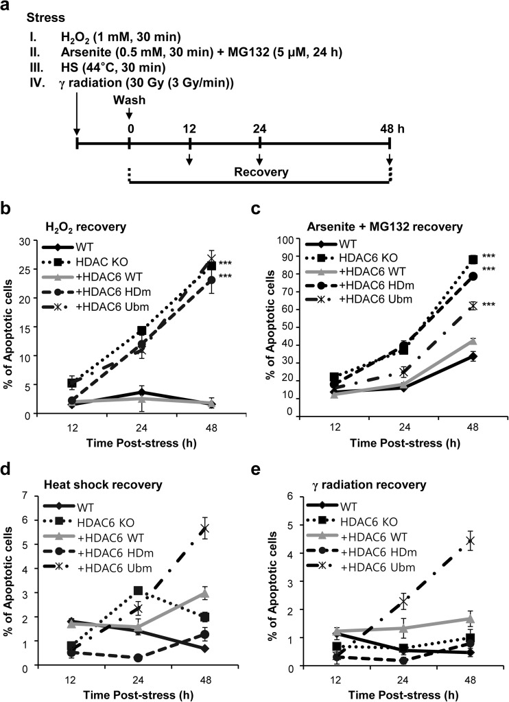
Histone deacetylase 6 (HDAC6) plays an important role in stress responses such as misfolded protein-induced aggresomes, autophagy, and stress granules. However, precisely how HDAC6 manages response during and after cellular stress remains largely unknown. This study aimed to investigate the effect of HDAC6 on various stress and post-stress recovery responses. We showed that HIF-1α protein levels were reduced in HDAC6 knockout (KO) MEFs compared to wild-type (WT) MEFs in hypoxia. Furthermore, under hypoxia, HIF-1α levels were also reduced following rescue with either a catalytically inactive or a ubiqiutin-binding mutant HDAC6. HDAC6 deacetylated and upregulated the stability of HIF-1α, leading to activation of HIF-1α function under hypoxia. Notably, both the deacetylase and ubiquitin-binding activities of HDAC6 contributed to HIF-1α stabilization, but only deacetylase activity was required for HIF-1α transcriptional activity. Suppression of HDAC6 enhanced the interaction between HIF-1α and HSP70 under hypoxic conditions. In addition to hypoxia, depletion of HDAC6 caused hypersensitivity to cell death during oxidative stress and post-stress recovery. However, HDAC6 depletion had no effect on cell death in response to heat shock or ionizing radiation. Overall, our data suggest that HDAC6 may serve as a critical stress regulator in response to different cellular stresses.

Keywords: Apoptosis; HIF-1α; Histone deacetylase 6; Hypoxia; Oxidative stress; Post-stress recovery.


Conflict of interest statement

The authors declare that they have no conflict of interest.

Figures

Fig. 1
Fig. 1
Deacetylation of HIF-1α by HDAC6 regulates its degradation and transcriptional activity under hypoxia. MEFs were incubated for 24 h in 21% O2 (a), in 1% O2 (b), or for 6 h with 500 μM CoCl2 to mimic hypoxia (c). Whole cell lysates were immunoprecipitated with an anti-HIF-1α antibody. Immunocomplexes were either probed for anti-HIF-1α or anti-acetylated lysine antibody. Whole cell lysates of 10% were used as input control. d MEFs were incubated for 6, 12, or 24 h with 500 μM CoCl2 to mimic hypoxia or in 21% O2. Immunoblotting analysis was performed with indicated antibodies. α-Tubulin is used as a loading control. e MEFs were co-transfected with an HRE-firefly luciferase vector and a Renilla vector as a control. Transfected cells were incubated for 24 h in 21% O2 then incubated for an additional 6 h with 500 μM CoCl2. Dual luciferase activity was measured, and the ratio of firefly to Renilla was used as the relative luciferase activity. f MEFs were incubated for 6 h with 500 μM CoCl2 to mimic hypoxia. mRNA expression of HIF-1α target genes was analyzed by qRT-PCR. mRNA values were normalized to that of actin. *P < 0.05 based on two-tailed unpaired Student’s t test
Fig. 2
Fig. 2
Deacetylation of HIF-1α by HDAC6 reduces the interaction between HIF-1α and HSP70. MEFs were incubated for 24 h in 1% O2. a Immunoblotting analysis was performed with indicated antibodies. Whole cell lysates of 10% were used as input control. b Whole cell lysates were immunoprecipitated with an anti-HIF-1α antibody. The precipitate was immunoblotted with anti-HIF-1α, anti-HSP70, or anti-HSP90 antibodies. α-Tubulin is used as a loading control
Fig. 3
Fig. 3
Loss of HDAC6 causes hypersensitivity to apoptosis in response to hypoxia. MEFs were exposed to normoxia, hypoxia (1% O2, 24 h), or reoxygenation (1% O2, 24 h, and 21% O2, 48 h). The number of apoptotic cells was counted and graphed following an Annexin-V/PI assay. *P < 0.05, ***P < 0.001 based on two-tailed unpaired Student’s t test
Fig. 4
Fig. 4
Depletion of HDAC6 results in reduced cell viability after stress release. a Schematic representation of the experimental procedure. MEFs of different genotypes were treated with 1 mM H2O2 for 30 min (b) or 5 μM MG 132 for 24 h then 0.5 mM arsenite for 30 min (c), heat shock at 44 °C for 30 min (d), or 30 Gy γ radiation (3 Gy/min) (e) and recovered at the indicated times after stress. The number of apoptotic cells was counted and graphed following an Annexin-V/PI assay. ***P < 0.001 based on two-tailed unpaired Student’s t test

References

    1. Anderson P, Kedersha N. RNA granules. J Cell Biol. 2006;172:803–808. doi: 10.1083/jcb.200512082. - DOI - PMC - PubMed
    1. Arnesen T, et al. Interaction between HIF-1 alpha (ODD) and hARD1 does not induce acetylation and destabilization of HIF-1 alpha. FEBS Lett. 2005;579:6428–6432. doi: 10.1016/j.febslet.2005.10.036. - DOI - PMC - PubMed
    1. Bardos JI, Ashcroft M. Negative and positive regulation of HIF-1: a complex network. Biochim Biophys Acta. 2005;1755:107–120. - PubMed
    1. Beere HM. Death versus survival: functional interaction between the apoptotic and stress-inducible heat shock protein pathways. J Clin Invest. 2005;115:2633–2639. doi: 10.1172/JCI26471. - DOI - PMC - PubMed
    1. Bilton R, et al. Arrest-defective-1 protein, an acetyltransferase, does not alter stability of hypoxia-inducible factor (HIF)-1alpha and is not induced by hypoxia or HIF. J Biol Chem. 2005;280:31132–31140. doi: 10.1074/jbc.M504482200. - DOI - PubMed

Publication types

MeSH terms