. 2017 Jun;29(6):10.1111/nmo.13031.
doi: 10.1111/nmo.13031. Epub 2017 Jan 23.

Brain networks associated with cognitive and hedonic responses to a meal

Affiliations

Brain networks associated with cognitive and hedonic responses to a meal

T Pribic et al. Neurogastroenterol Motil. 2017 Jun.

Abstract

Background: We recently reported interrelated digestive, cognitive, and hedonic responses to a meal. The aim of this study was to identify brain networks related to the hedonic response to eating.

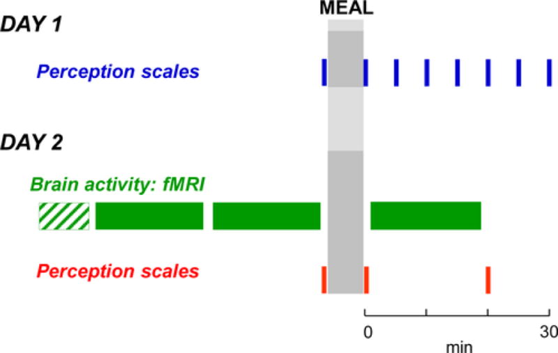
Methods: Thirty-eight healthy subjects (20-38 age range) were evaluated after a 5-hour fast and after ingestion of a test meal (juice and warm ham and cheese sandwich, 300 mL, 425 kcal). Perceptual and affective responses (satiety, abdominal fullness, digestive well-being, and positive mood), and resting scans of the brain using functional MRI (3T Trio, Siemens, Germany) were evaluated immediately before and after the test meal. A high-order group independent component analysis was performed to investigate ingestion-related changes in the intrinsic connectivity of brain networks, with a focus on thalamic and insular networks.

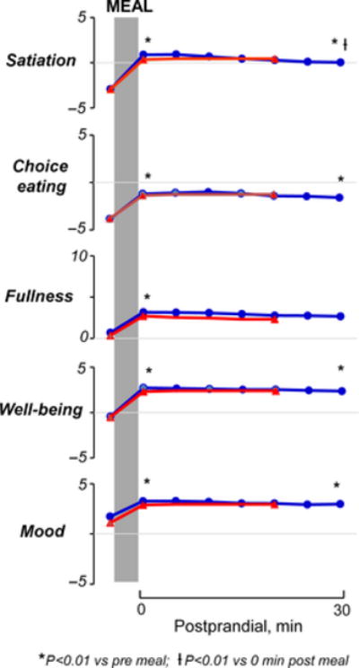
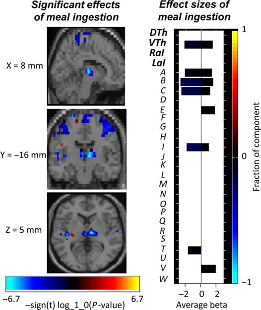
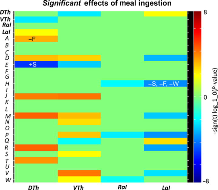
Key results: Ingestion induced satiation (3.3±0.4 score increase; P<.001) and abdominal fullness (2.4±0.3 score increase; P<.001). These sensations included an affective dimension involving digestive well-being (2.8±0.3 score increase; P<.001) and positive mood (1.8±0.2 score increase; P<.001). In general, thalamo-cortical connectivity increased with meal ingestion while insular-cortical connectivity mainly decreased. Furthermore, larger meal-induced changes (increase/decrease) in specific thalamic connections were associated with smaller changes in satiety/fullness. In contrast, a larger meal-induced decrease in insular-anterior cingulate cortex connectivity was associated with increased satiety, fullness, and digestive well-being.

Conclusions and inferences: Perceptual and emotional responses to food intake are related to brain connectivity in defined functional networks. Brain imaging may provide objective biomarkers of subjective effects of meal ingestion.

Keywords: brain imaging; hedonic response; meal ingestion; postprandial sensations.


Conflict of interest statement

Conflict of Interest

No competing interests declared by all investigators.

Figures

Figure 1
Figure 1
Experimental design. Perception in response to a probe meal was studied without (Day 1) and with brain imaging (Day 2: structural scan; functional scan)
Figure 2
Figure 2
Cognitive and hedonic responses to probe meal in separate experiments with and without brain imaging. No statistically significant differences between study days were detected
Figure 3
Figure 3
Composite image of the significant effects of meal ingestion on intra-network connectivity. The effect size for each component is shown to the right; primary networks of interest: dorsal thalamus (DTh), ventral thalamus (VTh), right anterior insula (RaI), left anterior insula (LaI); secondary networks of interest: bilateral secondary somatosensory cortex (A), right sensorimotor cortex (B), left sensorimotor cortex (C), bilateral subgenual anterior cingulate cortex (D), bilateral caudate (E), bilateral pallidum/putamen (F), bilateral hippocampus/parahippocampal gyrus/amygdala (G), bilateral anterior cingulate cortex/medial orbitofrontal cortex (H), bilateral paracentral lobule (I), bilateral primary somatosensory cortex (J), bilateral superior parietal (K), bilateral supramarginal (L), left temporoparietal junction (M), right superior/inferior parietal (N), bilateral ventrolateral prefrontal cortex (O), right left temporoparietal junction (P), bilateral medial cingulate cortex (Q), right inferior frontal operculum (R), bilateral precuneus (S), bilateral superior temporal (T), bilateral dorsal anterior cingulate cortex/medial cingulate cortex (U), bilateral medial prefrontal cortex (V), left dorsolateral prefrontal cortex (W)
Figure 4
Figure 4
Effects of meal ingestion on inter-ICN connectivity. Heatmap depicting significant effects between the primary networks of interest (thalamus and anterior insula) and the secondary networks of interest. The thalamus and anterior insula ICNs displayed significant changes in connectivity with sensorimotor, emotion/cognitive, and interoceptive ICNs. Significant correlations, positive (+) and negative (−) correlation, between meal-induced connectivity changes and meal-induced perceptual changes are also indicated: Δsatiety (S), Δfullness (F), Δwell-being (W). Considering the direction of meal-induced changes, a greater decrease in insula-ACC/mOFC connectivity is associated with a greater increase in satiety, fullness, and digestive well-being. In contrast, a greater meal-induced decrease in thalamus-caudate connectivity is associated with a smaller increase in satiety, and a greater meal-induced increase in thalamus-S2 connectivity is associated with a smaller increase in fullness. Primary networks of interest: dorsal thalamus (DTh), ventral thalamus (VTh), right anterior insula (RaI), left anterior insula (LaI); secondary networks of interest: bilateral secondary somatosensory cortex (A), right sensorimotor cortex (B), left sensorimotor cortex (C), bilateral subgenual anterior cingulate cortex (D), bilateral caudate (E), bilateral pallidum/putamen (F), bilateral hippocampus/parahippocampal gyrus/amygdala (G), bilateral anterior cingulate cortex/medial orbitofrontal cortex (H), bilateral paracentral lobule (I), bilateral primary somatosensory cortex (J), bilateral superior parietal (K), bilateral supramarginal (L), left temporoparietal junction (M), right superior/inferior parietal (N), bilateral ventrolateral prefrontal cortex (O), right left temporoparietal junction (P), bilateral medial cingulate cortex (Q), right inferior frontal operculum (R), bilateral precuneus (S), bilateral superior temporal (T), bilateral dorsal anterior cingulate cortex/medial cingulate cortex (U), bilateral medial prefrontal cortex (V), left dorsolateral prefrontal cortex (W)

Similar articles

Cited by

References

    1. Azpiroz F, Bouin M, Camilleri M, et al. Mechanisms of hypersensitivity in IBS and functional disorders. Neurogastroenterol Mot. 2007;19:62–88. - PubMed
    1. Kellow JE, Azpiroz F, Delvaux M, et al. Applied principles of neurogastroenterology: physiology/motility sensation. Gastroenterology. 2006;130:1412–1420. - PubMed
    1. Azpiroz F, Feinle C, Grundy D, Tack J. Gastric sensitivirty and reflexes: basic mechanism underlying clinical problems. J Gastroenterol. 2014;49:206–218. - PubMed
    1. Tack J, Caenepeel P, Piessevaux H, Cuomo R, Janssens J. Assessment of meal induced gastric accommodation by a satiety drinking test in health and in severe functional dyspepsia. Gut. 2003;52:1271–1277. - PMC - PubMed
    1. Feinle C, Azpiroz F. Dietary and life-style factors in funcional dyspepsia. Nat Rev Gastroenterol Hepatol. 2013;10:150–157. - PubMed