. 2017 Jan 24:7:41143.
doi: 10.1038/srep41143.

IL-22-producing CD4+T cells in the treatment response of rheumatoid arthritis to combination therapy with methotrexate and leflunomide

Affiliations

IL-22-producing CD4+T cells in the treatment response of rheumatoid arthritis to combination therapy with methotrexate and leflunomide

Wei Zhong et al. Sci Rep. .

Abstract

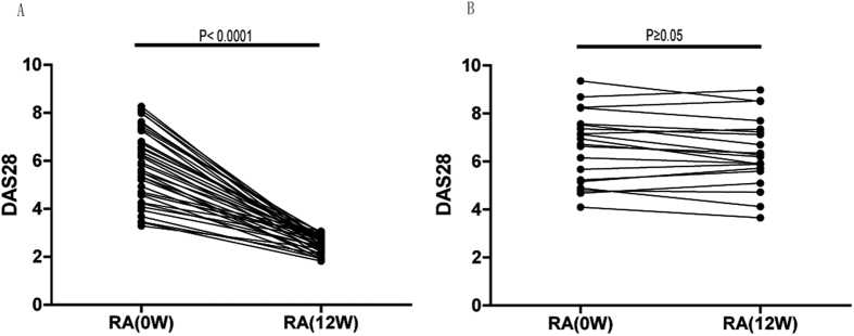
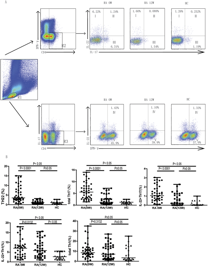
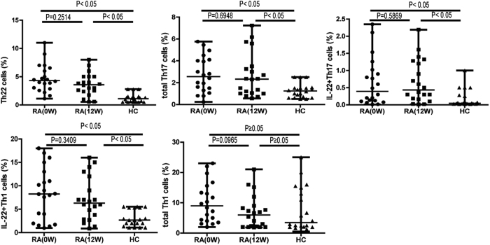
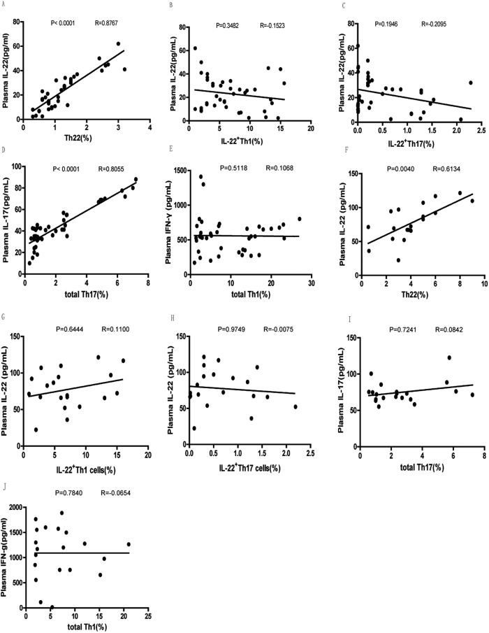
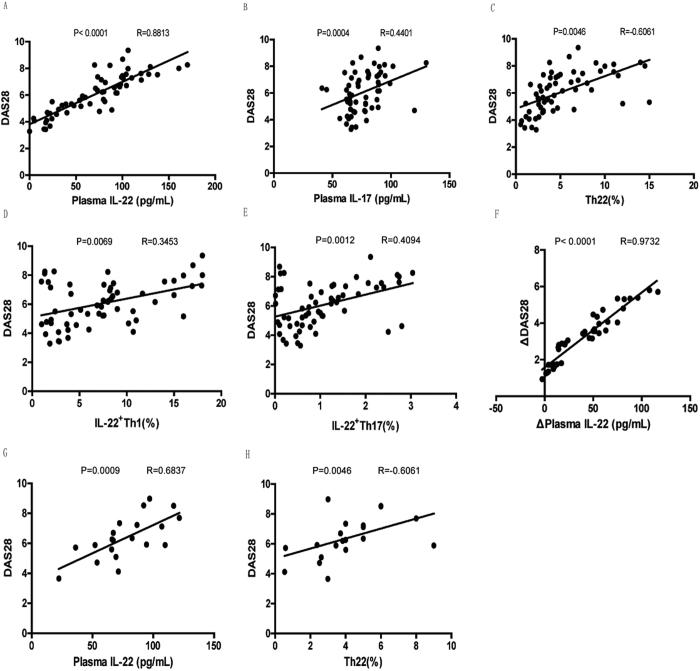
T cells are key players in immune-mediated rheumatoid arthritis (RA). We previously reported that interleukin (IL)-22+CD4+T helper (IL-22+ Th) cells and IL-22 critically control the pathogenesis of RA. Here we monitored circulating levels of different IL-22+ Th cell subsets and measured plasma levels of IL-22, IL-17, and interferon (IFN)-γ in 60 patients with active RA following 12-week combination methotrexate (MTX) and leflunomide (LEF) therapy (MTX+LEF) and 20 healthy individuals. We found the frequencies of circulating IFN-γ-IL-17-IL-22+ (Th22), IFN-γ-IL-17+ (total Th17), IFN-γ+IL-17-IL-22+ (IL-22+Th1) cells, and IFN-γ-IL-17+IL-22+ (IL-22+Th17) cells, as well as the plasma levels of IL-22, IL-17 and IFN-γ to be significantly reduced in RA patients that responded to treatment, but not in non-responders. Reductions in plasma IL-22 level significantly correlated with percentage of circulating Th22 cells and the decrease of plasma IL-22 level correlated with the reduction of DAS28 in responders. Our data suggests that circulating Th22 cells and plasma IL-22 level play a detrimental role in RA. The combination MTX+LEF therapy, by targeting Th22 cells and reducing IL-22 level, relieves the immune defects and ameliorates symptoms of RA. This study provides novel mechanistic understanding of the pathogenesis of RA, which may promote a design of better therapies for RA.


Conflict of interest statement

The authors declare that they have no competing financial interests.

Figures

Figure 1
Figure 1. DAS28 scores were significantly improved in RA responders (n = 40) but not in RA non-responders (n = 20) after the combination methotrexate (MTX)+ leflunomide (LEF) therapy.
DAS28 values were compared between pre- and post-treatment in each RA responder (A) or non-responder (B).
Figure 2
Figure 2. The percentages of circulating Th22, total Th17, and IL-22+Th17, IL22+Th1, but not total Th1 cells decreased following MTX+LEF therapy only in patients that exhibited clinical improvement (n = 40).
Peripheral blood mononuclear cells (PBMCs) were collected from RA patients responsive to treatment at baseline (before treatment, 0 week) and after treatment (12 weeks) with MTX+LEF, and analyzed by flow cytometry for the percentage of different Th subsets, including IFN-γIL-17IL-22+ (Th22), IFN-γIL-17+ (total Th17), IFN-γIL-17+IL-22+ (IL-22+Th17), IFN-γ+IL-17 (total Th1), and IFN-γ+IL-17IL-22+ (IL-22+Th1) cells. (A) The gating strategy for detecting different subsets of Th cells and representative flow images on samples from RA patients at 0 week (0 W), 12 week (12 W), or from healthy controls (HC; n = 20). R1, lymphoctyes; R2, IFN-γCD4+T cells; R3, IL-17CD4+T cells; I, Th22 cells; II, IL-22+Th17 cells; III, IL-22Th17 cells; IV; IL-22+Th1 cells; V; IL-22Th1 cells.
Figure 3
Figure 3. The percentages of circulating Th22, total Th17, IL-22+Th17, IL22+Th1, or total Th1 cells did not decrease following the combination MTX+ leflunomide LEF therapy in non-responding RA patients (n = 20).
PBMCs were collected from RA patients resistant to treatment at baseline (before treatment, 0 week) and after treatment (12 weeks) with MTX+LEF, and analyzed by flow cytometry for the percentage of different Th subsets, including IFN-γIL-17IL-22+ (Th22), IFN-γIL-17+ (total Th17), IFN-γIL-17+IL-22+ (IL-22+Th17), IFN-γ+IL-17 (total Th1), and IFN-γ+IL-17-IL-22+ (IL-22+Th1) cells.
Figure 4
Figure 4. Plasma levels of IL- 22, IL-17 and IFN-γ decreased following MTX+LEF treatment in RA responders.
The plasma levels of IL-22 (A), IL-17 (B), and IFN-γ (C) were measured by ELISA in RA responders and non-responders before treatment (0 W), after treatment (12 W), and in HC. R, response group; NR, non-response group. *P < 0.05 versus before treatment in response group. #P < 0.05 versus healthy control. P < 0.05 versus non-response group at the same time point.
Figure 5
Figure 5. Plasma levels of IL-22 and IL-17 positively correlate with the percentage of circulating Th22 and total Th17 cells, respectively in RA responders while the plasma levels of IL-22 positively correlated with the percentage of circulating Th22 cells in RA non-responders.
The correlation between the plasma IL-22 level and the percentage of circulating Th22 cells (A), the percentage of circulating IL-22+Th1 cells (B), or the percentage of circulating IL-22+Th17 cells (C) the correlation between the plasma level of IL-17 and the percentage of total circulatingTh17 cells (D), the correlation between the plasma level of IFN-γ and the percentage of total circulating Th1 cells (E) in RA responders; the correlation between the plasma IL-22 level and the percentage of circulating Th22 cells (F), the percentage of circulating IL-22+Th1 cells (G), or the percentage of circulating IL-22+Th17 cells (H) the correlation between the plasma level of IL-17 and the percentage of total circulatingTh17 cells (I); the correlation between the plasma level of IFN-γ and the percentage of total circulating Th1 cells (J) in RA non-responders after treatment were analyzed using Spearman’s rank correlation test.
Figure 6
Figure 6. DAS28 positively correlated with plasma IL-22, IL-17 levels, and IL-22-producing Th cells in untreated RA, only with plasma IL-22 level in RA responders, and with plasma IL-22 level as well as the percentage of circulating Th22 cells in RA non-responders after treatment.
The correlation between DAS28 and the plasma IL-22 level (A), the plasma level of IL-17 level (B), the percentage of circulating Th22 cells (C), the percentage of circulating IL-22+Th1 cells (D), or the percentage of circulating IL-22+Th17 cells (E) in untreated RA; the correlation between the decrease of DAS28 and the decrease of the plasma level of IL-22 (F) in RA responders after treatment; the correlation between the DAS28 and the plasma level of IL-22 (G), the percentage of circulating Th22 cells (H) in RA non-responders after treatment were analyzed using Spearman’s rank correlation test.

References

    1. Schett G. & Gravallese E. Bone erosion in rheumatoid arthritis: mechanisms, diagnosis and treatment. Nature reviews. Rheumatology 8, 656–664, doi: 10.1038/nrrheum.2012.153 (2012). - DOI - PMC - PubMed
    1. Zenewicz L. A. & Flavell R. A. Recent advances in IL-22 biology. International immunology 23, 159–163, doi: 10.1093/intimm/dxr001 (2011). - DOI - PubMed
    1. Rutz S., Eidenschenk C. & Ouyang W. IL-22, not simply a Th17 cytokine. Immunological reviews 252, 116–132, doi: 10.1111/imr.12027 (2013). - DOI - PubMed
    1. Duhen T., Geiger R., Jarrossay D., Lanzavecchia A. & Sallusto F. Production of interleukin 22 but not interleukin 17 by a subset of human skin-homing memory T cells. Nature immunology 10, 857–863, doi: 10.1038/ni.1767 (2009). - DOI - PubMed
    1. Patel D. D. & Kuchroo V. K. Th17 Cell Pathway in Human Immunity: Lessons from Genetics and Therapeutic Interventions. Immunity 43, 1040–1051, doi: 10.1016/j.immuni.2015.12.003 (2015). - DOI - PubMed

Publication types