. 2017 Jan 24:7:41254.
doi: 10.1038/srep41254.

Costunolide and dehydrocostuslactone combination treatment inhibit breast cancer by inducing cell cycle arrest and apoptosis through c-Myc/p53 and AKT/14-3-3 pathway

Affiliations

Costunolide and dehydrocostuslactone combination treatment inhibit breast cancer by inducing cell cycle arrest and apoptosis through c-Myc/p53 and AKT/14-3-3 pathway

Zhangxiao Peng et al. Sci Rep. .

Abstract

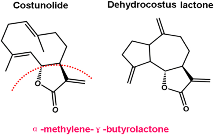
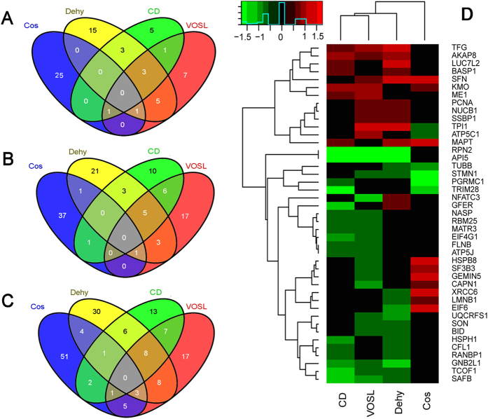
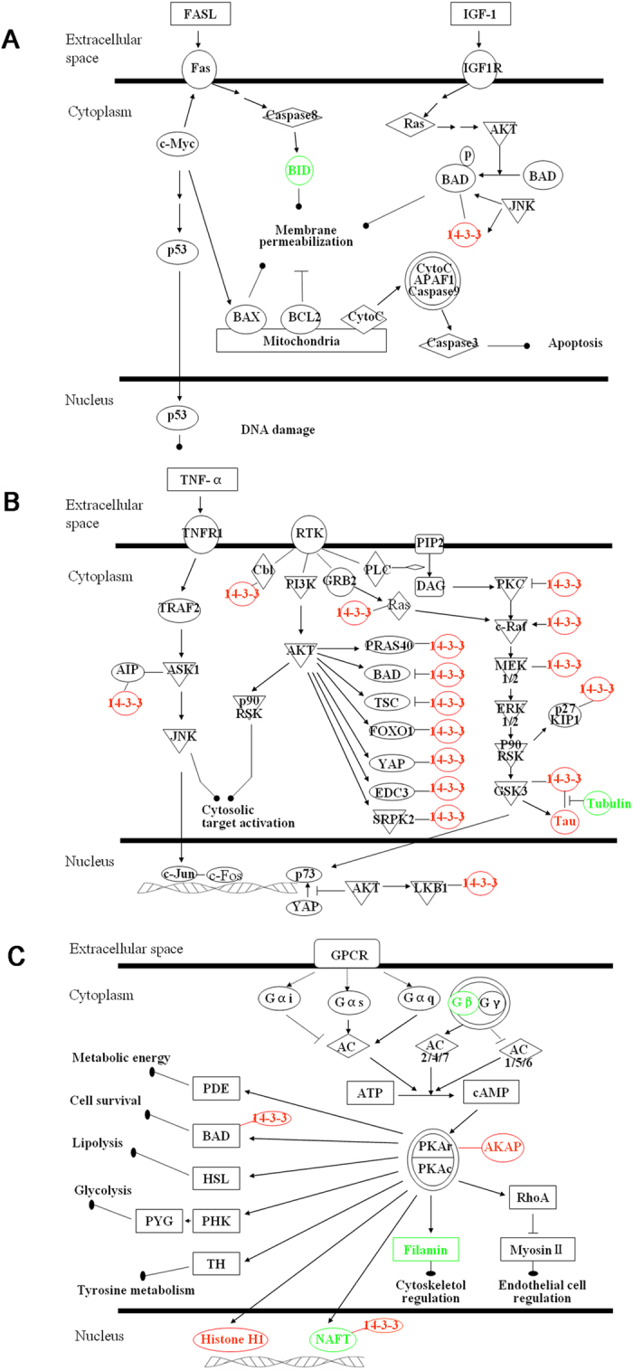
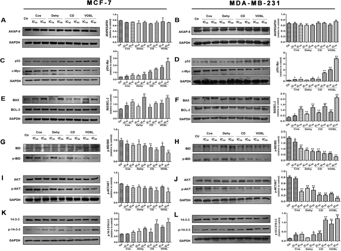
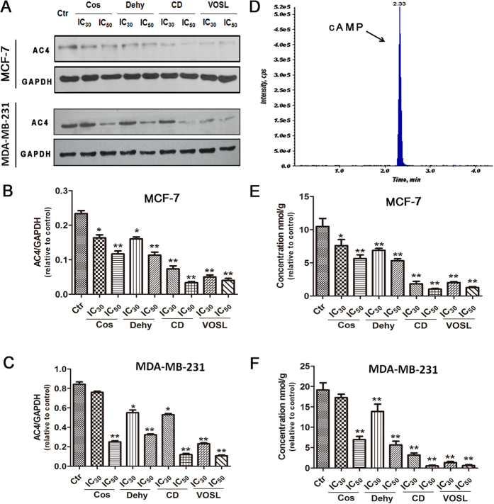
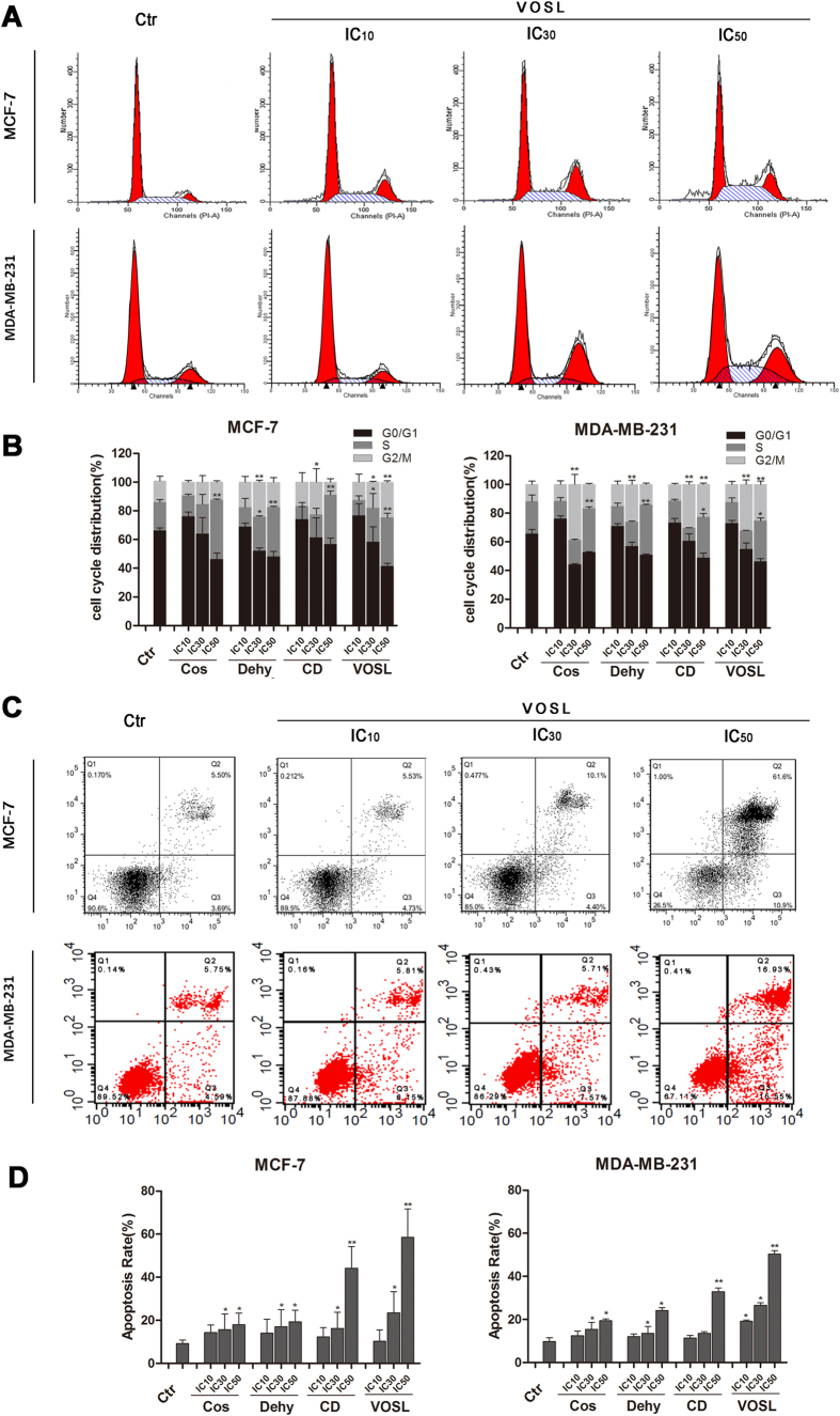
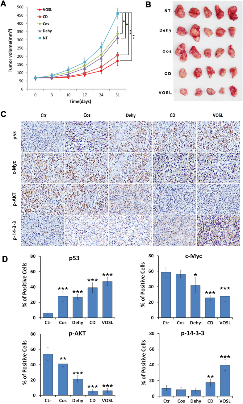
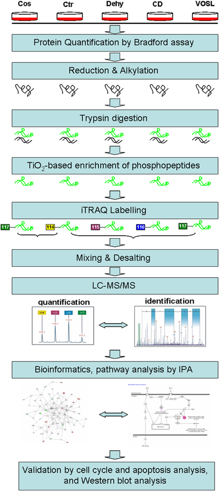
Our previous studies demonstrated that volatile oil from saussurea lappa root (VOSL), rich in two natural sesquiterpene lactones, costunolide (Cos) and dehydrocostuslactone (Dehy), exerts better anti-breast cancer efficacy and lower side effects than Cos or Dehy alone in vivo, however, their anti-cancer molecular mechanisms were still unknown. In this study, we investigated the underlying mechanisms of Cos and Dehy combination treatment (CD) on breast cancer cells through proteomics technology coupled with Western blot validation. Ingenuity Pathways Analysis (IPA) results based on the differentially expressed proteins revealed that both VOSL and CD affect the 14-3-3-mediated signaling, c-Myc mediated apoptosis signaling and protein kinase A (PKA) signaling. Western blot coupled with cell cycle and apoptosis analysis validated the results of proteomics analysis. Cell cycle arrest and apoptosis were induced in a dose-dependent manner, and the expressions of p53 and p-14-3-3 were significantly up-regulated, whereas the expressions of c-Myc, p-AKT, p-BID were significantly down-regulated, furthermore, the ratio of BAX/BCL-2 were significantly increased in breast cancer cells after CD and VOSL treatment. The findings indicated that VOSL and CD could induce breast cancer cell cycle arrest and apoptosis through c-Myc/p53 and AKT/14-3-3 signaling pathways and may be novel effective candidates for breast cancer treatment.


Figures

Figure 1
Figure 1. Chemical structures of Cos (C15H20O2) and Dehy (C15H18O2).
The α,β-unsaturated carbonyl group in the α-methylene-γ-butyrolactone moiety of Cos and Dehy is very important for exerting their various biological activities, such as anti-inflammatory, anti-cancer, anti-virus, anti-oxidant, anti-diabetes, anti-ulcer, and anthelmintic activities.
Figure 2
Figure 2. Venn diagram and heatmap of differentially expressed proteins.
The results from proteomics revealed that (A) there are 14 common up-regulated proteins, (B) 20 common down-regulated proteins, and (C) 43 common differentially expressed proteins at the VOSL, CD, Dehy, and Cos-treated groups. Moreover, (D) the heatmap and cluster analysis revealed that the VOLS-treated group shared the most differentially expressed proteins with the CD-treated group, which further demonstrated that CD is the most important anti-breast cancer ingredients in VOSL and share the same pharmacological mechanisms with VOSL.
Figure 3
Figure 3. The signaling pathways affected by Cos, Dehy, CD or VOSL.
(A) c-Myc mediated apoptosis signaling, (B) 14-3-3-mediated signaling, and (C) protein kinase A (PKA) signaling. Red and green colors represent up- and down- regulated, respectively.
Figure 4
Figure 4. Cos, Dehy, CD and VOSL regulated c-Myc mediated apoptosis signaling and 14-3-3-mediated signaling pathways in breast cancer cells.
MCF-7 cells (A,C,E,G,I,K) or MDA-MB-231 cells (B,D,F,H,J,L) were cultured in cell culture dishes, treated with 0 (Ctr), IC30 and IC50 of Cos, Dehy, CD, or VOSL, respectively for 48 h, then the expression of the indicated factors was examined by Western blot. Glyceraldehydes 3-phosphate dehydrogenase (GAPDH) was used as the loading control. The densitometry analysis of every factor was performed, and normalized with the corresponding GAPDH content. Values were presented as mean ± standard error (SE) of three independent experiments; *p < 0.05 and **p < 0.01 compared with the control group.
Figure 5
Figure 5. Expression levels of AC4 and cAMP in different treatment groups.
MCF-7 cells or MDA-MB-231 cells were cultured in cell culture dishes, treated with 0 (Ctr), IC30 and IC50 of Cos, Dehy, CD, or VOSL, respectively, for 48 h, then the expression of AC4 was examined by Western blot (A). GAPDH was used as the loading control. The densitometry analysis of AC4 was performed, and normalized with GAPDH content (B,C). Quantification of intracellular cAMP was performed on a Waters Acquity UPLC system using a Waters Acquity BEH C18 column (2.1 × 50 mm2, 1.7 μm) coupled to an AB Sciex Triple QuadTM 6500 mass spectrometer (D), and the concentrations of cAMP in different treatment groups were shown in (E,F). Values were presented as mean ± standard error (SE) of three independent experiments; *p < 0.05 and **p < 0.01 compared with the control group.
Figure 6
Figure 6. Effects on cell cycle and apoptosis of breast cancer cells after Cos, Dehy, CD, or VOSL treatment.
MCF-7 cells or MDA-MB-231 cells were planted into 6-well plates at 3 × 105 cells/well, incubated with the respective IC10, IC30, IC50 concentrations of Cos, Dehy, CD, or VOSL for 48 h, cells were harvested by trypsinisation, and then fixed by ice-cold ethanol (70%). After washing with PBS, the cell pellets were resuspended in propidium iodid (PI) staining buffer (50 μL/mL PI, RNase A). After 15 min of incubation at 37 °C, cell cycle distribution was analyzed by a FACScalibur System using ModFit software (A,B). MCF-7 or MDA-MB-231 cells were planted into 6-well plates at 2 × 105 cells per well, treated with the respective IC10, IC30, IC50 concentrations of Cos, Dehy, CD, or VOSL for 48 h, stained with Annexin V-FITC/PI, and then detected by a FACScalibur system (C,D). The cell cycle distribution and apoptotic percentages from three independent experiments were analyzed and compared, *p < 0.05 and **p < 0.01 compared with the control group.
Figure 7
Figure 7. VOSL and its main active ingredients suppress the growth of breast cancer MDA-MB-231 xenografts.
(A,B) The xenograft mouse models were randomly divided into five groups. The Cos, Dehy, CD and VOSL-treated groups were injected intraperitoneally at a dose of 20 mg/kg/day, respectively. The negative control (NT) was treated with an equal volume of vehicle. Tumor size was monitored at 0, 3, 10, 17, 24 and 31 days post-treatment and compared at 31 days post-treatment; *p < 0.05 and **p < 0.01 compared with the negative control (NT) group. (C,D) Tumor-bearing mice were sacrificed after 30 times of administrations and tumors were harvested and weighed, and then were cut into consecutive sections for examining the expression of p-AKT, p53, p-14-3-3 and c-Myc by immunohistochemistry. Original magnification 200×. The positive cells of the relevant factors in xenografts were presented as mean ± SD, *p < 0.05 and **p < 0.01 compared with the NT control group.
Figure 8
Figure 8. Workflow of this experiment.
The proteins from five experimental groups including the control group (Ctr), Cos treated group, Dehy treated group, CD treated group, and VOSL treated group, were harvested and quantified. The quantified proteins were reduced and alkylated, and then digested into peptides. After TiO2-based enrichment, the phosphopeptides in each group were labeled by 4-plex iTRAQ reagent separately. The labeled phosphopeptides were mixed into two pools, and the resulting phosphopeptide pools were desalted and then analyzed by liquid chromatography- tandem mass spectrometry (LC-MS/MS) system. Identified differential expression proteins were further analyzed using Ingenuity Pathways Analysis (IPA) (version 9.0) (Ingenuity® Systems, http://www.ingenuity.com) to statistically determine the functions and pathways most strongly associated with the protein list. Finally, the results of bioinformatics analysis were validated by cell cycle and apoptosis experiments, and Western blot experiments.

References

    1. Foo J. B. et al. Induction of cell cycle arrest and apoptosis by betulinicacid-rich fractionfrom Dillenia suffruticosa root in MCF-7 cells involved p53/p21 and mitochondrial signaling pathway. J. Ethnopharmacol. 166, 270–280 (2015). - PubMed
    1. Peng Z. X., Wang Y., Gu X., Guo X. J. & Yan C. Study on the pharmacokinetics andmetabolism of costunolide and dehydrocostus lactone in rats by HPLC-UV and UPLC-Q-TOF/MS. Biomed. Chromatogr. 28, 1325–1334 (2014). - PubMed
    1. Peng Z. X. et al. Metabolic transformation of breast cancer in a MCF-7 xenograft mouse model and inhibitory effect of volatile oil from Saussurea lappa Decne treatment. Metabolomics 11, 636–656 (2015).
    1. Peng Z. X., Wang Y., Gu X., Wen Y. Y. & Yan C. A platform for fast screening potential anti-breast cancer compounds in traditional Chinese medicines. Biomed. Chromatogr. 27, 1759–1766 (2013). - PubMed
    1. Lin X., Peng Z. & Su C. Potential Anti-Cancer Activities and Mechanism of Costunolide and Dehydrocostuslactone. Int. J. Mol. Sci. 16, 10888–10906 (2015). - PMC - PubMed

Publication types

MeSH terms