

Novel Approach for Isolation and Identification of Porcine Epidemic Diarrhea Virus (PEDV) Strain NJ Using Porcine Intestinal Epithelial Cells
- PMID: 28117718
- PMCID: PMC5294988
- DOI: 10.3390/v9010019
Novel Approach for Isolation and Identification of Porcine Epidemic Diarrhea Virus (PEDV) Strain NJ Using Porcine Intestinal Epithelial Cells
Abstract
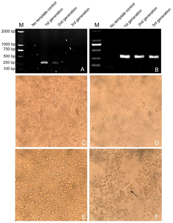
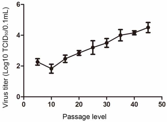
Porcine epidemic diarrhea virus (PEDV), which is the causative agent of porcine epidemic diarrhea in China and other countries, is responsible for serious economic losses in the pork industry. Inactivated PEDV vaccine plays a key role in controlling the prevalence of PEDV. However, consistently low viral titers are obtained during the propagation of PEDV in vitro; this represents a challenge to molecular analyses of the virus and vaccine development. In this study, we successfully isolated a PEDV isolate (strain NJ) from clinical samples collected during a recent outbreak of diarrhea in piglets in China, using porcine intestinal epithelial cells (IEC). We found that the isolate was better adapted to growth in IECs than in Vero cells, and the titer of the IEC cultures was 104.5 TCID50/0.1 mL at passage 45. Mutations in the S protein increased with the viral passage and the mutations tended towards attenuation. Viral challenge showed that the survival of IEC-adapted cultures was higher at the 45th passage than at the 5th passage. The use of IECs to isolate and propagate PEDV provides an effective approach for laboratory-based diagnosis of PEDV, as well as studies of the epidemiological characteristics and molecular biology of this virus.
Keywords: isolation and identification; porcine epidemic diarrhea virus (PEDV) NJ strain; porcine intestinal epithelial cells.
Conflict of interest statement
The authors declare no conflict of interest.
Figures
Similar articles
-
Deletion of a 197-Amino-Acid Region in the N-Terminal Domain of Spike Protein Attenuates Porcine Epidemic Diarrhea Virus in Piglets.J Virol. 2017 Jun 26;91(14):e00227-17. doi: 10.1128/JVI.00227-17. Print 2017 Jul 15. J Virol. 2017. PMID: 28490591 Free PMC article.
-
Epidemic strain YC2014 of porcine epidemic diarrhea virus could provide piglets against homologous challenge.Virol J. 2016 Apr 22;13:68. doi: 10.1186/s12985-016-0529-z. Virol J. 2016. PMID: 27103490 Free PMC article.
-
Porcine epidemic diarrhea vaccine evaluation using a newly isolated strain from Korea.Vet Microbiol. 2018 Jul;221:19-26. doi: 10.1016/j.vetmic.2018.05.012. Epub 2018 May 24. Vet Microbiol. 2018. PMID: 29981703
-
Epidemiology of porcine epidemic diarrhea virus among Chinese pig populations: A meta-analysis.Microb Pathog. 2019 Apr;129:43-49. doi: 10.1016/j.micpath.2019.01.017. Epub 2019 Jan 23. Microb Pathog. 2019. PMID: 30682525
-
[Advances in reverse genetics to treat porcine epidemic diarrhea virus].Sheng Wu Gong Cheng Xue Bao. 2017 Feb 25;33(2):205-216. doi: 10.13345/j.cjb.160282. Sheng Wu Gong Cheng Xue Bao. 2017. PMID: 28956377 Review. Chinese.
Cited by
-
Dual priming oligonucleotide (DPO)-based real-time RT-PCR assay for accurate differentiation of four major viruses causing porcine viral diarrhea.Mol Cell Probes. 2019 Oct;47:101435. doi: 10.1016/j.mcp.2019.101435. Epub 2019 Aug 12. Mol Cell Probes. 2019. PMID: 31415867 Free PMC article.
-
Susceptibility of porcine IPEC-J2 intestinal epithelial cells to infection with porcine deltacoronavirus (PDCoV) and serum cytokine responses of gnotobiotic pigs to acute infection with IPEC-J2 cell culture-passaged PDCoV.Vet Microbiol. 2018 Jul;221:49-58. doi: 10.1016/j.vetmic.2018.05.019. Epub 2018 May 30. Vet Microbiol. 2018. PMID: 29981708 Free PMC article.
-
Maternal immunization and vitamin A sufficiency impact sow primary adaptive immunity and passive protection to nursing piglets against porcine epidemic diarrhea virus infection.Front Immunol. 2024 May 15;15:1397118. doi: 10.3389/fimmu.2024.1397118. eCollection 2024. Front Immunol. 2024. PMID: 38812505 Free PMC article.
-
Replicative capacity of porcine deltacoronavirus and porcine epidemic diarrhea virus in primary bovine mesenchymal cells.Vet Microbiol. 2020 May;244:108660. doi: 10.1016/j.vetmic.2020.108660. Epub 2020 Apr 1. Vet Microbiol. 2020. PMID: 32402338 Free PMC article.
-
Immunogenicity of eGFP-Marked Recombinant Lactobacillus casei against Transmissible Gastroenteritis Virus and Porcine Epidemic Diarrhea Virus.Viruses. 2017 Sep 25;9(10):274. doi: 10.3390/v9100274. Viruses. 2017. PMID: 28946696 Free PMC article.
References
-
- Debouck P., Pensaert M. Experimental infection of pigs with a new porcine enteric coronavirus, CV 777. Am. J. Vet. Res. 1980;41:219–223. - PubMed
-
- Chen Q., Li G., Stasko J., Thomas J.T., Stensland W.R., Pillatzki A.E., Gauger P.C., Schwartz K.J., Madson D., Yoon K.-J. Isolation and characterization of porcine epidemic diarrhea viruses associated with the 2013 disease outbreak among swine in the United States. J. Clin. Microbiol. 2014;52:234–243. doi: 10.1128/JCM.02820-13. - DOI - PMC - PubMed
-
- Kim S.H., Kim I.J., Pyo H.M., Tark D.S., Song J.Y., Hyun B.H. Multiplex real-time RT-PCR for the simultaneous detection and quantification of transmissible gastroenteritis virus and porcine epidemic diarrhea virus. J. Virol. Methods. 2007;146:172–177. doi: 10.1016/j.jviromet.2007.06.021. - DOI - PMC - PubMed
Publication types
MeSH terms
LinkOut - more resources
Full Text Sources
Other Literature Sources
