Comparative Study
. 2017 Jul;17(7):1778-1790.
doi: 10.1111/ajt.14210. Epub 2017 Mar 3.

Role of Intrinsic (Graft) Versus Extrinsic (Host) Factors in the Growth of Transplanted Organs Following Allogeneic and Xenogeneic Transplantation

Affiliations
Comparative Study

Role of Intrinsic (Graft) Versus Extrinsic (Host) Factors in the Growth of Transplanted Organs Following Allogeneic and Xenogeneic Transplantation

T Tanabe et al. Am J Transplant. 2017 Jul.

Abstract

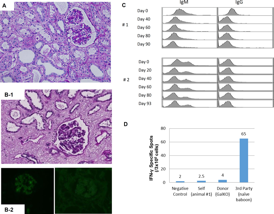
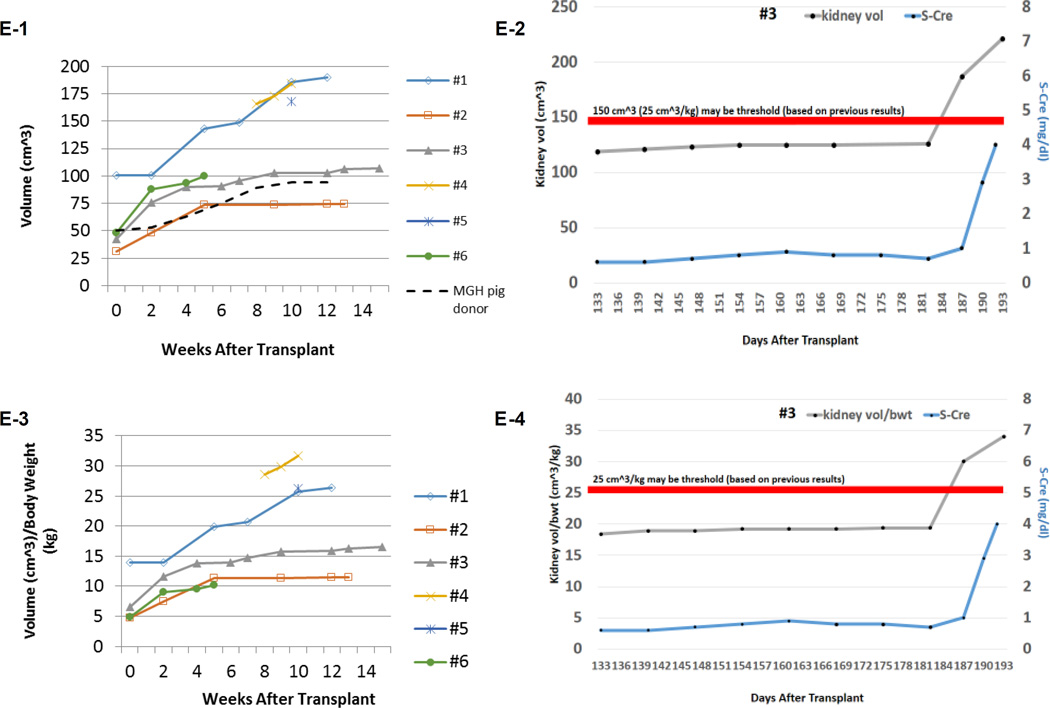
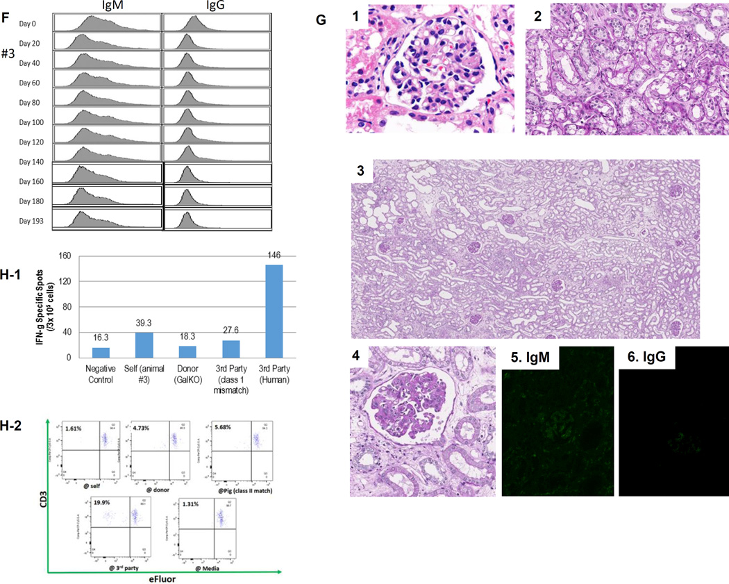
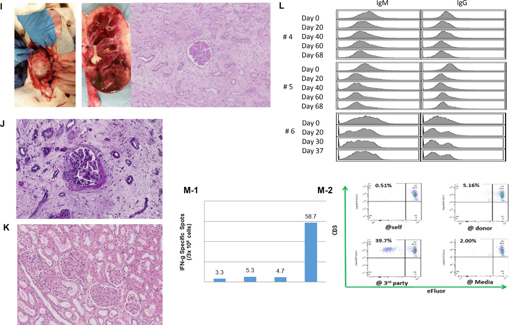
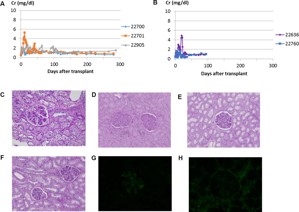
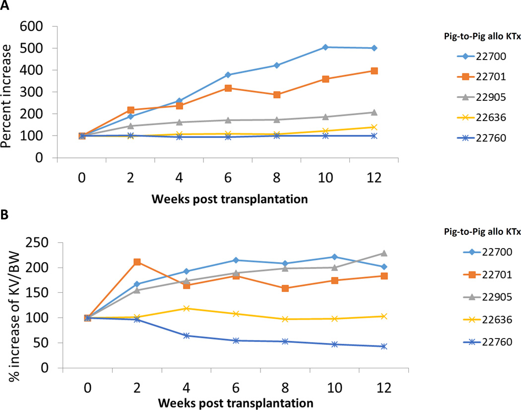
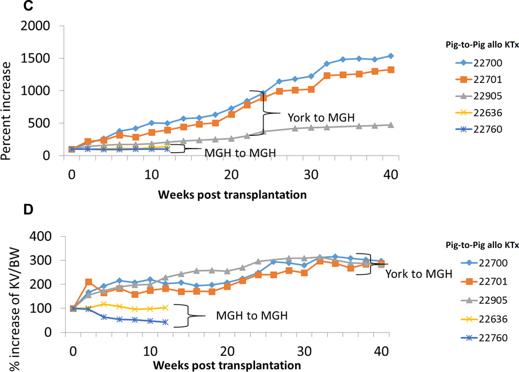
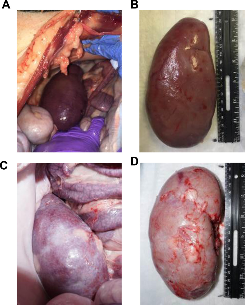
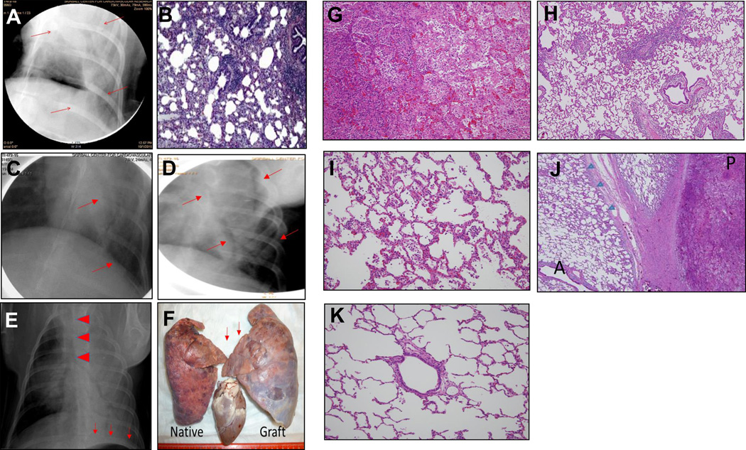
In our studies of life-supporting α-1,3-galactocyltransferase knockout (GalT-KO) pig-to-baboon kidneys, we found that some recipients developed increased serum creatinine with growth of the grafts, without histological or immunological evidence of rejection. We hypothesized that the rapid growth of orthotopic pig grafts in smaller baboon recipients may have led to deterioration of organ function. To test this hypothesis for both kidneys and lungs, we assessed whether the growth of outbred (Yorkshire) organ transplants in miniature swine was regulated by intrinsic (graft) or extrinsic (host environment) factors. Yorkshire kidneys exhibited persistent growth in miniature swine, reaching 3.7 times their initial volume over 3 mo versus 1.2 times for miniature swine kidneys over the same time period. Similar rapid early growth of lung allografts was observed and, in this case, led to organ dysfunction. For xenograft kidneys, a review of our results suggests that there is a threshold for kidney graft volume of 25 cm3 /kg of recipient body weight at which cortical ischemia is induced in transplanted GalT-KO kidneys in baboons. These results suggest that intrinsic factors are responsible, at least in part, for growth of donor organs and that this property should be taken into consideration for growth-curve-mismatched transplants, especially for life-supporting organs transplanted into a limited recipient space.

Keywords: growth and development; kidney (allograft) function/dysfunction; kidney transplantation/nephrology; lung failure/injury; lung transplantation/pulmonology; organ allocation; translational research/science; xenotransplantation.


Conflict of interest statement

Disclosure

The authors of this manuscript have no conflicts of interest to disclose as described by the American Journal of Transplantation.

Figures

Figure 1
Figure 1
(A) Periodic acid-Schiff (PAS) staining of a kidney graft for baboon #2 on day 93. No sign of rejection or interstitial fibrosis. (B-1) PAS staining of a kidney graft for baboon #1 on day 90 showed interstitial fibrosis and tubular atrophy but no evidence of glomerulitis, peritubular capillaritis or a mononuclear cell infiltration. (B-2) Immunofluorescence staining for baboon #1 on POD 90. IgM staining showed mild mesangial deposition (left) while IgG deposition wasn’t seen (right). (C) Anti-non-Gal natural antibody for baboons #1 and #2 assessed by flow-cytometry demonstrating neither elicit anti-pig IgM nor IgG developed.(D) ELISPOT assay for IFN-γ at the time of euthanasia (POD 90) for baboon #1. Many spots could be seen in the wells stimulated by 3rd party while only a small number of spots against GalTKO pig cells similar to self were seen. (E-1) Kidney volume (cm3) of all xenoTK Tx recipients against weeks after Tx (solid lines). The growth of the TKs (solid lines) was similar to what was observed in a remaining kidney of a miniature swine donor who was kept alive after kidney donation in order to measure autologous kidney growth (dashed line).(E-2) Kidney volume and serum Cre levels between 19 and 27 weeks after Tx for baboon #3. (E-3) Ratio of kidney volume (cm3) / recipient’s body weight (kg) against weeks after Tx. (E-4) Ratio of kidney volume (cm3) / recipient’s body weight (kg) and serum Cre levels for baboon #3 between 19 and 27 weeks after Tx.(F) Anti-non-Gal natural antibody for baboon #3 assessed by flow-cytometry demonstrating neither elicit anti-pig IgM nor IgG developed.(G-1 and 2) The graft biopsy at POD 121 from baboon #3 showed no interstitial edema and normal appearance of glomeruli. (G-3 and 4) Biopsy at POD 193 from baboon #3 showed well preserved glomeruli with no glomerulitis, no endoarteritis, and no apparent interstitial cell infiltration. However, widening of interstitial area was noted with interstitial edema. (G-5 and 6) Neither IgM nor IgG deposition was observed in the excised grafts from baboon #3. (H-1 and 2) ELISPOT assay for IFN-γ and MLR at the time of euthanasia for baboon #3 demonstrated that animal was anti-pig specific unresponsive. (I) Necropsy sample of kidney graft (left) and PAS staining of kidney graft for baboon #4 on POD 68 (right). Glomerular capillaries were relatively preserved with diffuse tubular necrosis and absence of thrombi in the small interlobular arteries. (J) PAS staining of a kidney graft for baboon #5 on day 68 showed interstitial fibrosis and tubular atrophy but glomerular capillaries were relatively preserved. (K) Kidney graft for baboon #6 on day 37 showed no sign of rejection. (L) Anti-non-Gal natural antibody for baboon #4, #5 and #6 assessed by FACS demonstrating neither elicit anti-pig IgM nor IgG developed. (M-1) INF-γ ELISPOT assay for baboon #4 at the end of study (POD68). (M-2) MLR for baboon #4 on day 59.
Figure 1
Figure 1
(A) Periodic acid-Schiff (PAS) staining of a kidney graft for baboon #2 on day 93. No sign of rejection or interstitial fibrosis. (B-1) PAS staining of a kidney graft for baboon #1 on day 90 showed interstitial fibrosis and tubular atrophy but no evidence of glomerulitis, peritubular capillaritis or a mononuclear cell infiltration. (B-2) Immunofluorescence staining for baboon #1 on POD 90. IgM staining showed mild mesangial deposition (left) while IgG deposition wasn’t seen (right). (C) Anti-non-Gal natural antibody for baboons #1 and #2 assessed by flow-cytometry demonstrating neither elicit anti-pig IgM nor IgG developed.(D) ELISPOT assay for IFN-γ at the time of euthanasia (POD 90) for baboon #1. Many spots could be seen in the wells stimulated by 3rd party while only a small number of spots against GalTKO pig cells similar to self were seen. (E-1) Kidney volume (cm3) of all xenoTK Tx recipients against weeks after Tx (solid lines). The growth of the TKs (solid lines) was similar to what was observed in a remaining kidney of a miniature swine donor who was kept alive after kidney donation in order to measure autologous kidney growth (dashed line).(E-2) Kidney volume and serum Cre levels between 19 and 27 weeks after Tx for baboon #3. (E-3) Ratio of kidney volume (cm3) / recipient’s body weight (kg) against weeks after Tx. (E-4) Ratio of kidney volume (cm3) / recipient’s body weight (kg) and serum Cre levels for baboon #3 between 19 and 27 weeks after Tx.(F) Anti-non-Gal natural antibody for baboon #3 assessed by flow-cytometry demonstrating neither elicit anti-pig IgM nor IgG developed.(G-1 and 2) The graft biopsy at POD 121 from baboon #3 showed no interstitial edema and normal appearance of glomeruli. (G-3 and 4) Biopsy at POD 193 from baboon #3 showed well preserved glomeruli with no glomerulitis, no endoarteritis, and no apparent interstitial cell infiltration. However, widening of interstitial area was noted with interstitial edema. (G-5 and 6) Neither IgM nor IgG deposition was observed in the excised grafts from baboon #3. (H-1 and 2) ELISPOT assay for IFN-γ and MLR at the time of euthanasia for baboon #3 demonstrated that animal was anti-pig specific unresponsive. (I) Necropsy sample of kidney graft (left) and PAS staining of kidney graft for baboon #4 on POD 68 (right). Glomerular capillaries were relatively preserved with diffuse tubular necrosis and absence of thrombi in the small interlobular arteries. (J) PAS staining of a kidney graft for baboon #5 on day 68 showed interstitial fibrosis and tubular atrophy but glomerular capillaries were relatively preserved. (K) Kidney graft for baboon #6 on day 37 showed no sign of rejection. (L) Anti-non-Gal natural antibody for baboon #4, #5 and #6 assessed by FACS demonstrating neither elicit anti-pig IgM nor IgG developed. (M-1) INF-γ ELISPOT assay for baboon #4 at the end of study (POD68). (M-2) MLR for baboon #4 on day 59.
Figure 2
Figure 2
Creatinine (Cre) levels following Tx of (A) Yorkshire kidneys and (B) miniature swine kidneys in miniature swine recipients (x-axis represents days after kidney Tx). Stable Cre levels were maintained until euthanasia in both groups. Histologic findings in the Yorkshire kidney graft for (C) 22905(POD 91), (D) 22700, (E) 22701(POD 350) and (F) animal 22905(POD 306). Minimal interstitial cell infiltration was seen on PAS staining (C), and neither IgM (G) nor IgG (H) deposition was observed within the graft. No evidence of rejection was observed in York-to-mini kidney Tx models 300 days following kidney Tx (Figs. D, E and F).
Figure 3
Figure 3
(A) % increase in kidney graft volume after fully MHC mismatched kidney Tx in first 12 weeks. (B) % increase of kidney volume (cm3) / recipent’s body weight (kg) in first 12 weeks. (C) % increase in kidney graft volume after fully MHC mismatched kidney Tx 40 weeks. (D) % increase of kidney volume (cm3) / recipient’s body weight (kg) 40 weeks.
Figure 3
Figure 3
(A) % increase in kidney graft volume after fully MHC mismatched kidney Tx in first 12 weeks. (B) % increase of kidney volume (cm3) / recipent’s body weight (kg) in first 12 weeks. (C) % increase in kidney graft volume after fully MHC mismatched kidney Tx 40 weeks. (D) % increase of kidney volume (cm3) / recipient’s body weight (kg) 40 weeks.
Figure 4
Figure 4
(A) Representative image of a kidney graft at the time of euthanasia and (B) explanted kidney graft for 22700. (C) Picture at time of euthanasia and (D) explanted kidney graft for 22701.
Figure 5
Figure 5
(A) POD 8 chest X-ray (1st recipient) following allogeneic lung Tx. The arrows demonstrate areas of consolidation within the graft. (B) Pathological examination revealed infiltration of the graft with inflammatory cells and lymphocytes, suggesting pneumonia and cellular rejection. (C,D): POD 15 chest X-ray (C, 2nd recipient) and POD 16 chest x-ray (D, 3rd recipient) demonstrating clear aeration of the lung allografts. (E) POD 29 chest x-ray (2nd recipient). Arrows depict caudal displacement of the left hemi-diaphragm, suggesting enlargement of the graft. Arrowheads demonstrate displacement of the trachea to the right. Consolidation was found diffusely, especially in the left upper lung, suggesting atelectasis and subsequent pneumonia. (F) Necropsy findings of the native (right) and lung graft (left). The graft enlarged more than that of native lung, especially caudally. Arrows indicate the compressed upper lobe of the graft, which enlarged toward the mediastinum. (G-K) Representative pathological findings of the native and grafted lung: (G) Bleeding, neutrophil infiltration and cellular debris were seen, indicating pneumonia. (H) Mild bronchiolitis in the lower lobe of the graft. (I) Mild pneumonia in the upper lobe of the native lung. (J) There were well-aerated regions (A) and lobular pneumonia regions (P) in the upper lobe of the graft. Evidence of pneumonia extended from the pleural region (arrow heads). (K) No evidence of cellular rejection was found in the graft.

Similar articles

Cited by

References

    1. Klassen DK, Edwards LB, Stewart DE, Glazier AK, Orlowski JP, Berg CL. The OPTN Deceased Donor Potential Study: Implications for Policy and Practice. American journal of transplantation : official journal of the American Society of Transplantation and the American Society of Transplant Surgeons. 2016;16(6):1707–1714. - PubMed
    1. Sachs DH. The pig as a xenograft donor. Pathologie-biologie. 1994;42(3):217–219. - PubMed
    1. Sato M, Kagoshima A, Saitoh I, Inada E, Miyoshi K, Ohtsuka M, et al. Generation of alpha-1,3-Galactosyltransferase-Deficient Porcine Embryonic Fibroblasts by CRISPR/Cas9-Mediated Knock-in of a Small Mutated Sequence and a Targeted Toxin-Based Selection System. Reproduction in domestic animals = Zuchthygiene. 2015;50(5):872–880. - PubMed
    1. Hai T, Teng F, Guo R, Li W, Zhou Q. One-step generation of knockout pigs by zygote injection of CRISPR/Cas system. Cell research. 2014;24(3):372–375. - PMC - PubMed
    1. Mohiuddin MM, Singh AK, Corcoran PC, Thomas Iii ML, Clark T, Lewis BG, et al. Chimeric 2C10R4 anti-CD40 antibody therapy is critical for long-term survival of GTKO.hCD46.hTBM pig-to-primate cardiac xenograft. Nature communications. 2016;7:11138. - PMC - PubMed

Publication types

Substances