

FEM1 proteins are ancient regulators of SLBP degradation
- PMID: 28118078
- PMCID: PMC5384579
- DOI: 10.1080/15384101.2017.1284715
FEM1 proteins are ancient regulators of SLBP degradation
Abstract
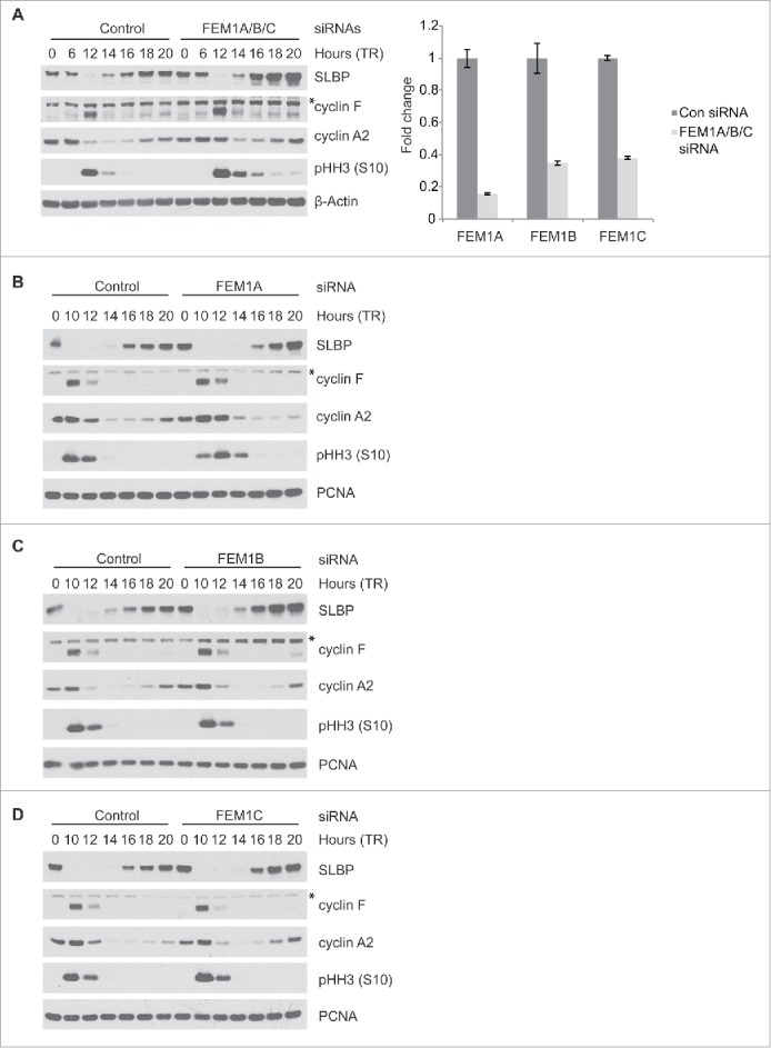
FEM1A, FEM1B, and FEM1C are evolutionarily-conserved VHL-box proteins, the substrate recognition subunits of CUL2-RING E3 ubiquitin ligase complexes. Here, we report that FEM1 proteins are ancient regulators of Stem-Loop Binding Protein (SLBP), a conserved protein that interacts with the stem loop structure located in the 3' end of canonical histone mRNAs and functions in mRNA cleavage, translation and degradation. SLBP levels are highest during S-phase coinciding with histone synthesis. The ubiquitin ligase complex SCFcyclin F targets SLBP for degradation in G2 phase; however, the regulation of SLBP during other stages of the cell cycle is poorly understood. We provide evidence that FEM1A, FEM1B, and FEM1C interact with and mediate the degradation of SLBP. Cyclin F, FEM1A, FEM1B and FEM1C all interact with a region in SLBP's N-terminus using distinct degrons. An SLBP mutant that is unable to interact with all 4 ligases is expressed at higher levels than wild type SLBP and does not oscillate during the cell cycle. We demonstrate that orthologues of SLBP and FEM1 proteins interact in C. elegans and D. melanogaster, suggesting that the pathway is evolutionarily conserved. Furthermore, we show that FEM1 depletion in C. elegans results in the upregulation of SLBP ortholog CDL-1 in oocytes. Notably, cyclin F is absent in flies and worms, suggesting that FEM1 proteins play an important role in SLBP targeting in lower eukaryotes.
Keywords: CDL-1; CRL2; Cullin-RING Ligase; Cyclin F; DNA replication; FEM1; FEM1A; FEM1B; FEM1C; SLBP; histone; ubiquitin.
Figures


Comment in
-
"FEM1"nism controls SLBP stability during cell cycle.Cell Cycle. 2017 Apr 3;16(7):597-598. doi: 10.1080/15384101.2017.1302226. Cell Cycle. 2017. PMID: 28324668 Free PMC article. No abstract available.
Similar articles
-
Cyclin F-Mediated Degradation of SLBP Limits H2A.X Accumulation and Apoptosis upon Genotoxic Stress in G2.Mol Cell. 2016 Nov 3;64(3):507-519. doi: 10.1016/j.molcel.2016.09.010. Epub 2016 Oct 20. Mol Cell. 2016. PMID: 27773672 Free PMC article.
-
Structural insights into SMCR8 C-degron recognition by FEM1B.Biochem Biophys Res Commun. 2021 Jun 11;557:236-239. doi: 10.1016/j.bbrc.2021.04.046. Epub 2021 Apr 20. Biochem Biophys Res Commun. 2021. PMID: 33892462
-
DDB1 and CUL4 associated factor 11 (DCAF11) mediates degradation of Stem-loop binding protein at the end of S phase.Cell Cycle. 2016 Aug 2;15(15):1986-96. doi: 10.1080/15384101.2016.1191708. Epub 2016 Jun 2. Cell Cycle. 2016. PMID: 27254819 Free PMC article.
-
Stem-loop binding protein and metal carcinogenesis.Semin Cancer Biol. 2021 Nov;76:38-44. doi: 10.1016/j.semcancer.2021.08.006. Epub 2021 Aug 18. Semin Cancer Biol. 2021. PMID: 34416372 Free PMC article. Review.
-
Structure-specific nucleic acid recognition by L-motifs and their diverse roles in expression and regulation of the genome.Biochim Biophys Acta. 2015 Jun;1849(6):677-87. doi: 10.1016/j.bbagrm.2015.02.006. Epub 2015 Mar 4. Biochim Biophys Acta. 2015. PMID: 25748361 Free PMC article. Review.
Cited by
-
Hominini-specific regulation of the cell cycle by stop codon readthrough of FEM1B.J Cell Sci. 2024 Aug 15;137(16):jcs261921. doi: 10.1242/jcs.261921. Epub 2024 Aug 29. J Cell Sci. 2024. PMID: 39140134 Free PMC article.
-
Molecular basis for ubiquitin ligase CRL2FEM1C-mediated recognition of C-degron.Nat Chem Biol. 2021 Mar;17(3):263-271. doi: 10.1038/s41589-020-00703-4. Epub 2021 Jan 4. Nat Chem Biol. 2021. PMID: 33398170
-
Novel Class of Viral Ankyrin Proteins Targeting the Host E3 Ubiquitin Ligase Cullin-2.J Virol. 2018 Nov 12;92(23):e01374-18. doi: 10.1128/JVI.01374-18. Print 2018 Dec 1. J Virol. 2018. PMID: 30258003 Free PMC article.
-
Structural basis and regulation of the reductive stress response.Cell. 2021 Oct 14;184(21):5375-5390.e16. doi: 10.1016/j.cell.2021.09.002. Epub 2021 Sep 24. Cell. 2021. PMID: 34562363 Free PMC article.
-
Downregulation of FEM1C enhances metastasis and proliferation in colorectal cancer.Ann Transl Med. 2021 Sep;9(17):1391. doi: 10.21037/atm-21-4244. Ann Transl Med. 2021. PMID: 34733943 Free PMC article.
References
-
- Lydeard JR, Schulman BA, Harper JW. Building and remodelling Cullin-RING E3 ubiquitin ligases. EMBO Rep 2013; 14:1050-61; PMID:24232186; http://dx.doi.org/10.1038/embor.2013.173 - DOI - PMC - PubMed
-
- Kamura T, Maenaka K, Kotoshiba S, Matsumoto M, Kohda D, Conaway RC, Conaway JW, Nakayama KI. VHL-box and SOCS-box domains determine binding specificity for Cul2-Rbx1 and Cul5-Rbx2 modules of ubiquitin ligases. Genes Dev 2004; 18:3055-65; PMID:15601820; http://dx.doi.org/10.1101/gad.1252404 - DOI - PMC - PubMed
-
- Skaar JR, Pagan JK, Pagano M. Mechanisms and function of substrate recruitment by F-box proteins. Nat Rev Mol Cell Biol 2013; 14:369-81; PMID:23657496; http://dx.doi.org/10.1038/nrm3582 - DOI - PMC - PubMed
-
- Starostina NG, Lim JM, Schvarzstein M, Wells L, Spence AM, Kipreos ET. A CUL-2 ubiquitin ligase containing three FEM proteins degrades TRA-1 to regulate C. elegans sex determination. Dev Cell 2007; 13:127-39; http://dx.doi.org/10.1016/j.devcel.2007.05.008 - DOI - PMC - PubMed
-
- Doniach T, Hodgkin J. A sex-determining gene, fem-1, required for both male and hermaphrodite development in Caenorhabditis elegans. Dev Biol 1984; 106:223-35; PMID:6541600; http://dx.doi.org/10.1016/0012-1606(84)90077-0 - DOI - PubMed
MeSH terms
Substances
Grants and funding
LinkOut - more resources
Full Text Sources
Other Literature Sources
Molecular Biology Databases
Research Materials