. 2017 Feb;13(2):271-282.
doi: 10.1080/21645515.2017.1264766. Epub 2017 Jan 24.

A plant protein signal sequence improved humoral immune response to HPV prophylactic and therapeutic DNA vaccines

Affiliations

A plant protein signal sequence improved humoral immune response to HPV prophylactic and therapeutic DNA vaccines

Silvia Massa et al. Hum Vaccin Immunother. 2017 Feb.

Abstract

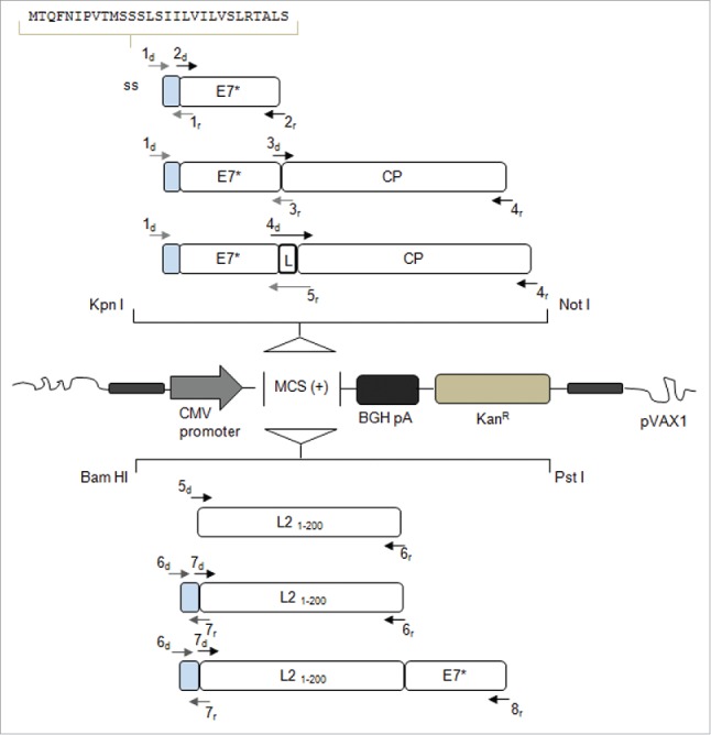
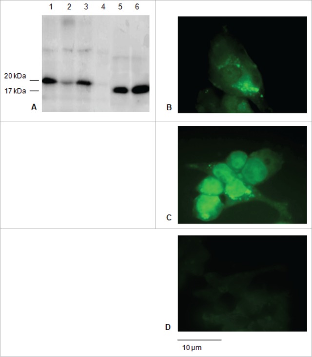
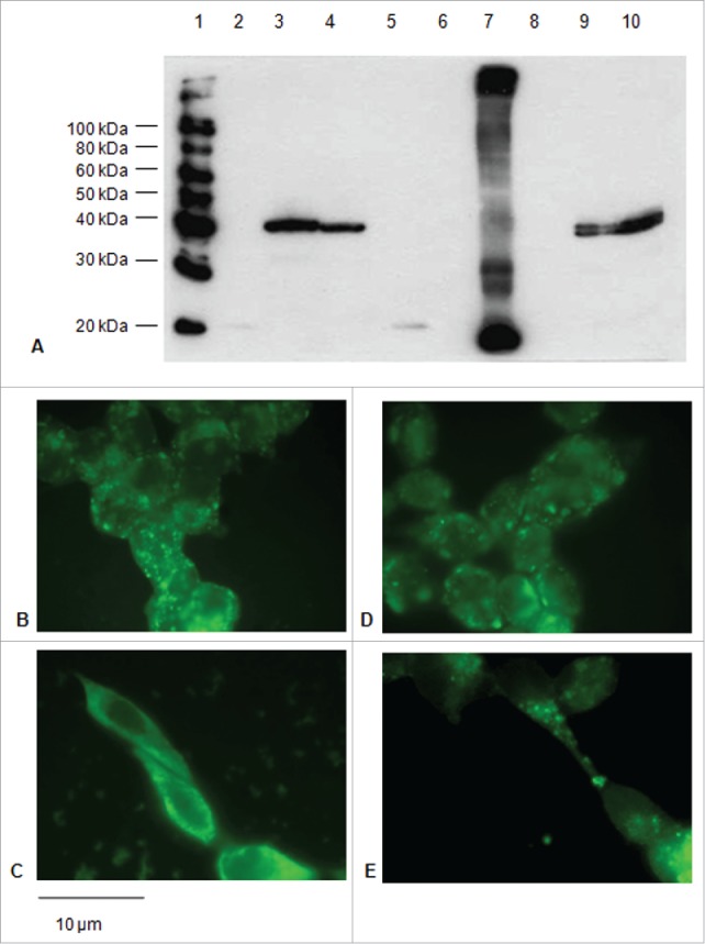
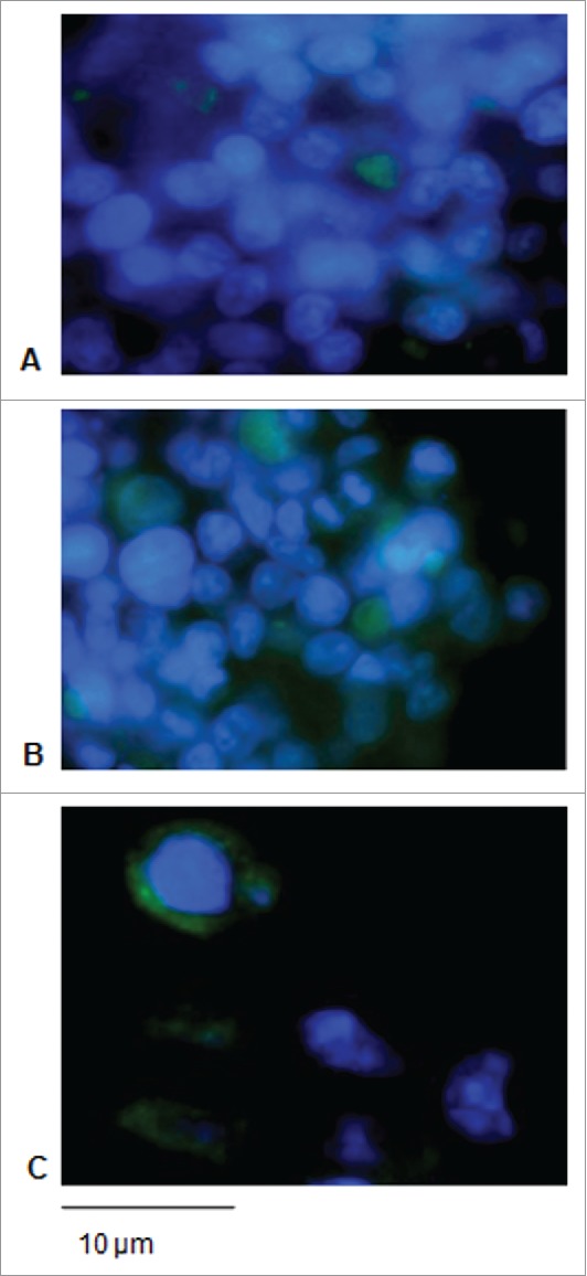
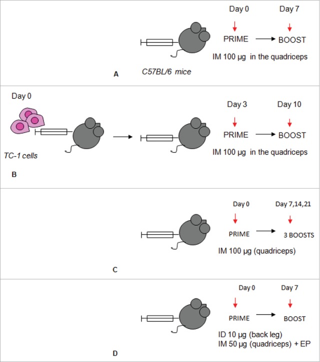
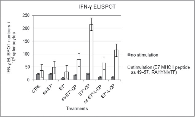
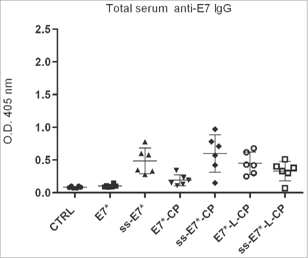
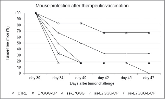
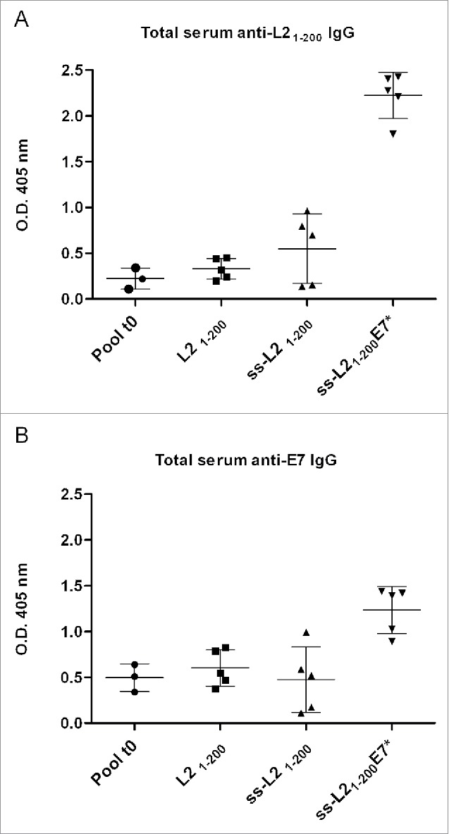
Signal sequences (ss) play a critical role in the sorting of nascent secretory and membrane proteins. This function has been conserved from bacteria through eukaryotes, although ss appear diverse in length and amino acid composition. Sorting of proteins is also critical to instruct antigens for a proper immunological response. Thus, a plant ss was used to drive Human Papillomavirus (HPV) model antigens into the human secretory pathway: the HPV16 E7 oncoprotein, its chimera with the coat protein (CP) of the Potato Virus X (PVX), the first 200 amino acids of the HPV16 minor capsid protein L2 (known to harbour cross-reacting epitopes) and its chimera with E7 gene. These genes were used to transfect HEK-293 cells and to immunize C57BL/6 mice. The ss-provided genes were expressed, and proteins detected by immunofluorescence and immunoblotting. Mouse immunization with DNA constructs carrying the ss elicited a strong humoral response against both E7 and L2 and a weak cell-mediated immunity. To our knowledge this is the first demonstration that a signal sequence derived from a plant can modulate the sorting of a heterologous protein in mammalian cells. This activity in mammalian cells may be responsible for the observed increased humoral response to DNA-based vaccines that are generally weak inducers of IgG response. This might open new perspectives in the design of DNA vaccines, especially to counteract infections where a strong humoral response is needed.

Keywords: HPV; humoral immune response; infectious diseases; plant signal sequences; vaccines.


Figures

Figure 1.
Figure 1.
Schematic representation of the genes introduced into the mammalian vector pVAX1 with indication of the oligonucleotides used for PCR amplification and assembly (see Table 1). The recombinant genes are under the control of the CMV promoter and the BGH pA signal constituting the expression cassette, while the selectable marker is KanR. BGH pA: Bovine Growth Hormone poly-adenilation signal; KanR: Kanamycin Resistance; ss (light gray boxes): signal sequence of the gene encoding the Polygalacturonase Inhibiting Protein of Phaseolus vulgaris; E7*: attenuated E7 gene of HPV16; CP: Potato Virus-X coat protein gene; L: linker sequence encoding the Gly-Pro-Gly-Pro tetrapeptide; L21–200: nucleotide sequence corresponding to aa 1-200 of the L2 minor capsid protein of HPV type 16.
Figure 2.
Figure 2.
Expression of the E7* protein in HEK-293 cells upon transfection. A: Western blot of total soluble proteins from 1×10⁶ cells transfected with the E7* +/− ss and of culture media. 1) ss-E7* (cell lysate); 2) ss-E7* culture medium; 3) E7* (cell lysate); 4) E7* culture medium; 5), 6) purified His6-E7* from E. coli 25 ng and 50 ng, respectively. The E7* protein runs as a 20 kDa protein in HEK-293 cells, while His6-E7* shows a slightly lower molecular weight in bacteria. B, C, D: Immunofluorescence (FITC) detected in cells transiently transfected with ss-E7* (b), with E7* (c) and non-transfected HEK-293 cells used as a negative control (d) (original magnification, 100x; scale bar, 10 µm).
Figure 3.
Figure 3.
Expression of the E7*-CP proteins in HEK-293 cells upon transfection. A: Immunoblotting of proteins from lysates of 1×10⁶ cells transfected with the E7*-CP fusions with or without ss. 1) Biotinylated Protein Ladder NEB; 2) E7* (cell lysate); 3) E7*-CP (cell lysate); 4) E7*-L-CP (cell lysate); 5) ss-E7* (cell lysate); 6) ss-E7*-CP (culture medium); 7) ss-E7 plant extract; 8) ss-E7*-L-CP (culture medium); 9) ss-E7*-CP (cell lysate); 10) ss-E7*-L-CP (cell lysate). B, C, D, E: Immunofluorescence (FITC) detected in cells transfected with E7*-CP (B, C) and E7*-L-CP (D,E) plasmids without or with the ss, respectively (original magnification, 100x; scale bar, 10 µm).
Figure 4.
Figure 4.
Expression of the L21–200 proteins in HEK-293 cells upon transfection. Immunofluorescence in cells transfected with L21–200 (A), ss- L21–200 (B) and ss-L21–200-E7* (C) constructs. FITC fluorescence was coupled with DAPI nuclei blue counter-staining (original magnification, 100x; scale bar, 10 µm).
Figure 5.
Figure 5.
Immunization schedules. Immunization with the ss-E7* +/−CP series in absence (A) or in presence of tumor challenge (B) to evaluate immune response and tumor rejection, respectively. Long-term (C) and short-term (D) immunization protocols used to induce antibody response in mice vaccinated with the the L21–200 +/− E7* series. ID: Intra-dermal injection; IM: Intra-muscular injection; EP: Electroporation.
Figure 6.
Figure 6.
ELISPOT assay for IFN-γ-secreting splenocytes after vaccination with ss-E7* +/− CP in C57BL/6 mice. Data are presented as mean spot number ± SD per 1×10⁶ splenocytes from triplicate wells. Cells were stimulated with an E7 specific H-2Db cytotoxic T lymphocyte MHC class I epitope. Gray columns represent non-stimulated splenocytes. The presence of IFN-γ producing E7 specific T-cell precursors was determined using an anti-IFN-γ antibody, as described in Material and Methods.
Figure 7.
Figure 7.
Humoral immunological response in mice after vaccination with ss-E7* +/− CP. Data for ELISA test are reported from a representative experiment. Humoral E7-specific serum IgG responses are presented as optical density values at 405 nm of 1:100 diluted sera. The mean titer value from each group is marked with a dash. Means and variances are significantly different (p< 0,05) according to the One-way ANOVA test and to the Bonferroni's multiple comparison test.
Figure 8.
Figure 8.
Growth inhibition of experimental TC-1-induced tumors after therapeutic vaccination of mice with ss-E7*, ss-E7*-CP and ss-E7*-L-CP. Results are presented as the percentage of tumor-free mice. The presence of tumors was monitored by palpation twice per week. The animals were sacrificed on day 47 after tumor challenge, when all the animals with tumors were euthanized for ethical reasons. E7*-CP was administered for comparison of efficacy data.
Figure 9.
Figure 9.
L21–200 expression in E. Coli. Lane 1: L21–200 purified under native conditions; lane 2: L21–200 purified under denaturing conditions; lane 3 Molecular weight Marker Precision Plus Protein™ Dual Color Standards (Bio-rad Milan, Italy).
Figure 10.
Figure 10.
Humoral immunological response to L21–200 and E7* in mice after vaccination with the L21–200, ss-L21–200 and ss-L21–200-E7*. C57BL/6 mice were immunized as described in Fig. 5D. Data for ELISA test were generated for sera collected from every mouse (m 1-5: mouse 1-5) of each vaccination group after coating with either His6-L21–200 (Fig. 10A) or His6-E7 (Fig. 10B) and are reported from one representative experiment. Humoral specific serum IgG responses are presented as optical density values at 405 nm of 1:100 diluted sera. The mean titer value from each group is marked with a dash. Pool t0: average of pool of pre immune sera for each treatments. Means and variances are significantly different (p < 0,05) according to the One-way ANOVA test and to the Bonferroni's multiple comparison test.
Figure 11.
Figure 11.
Reactivity of sera of immunized mice against the L21–200 peptide 6 months post-immunization, as described in Fig. 5D. 50ng of L21–200 were loaded for each lane and revealed with RG-1 monoclonal antibody (strip 1) or 1:100 dilutions of pooled sera from ss-L21–200 (strip 2), L21–200 (strip 3), and ss-L21–200-E7* (strip 4) mouse groups.

References

    1. Vici P, Mariani L, Pizzuti L, Sergi D, Di Lauro L, Vizza E, Tomao F, Tomao S, Mancini E, Vincenzoni C, et al.. Emerging biological treatments for uterine cervical carcinoma. J Cancer 2014; 5:86-97; PMID:24494026; http://dx.doi.org/10.7150/jca.7963 - DOI - PMC - PubMed
    1. Mariani L, Venuti A. HPV vaccine: an overview of immune response, clinical protection, and new approaches for the future. J Transl Med 2010; 8:105; PMID:20979636; http://dx.doi.org/10.1186/1479-5876-8-105 - DOI - PMC - PubMed
    1. Tyler M, Tunban E, Chackerian B. Second-generation prophylactic HPV vaccines: successes and challenges. Expert Rev Vaccines 2014; 13:247-55; PMID:24350614; http://dx.doi.org/10.1586/14760584.2014.865523 - DOI - PMC - PubMed
    1. Whang DW, Zhihai L, Jieqiong X, Junqi W, Li Z, Yajing L, Fei F, Lu X, Minxi W, Zhibo K, et al.. Identification of broad-genotype HPV L2 neutralization site for pan-HPV vaccine development by a cross-neutralizing antibody. PLoS One 2015; 10:1-18 - PMC - PubMed
    1. Ghambira R, Karanam B, Jagu S, Roberts JN, Buck CB, Bossis I, Alphs H, Culp T, Christensen ND, Roden RB. A protective and broadly cross-neutralizing epitope of human papillomavirus L2. J Virol 2007; 81:13927-31; PMID:17928339; http://dx.doi.org/10.1128/JVI.00936-07 - DOI - PMC - PubMed

Publication types

MeSH terms