. 2017 Jan 24;12(1):e0170599.
doi: 10.1371/journal.pone.0170599. eCollection 2017.

Chlorin e6-Mediated Photodynamic Therapy Suppresses P. acnes-Induced Inflammatory Response via NFκB and MAPKs Signaling Pathway

Affiliations

Chlorin e6-Mediated Photodynamic Therapy Suppresses P. acnes-Induced Inflammatory Response via NFκB and MAPKs Signaling Pathway

Yoon-Young Wang et al. PLoS One. .

Abstract

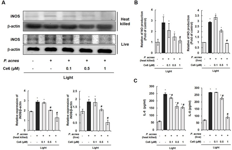
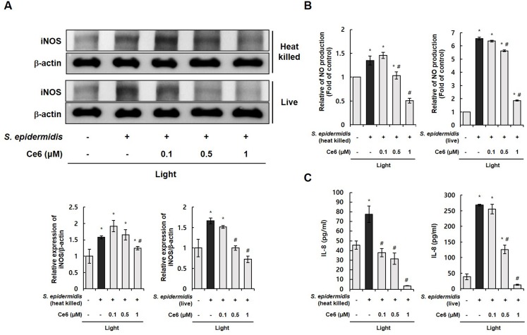
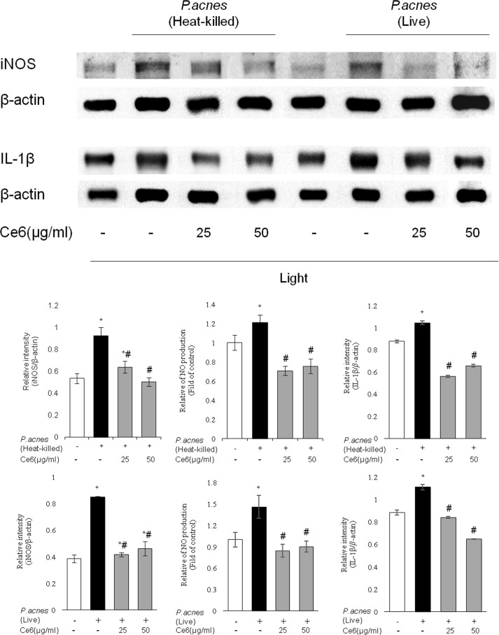
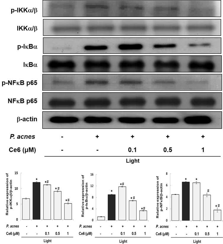
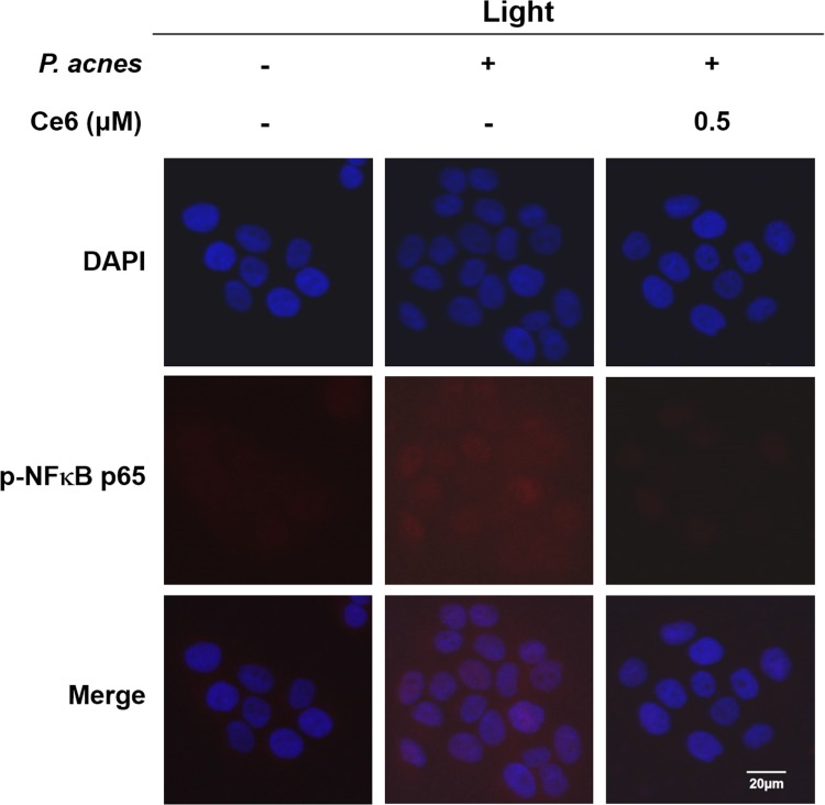
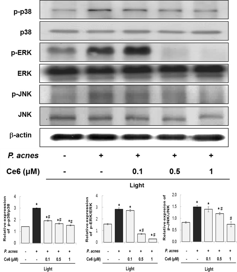
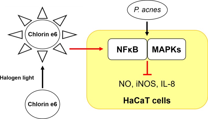
Photodynamic therapy (PDT), consisting of photosensitizer, light, and oxygen has been used for the treatment of various diseases including cancers, microbial infections and skin disorders. In this study, we examined the anti-inflammatory effect of chlorin e6-mediated PDT in P. acnes-infected HaCaT cells using photosensitizer chlorin e6 (Ce6) and halogen light. The live and heat-killed P. acnes triggered an upregulation of inflammatory molecules such as iNOS, NO, and inflammatory cytokine in HaCaT cells and mouse model. Ce6-mediated PDT notably downregulated the expression of these inflammatory molecules in vitro and in vivo. Similarly, chlorin e6-mediated PDT was capable of regulating inflammatory response in both live and heat killed S. epidermidis exposed HaCaT cells. Moreover, phosphorylation of p38, JNK, and ERK were reduced by Ce6-mediated PDT. Ce6-mediated PDT also reduced the phosphorylation of IKKα/β, IĸBα and NFκB p65 in P. acnes-stimulated HaCaT cells. In addition, the dramatic increase in the nuclear translocation of NFκB p65 observed upon stimulation with P. acnes was markedly impaired by Ce6-based PDT. This is the first suggestion that Ce6-mediated PDT suppresses P. acnes-induced inflammation through modulating NFκB and MAPKs signaling pathways.


Conflict of interest statement

The authors have declared that no competing interests exist.

Figures

Fig 1
Fig 1. Ce6-mediated PDT effectively inhibited iNOS, NO, and inflammatory cytokine IL-8 in P. acnes-triggered inflammation
(A) Western blot results demonstrate that Ce6-mediated PDT suppressed the expression iNOS in live and heat-killed P. acnes-infected HaCaT cells. The ratio of immunointensity between the iNOS and β-actin was calculated. (B) Ce6-mediated PDT suppressed NO production in live and heat-killed P. acnes-stimulated HaCaT cells. (C) ELISA results demonstrate that Ce6-mediated PDT reduced IL-8 in live and heat-killed P. acnes-infected HaCaT cells. Results are expressed as mean ± SEM. *P<0.05 compared with the non-treated control group. #P<0.05 compared with P. acnes treated cells only.
Fig 2
Fig 2. Ce6-mediated PDT effectively inhibited iNOS, NO, and inflammatory cytokine IL-8 in S. epidermidis-triggered inflammation
(A) Western blot results demonstrate that Ce6-mediated PDT suppressed the expression iNOS in live and heat-killed S. epidermidis-infected HaCaT cells. The ratio of immunointensity between the iNOS and β-actin was calculated. (B) Ce6-mediated PDT suppressed NO production in live and heat-killed S. epidermidis-stimulated HaCaT cells. (C) ELISA results demonstrate that Ce6-mediated PDT reduced IL-8 in live and heat-killed S. epidermidis-infected HaCaT cells. Results are expressed as mean ± SEM. *P<0.05 compared with the non-treated control group. #P<0.05 compared with S. epidermidis treated cells only.
Fig 3
Fig 3. In vivo therapeutic effects of Ce6-mediated PDT on live and heat-killed P. acnes-induced inflammation.
The left ear of ICR mice was injected with live and heat-killed P. acnes. Right ear received an equal amount of PBS serving as a control. Ce6 (1, 25 and 50 μg/ml in PBS) was epicutaneously applied on the left ear. Ce6-mediated PDT markedly decreased the expression of iNOS, NO and pro-inflammatory cytokine IL-1β induced by live and heat-killed P. acnes. Results are expressed as mean ± SEM. *P<0.05 compared with the non-stimulated control group. #P<0.05 compared with P. acnes stimulated mice only.
Fig 4
Fig 4. Ce6-mediated PDT suppressed NFκB signaling pathways in heat-killed P. acnes-stimulated HaCaT cells.
Western blot analysis shows that phosphorylation of IKKα/β, IκBα and NFκB were suppressed by Ce6-mediated PDT. Results are expressed as mean ± SEM. *P<0.05 compared with the non-treated control group. #P<0.05 compared with the heat-killed P. acnes treated cells only.
Fig 5
Fig 5. Effect of Ce6-mediated PDT on NFκB p65 translocation in heat-killed P. acnes-infected HaCaT cells.
Representative photomicrographs of immune-labelled NFκB (red) and nuclear counterstaining with DAPI (blue) in P. acnes-exposed HaCaT cells without and with 0.5 μM Ce6 mediated PDT. P. acnes infection induced increased co-localization of NFκB and DAPI (purple) in HaCaT cells. However, Ce6 mediated PDT reduced the nuclear translocation of NFκB. Scale bar = 20 μm.
Fig 6
Fig 6. Effects of Ce6-mediated PDT on MAPKs signaling pathways in heat-killed P. acnes-infected HaCaT cells.
Western blot analysis shows that the phosphorylation of p38, JNK and ERK were suppressed by Ce6-mediated PDT. Results are expressed as mean ± SEM. *P<0.05 compared with the non-treated control group. #P<0.05 compared with the heat-killed P. acnes treated cells only.
Fig 7
Fig 7. Schematic diagram of the suppressive effect of chlorin e6-based photodynamic therapy on the inflammatory response via NFκB and MAPKs in P. acnes-infected HaCaT cells.

References

    1. Global Burden of Disease Study 2013 Collaborators. Global, regional, and national incidence, prevalence, and years lived with disability for 301 acute and chronic diseases and injuries in 188 countries, 1990–2013: a systematic analysis for the global burden of disease study 2013. Lancet. 2015; 386:743–800. 10.1016/S0140-6736(15)60692-4 - DOI - PMC - PubMed
    1. Hay RJ, Johns NE, Williams HC, Bolliger IW, Dellavalle RP, Margolis DJ et al. The global burden of skin disease in 2010: An analysis of the prevalence and impact of skin conditions. J Invest Dermatol. 2014; 134:1527–1534. 10.1038/jid.2013.446 - DOI - PubMed
    1. Tanghetti EA. The role of inflammation in the pathology of acne. J Clin Aesthet Dermatol. 2013; 6:27–35. - PMC - PubMed
    1. Kim J. Review of the innate immune response in acne vulgaris: activation of Toll-like receptor 2 in acne triggers inflammatory cytokine responses. Dermatol. 2005; 211:193–198. - PubMed
    1. Mogensen TH. Pathogen recognition and inflammatory signaling in innate immune defenses. Clin Microbiol Rev. 2009; 22:240–273. 10.1128/CMR.00046-08 - DOI - PMC - PubMed

MeSH terms