. 2017 Jan 23;40(2):185-192.
doi: 10.1016/j.devcel.2016.12.002.

The Spatiotemporal Limits of Developmental Erk Signaling

Affiliations

The Spatiotemporal Limits of Developmental Erk Signaling

Heath E Johnson et al. Dev Cell. .

Abstract

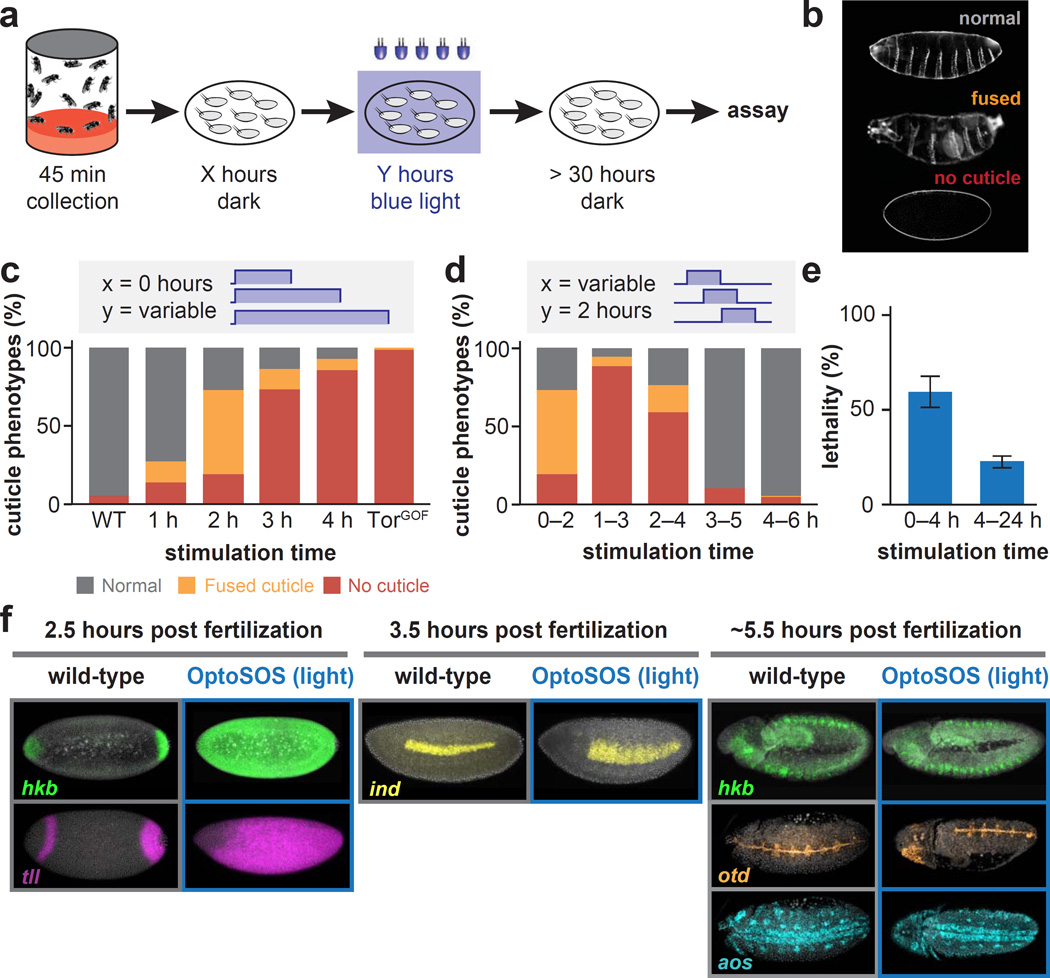
Animal development is characterized by signaling events that occur at precise locations and times within the embryo, but determining when and where such precision is needed for proper embryogenesis has been a long-standing challenge. Here we address this question for extracellular signal regulated kinase (Erk) signaling, a key developmental patterning cue. We describe an optogenetic system for activating Erk with high spatiotemporal precision in vivo. Implementing this system in Drosophila, we find that embryogenesis is remarkably robust to ectopic Erk signaling, except from 1 to 4 hr post-fertilization, when perturbing the spatial extent of Erk pathway activation leads to dramatic disruptions of patterning and morphogenesis. Later in development, the effects of ectopic signaling are buffered, at least in part, by combinatorial mechanisms. Our approach can be used to systematically probe the differential contributions of the Ras/Erk pathway and concurrent signals, leading to a more quantitative understanding of developmental signaling.

Keywords: Drosophila; MAP kinase; embryogenesis; optogenetics; signal transduction.


Figures

Figure 1
Figure 1. Light-mediated activation of signaling pathways in vivo
(A) Schematic of optogenetic control of Erk signaling. An upstream activation sequence drives tissue-specific expression of both optogenetic components, tRFP-SSPB-SOScat and iLID-CAAX, which are cleaved by a P2A sequence into separate peptides. Recruiting SOScat to the membrane with light activates the Ras/Erk cascade. (B) Quantification of membrane SOScat recruitment over time for varying light intensities. (C) Local illumination (left panel) can be used to generate spatially precise patterns of membrane SOScat recruitment (middle and right panels). See also Figure S1.
Figure 2
Figure 2. Light stimulation induces global Erk activity and downstream gene expression
(A) Comparison of Erk activity in wild-type, unstimulated and light-stimulated OptoSOS embryos at nuclear cycle 14. Embryos were stained for dpErk (red) and DAPI (white). (B) Quantification of dpErk levels from embryos stimulated as in panel a (mean +/− SEM). (C) Fluorescence in situ hybridization for tailless (til) in wild-type or light-simulated OptoSOS embryos at nuclear cycle 14. (D) dpErk activation in 12 hour old OptoSOS embryos stimulated for 1 h with light, compared with similar age wild-type embryos. Scale bar is 100 urn in all panels. (E) Quantified dpErk intensity along line scans indicated by the dashed lines in (D). See Table S1 for numbers of embryos/replicates; see also Figure S2.
Figure 3
Figure 3. Consequences of perturbing the level and location of Erk activity
(A) Schematic of our setup where a digital micromirror device was used to apply spatially-patterned light to each embryo. (B) Lethality of local Erk activation from 1–3 hours post fertilization (mean + SD). Light was applied to ∼15% of the embryo at a pole, or the middle ∼45% of the embryo. The number of hatched larvae was counted. (C) Lethality after illuminating progressively decreasing stimulation areas in the middle of the embryo (mean + SD). (D) Cuticle phenotypes from GOF mutations in the Erk pathway (TorGOF and MekGOF). (E) Quantification of cuticle phenotypes from the experiment shown in (A). (F) Capicua staining in WT and OptoSOS embryos after 1 hr of illumination. (G) Quantification of Capicua staining as shown in (F) (mean +/− SEM). (H) Left panels: schematic of experiment: OptoSOS embryos were spatially illuminated during nuclear cycles 13–14, and observed through gastrulation. Right panels: still images of OptoSOS embryos exhibiting tissue contraction during gastrulation. See Table S1 for numbers of embryos/replicates; see also Figure S3.
Figure 4
Figure 4. A temporal window of sensitivity to ectopic Erk activation
(A) Embryos are collected for 45 minutes after which they are placed in the dark for X hours before being stimulated globally for Y hours under blue light. After allowing sufficient time to hatch, embryos are then assayed for lethality and segmentation defects. (B) Cuticle phenotypes obtained by varying light exposure duration and start time. (C-D) Quantification of cuticle defects varying light duration (C) or start time (D), compared to wild-type and a TorGOF mutant. (E) Lethality after stimulation with an intermediate light intensity from 0–4 hours after collection versus from 4 hours until hatching (mean +/− SEM). (F) Fluorescence in situ hybridization for hkb, tll, ind, otd , and aos at the indicated developmental stages for wild-type and light illuminated OptoSOS embryos. See Table S1 for numbers of embryos/replicates; see also Figure S4.

Comment in

  • Lighting Up ERK Activity.
    Shilo BZ, Barkai N. Shilo BZ, et al. Dev Cell. 2017 Jan 23;40(2):115-116. doi: 10.1016/j.devcel.2016.12.016. Dev Cell. 2017. PMID: 28118596

References

    1. Bischof J, Maeda RK, Hediger M, Karch F, Basler K. An optimized transgenesis system for Drosophila using germ-line-specific phiC31 integrases. Proceedings of the National Academy of Sciences of the United States of America. 2007;104:3312–3317. - PMC - PubMed
    1. Brunner D, Oellers N, Szabad J, Biggs WH, 3rd, Zipursky SL, Hafen E. A gain-of-function mutation in Drosophila MAP kinase activates multiple receptor tyrosine kinase signaling pathways. Cell. 1994;76:875–888. - PubMed
    1. Buckley CE, Moore RE, Reade A, Goldberg AR, Weiner OD, Clarke JD. Reversible Optogenetic Control of Subcellular Protein Localization in a Live Vertebrate Embryo. Developmental cell. 2016;36:117–126. - PMC - PubMed
    1. Corson LB, Yamanaka Y, Lai KM, Rossant J. Spatial and temporal patterns of ERK signaling during mouse embryogenesis. Development. 2003;130:4527–4537. - PubMed
    1. Daniels RW, Rossano AJ, Macleod GT, Ganetzky B. Expression of multiple transgenes from a single construct using viral 2A peptides in Drosophila. PloS one. 2014;9:e100637. - PMC - PubMed

Publication types

MeSH terms

Substances