. 2017 Feb 7;8(6):9053-9066.
doi: 10.18632/oncotarget.14754.

Middle east respiratory syndrome corona virus spike glycoprotein suppresses macrophage responses via DPP4-mediated induction of IRAK-M and PPARγ

Affiliations

Middle east respiratory syndrome corona virus spike glycoprotein suppresses macrophage responses via DPP4-mediated induction of IRAK-M and PPARγ

Ahmed A Al-Qahtani et al. Oncotarget. .

Abstract

Middle East Respiratory Syndrome Corona Virus (MERS-CoV) is transmitted via the respiratory tract and causes severe Acute Respiratory Distress Syndrome by infecting lung epithelial cells and macrophages. Macrophages can readily recognize the virus and eliminate it. MERS-CoV infects cells via its Spike (S) glycoprotein that binds on Dipeptidyl-Peptidase 4 (DPP4) receptor present on macrophages. Whether this Spike/DPP4 association affects macrophage responses remains unknown. Herein we demonstrated that infection of macrophages with lentiviral particles pseudotyped with MERS-CoV S glycoprotein results in suppression of macrophage responses since it reduced the capacity of macrophages to produce TNFα and IL-6 in naive and LPS-activated THP-1 macrophages and augmented LPS-induced production of the immunosuppressive cytokine IL-10. MERS-CoV S glycoprotein induced the expression of the negative regulator of TLR signaling IRAK-M as well as of the transcriptional repressor PPARγ. Inhibition of DPP4 by its inhibitor sitagliptin or siRNA abrogated the effects of MERS-CoV S glycoprotein on IRAK-M, PPARγ and IL-10, confirming that its immunosuppressive effects were mediated by DPP4 receptor. The effect was observed both in THP-1 macrophages and human primary peripheral blood monocytes. These findings support a DPP4-mediated suppressive action of MERS-CoV in macrophages and suggest a potential target for effective elimination of its pathogenicity.

Keywords: DPP4; IRAK-M; Immune response; Immunity; Immunology and Microbiology Section; MERS CoV; cytokines; macrophages.


Conflict of interest statement

CONFLICTS OF INTEREST

The authors have no conflicts of interest to declare.

Figures
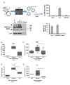
Figure 1
Figure 1. MERS-CoV S glycoprotein suppressed LPS-induced cytokine production in THP1 cells
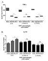
A. Lentiviral particles pseudotyped with MERS S glycoprotein were generated in HEK293T cells following transfection with plasmid pNL4.3R-E-luciferase and pcDNA3.1+ containing the MERS-CoV wild-type or D510A mutant spike glycoprotein. Normalized supernatants were used to infect THP-1 cells and infection was monitored by luciferase assay. The C9-tagged MERS-CoV Spike glycoprotein was detected by Western blot in purified lentiviral particles. HIV-1 p24 envelope protein was used as a normalizing control for the semi-quantitative analysis of MERS-CoV S protein among different types of pseudotyped viruses. THP-1 macrophages were infected with lentiviral particles pseudotyped with MERS wild type (wt) or the D510 mutant S glycoprotein (D510A) of MERS-CoV, or left uninfected (mock), and were stimulated with LPS. TNFα was measured in the supernatants at the naïve state B. and following LPS stimulation C. IL-6 was also measured in naïve and LPS-treated THP1 macrophages D., E. * p < 0.05, **p < 0.01
Figure 2
Figure 2. MERS-CoV S glycoprotein augmented LPS-induced IL-10 production
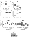
A., B. THP-1 macrophages were infected with lentiviral particles pseudotyped with MERS wild type (wt) or the D510A mutant S glycoprotein (D510A) of MERS-CoV, or left uninfected (mock), and were stimulated with LPS. IL-10 mRNA was measured in the cell extracts. C. IL-10 protein was measured in the supernatants of infected THP1 macrophages at the naïve state and following LPS stimulation. ns: not significant, * p < 0.05, ***p < 0.001
Figure 3
Figure 3. Interaction of MERS-CoV S glycoprotein with DPP4 induced IRAK-M and PPARγ expression
A., B. THP-1 macrophages were infected with lentiviral particles pseudotyped with MERS wild type (wt) or the D510A mutant S glycoprotein (D510A) of MERS-CoV, or left uninfected (mock) in the presence or absence of the DPP4 inhibitor sitagliptin or siRNA targeting DPP4. 24 hours post-infection RNA was isolated and the levels of IRAK-M A., B. and PPARγ C., D. and IL-10 E., F. were measured. ns: not significant, * p < 0.05, **p < 0.01
Figure 4
Figure 4. Time-course of IRAK-M and PPARγ expression following infection
THP-1 macrophages were infected with lentiviral particles pseudotyped with MERS CoV wild type S glycoprotein (wt) or the D510A mutant S glycoprotein (D510A) of MERS-CoV, or left uninfected (mock) for 12, 24 and 48 hours. Timepoint 0 corresponds to mock-treated cells. RNA was isolated at the respective time-points and the levels of IRAK-M A.-C. and PPARγ D.-F. were measured by RT-PCR. Normalization was performed against mock-treated cells in each time point. ns: not significant, * p < 0.05.
Figure 5
Figure 5. Suppression of TNFα and induction of IL-10 by MERS-CoV S glycoprotein is mediated by IRAK-M and PPARγ
THP1 macrophages were transfected with siRNAs targeting IRAK-M or PPARγ and subsequently infected with lentiviral particles pseudotyped with MERS CoV wild type S glycoprotein (wt) or the D510A mutant S glycoprotein (D510A) of MERS-CoV, or left uninfected (mock). Production of TNFα A. and IL-10 B. in response to LPS was measured in culture supernatants. ns: not significant, * p < 0.05, **p < 0.01
Figure 6
Figure 6. Infection of primary human monocytes with MERS-CoV S glycoprotein pseudotyped particles
Primary human monocytes were isolated from buffy coats of healthy human donors and were infected with lentiviral particles pseudotyped with MERS wild type (wt S) or the D510A mutant S glycoprotein (D510A) of MERS-CoV, or left uninfected (mock) for 24 hours, in the presence or absence of the DPP4 inhibitor sitagliptin, and were stimulated with LPS. TNFa A.,B. and IL-10 C.,D. secretion was quantified by ELISA. Expression of IRAK-M E. and PPARγ F. mRNA and protein G. in naïve human macrophages quantified by RT-PCR and protein levels by Western blot, respectively. Results are representative of three independent experiments. ns: not significant, * p < 0.05, **p < 0.01, ***p < 0.001
Figure 7
Figure 7. Model of MERS-CoV S glycoprotein immunosuppressive action

References

    1. Zaki AM, van Boheemen S, Bestebroer TM, Osterhaus AD, Fouchier RA. Isolation of a novel coronavirus from a man with pneumonia in Saudi Arabia. N Engl J Med. 2012;367:1814–20. - PubMed
    1. Kucharski AJ, Althaus CL. The role of superspreading in Middle East respiratory syndrome coronavirus (MERS-CoV) transmission. Euro Surveill. 2015;20 - PubMed
    1. Assiri A, Al-Tawfiq JA, Al-Rabeeah AA, Al-Rabiah FA, Al-Hajjar S, Al-Barrak A, Flemban H, Al-Nassir WN, Balkhy HH, Al-Hakeem RF, Makhdoom HQ, Zumla AI, Memish ZA. Epidemiological, demographic, and clinical characteristics of 47 cases of Middle East respiratory syndrome coronavirus disease from Saudi Arabia: a descriptive study. Lancet Infect Dis. 2013;13:752–61. - PMC - PubMed
    1. Drosten C, Seilmaier M, Corman VM, Hartmann W, Scheible G, Sack S, Guggemos W, Kallies R, Muth D, Junglen S, Muller MA, Haas W, Guberina H, et al. Clinical features and virological analysis of a case of Middle East respiratory syndrome coronavirus infection. Lancet Infect Dis. 2013;13:745–51. - PMC - PubMed
    1. Assiri A, McGeer A, Perl TM, Price CS, Al Rabeeah AA, Cummings DA, Alabdullatif ZN, Assad M, Almulhim A, Makhdoom H, Madani H, Alhakeem R, Al-Tawfiq JA, et al. Hospital outbreak of Middle East respiratory syndrome coronavirus. N Engl J Med. 2013;369:407–16. - PMC - PubMed

MeSH terms