. 2017 Feb 7;8(6):9035-9052.
doi: 10.18632/oncotarget.14764.

The involvement of endoplasmic reticulum stress response in immune dysfunction of dendritic cells after severe thermal injury in mice

Affiliations

The involvement of endoplasmic reticulum stress response in immune dysfunction of dendritic cells after severe thermal injury in mice

Xiao-Mei Zhu et al. Oncotarget. .

Abstract

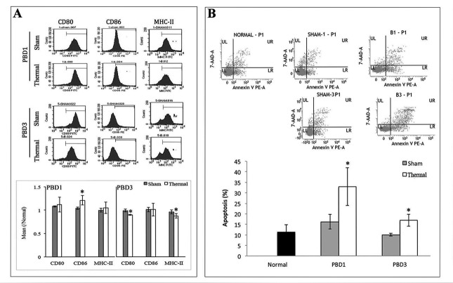
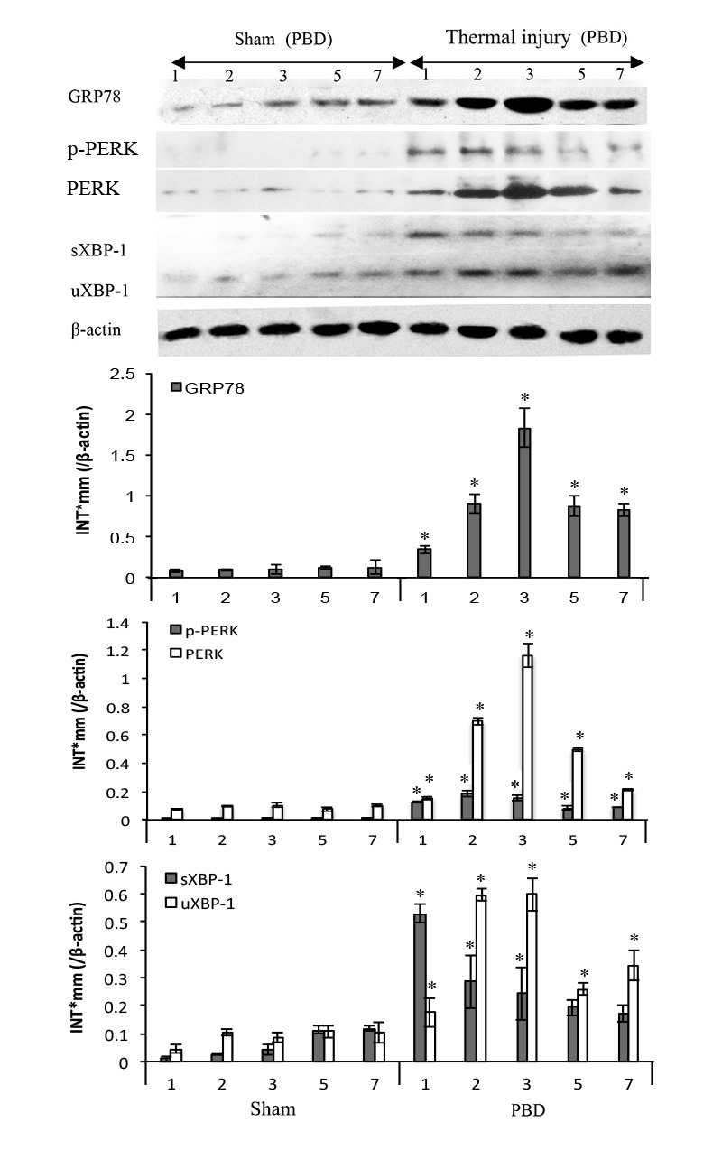
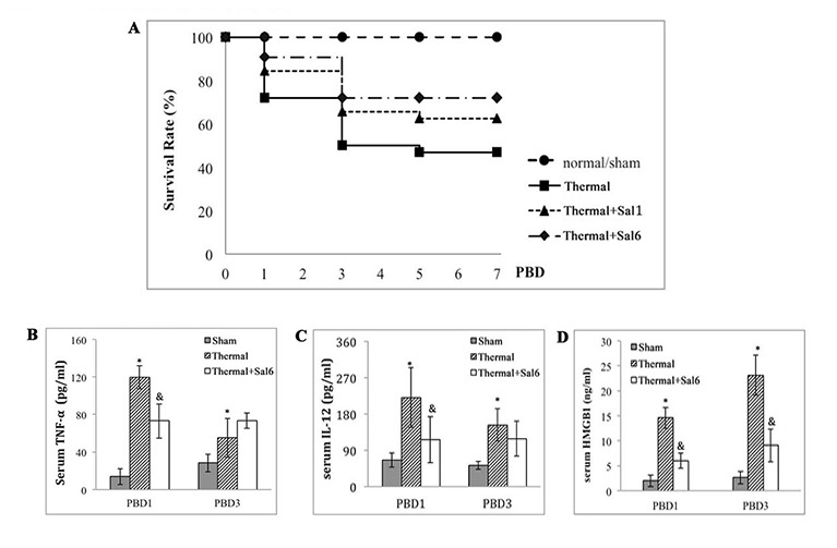
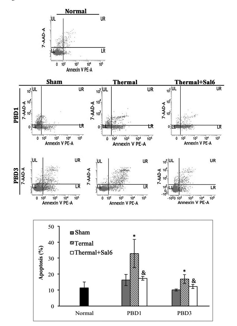
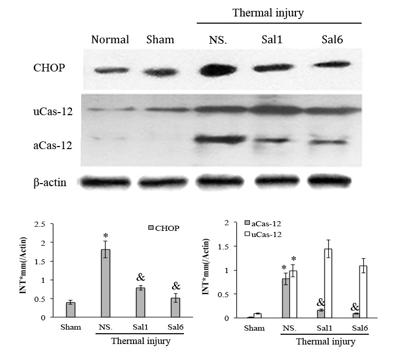
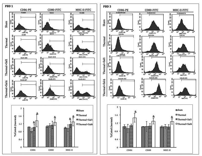
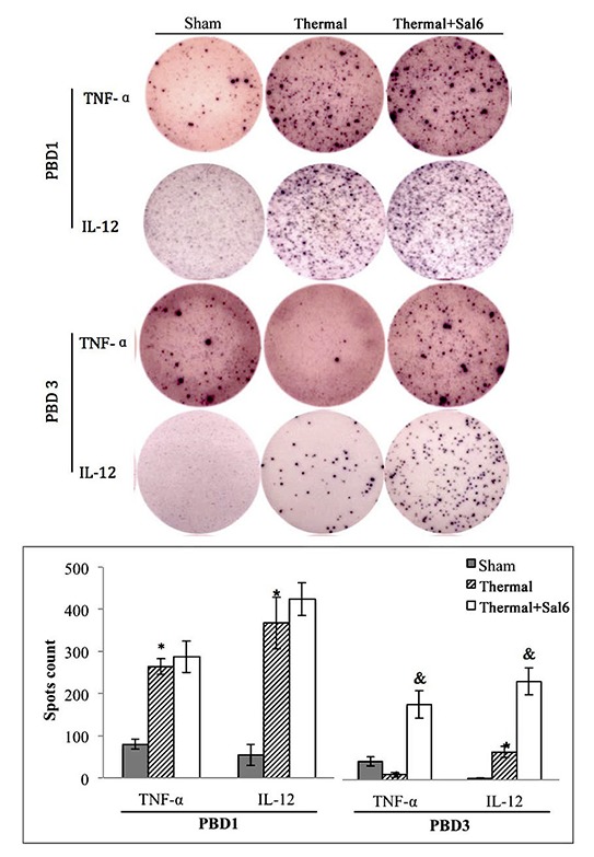
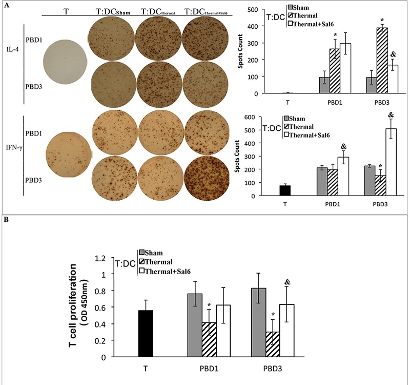
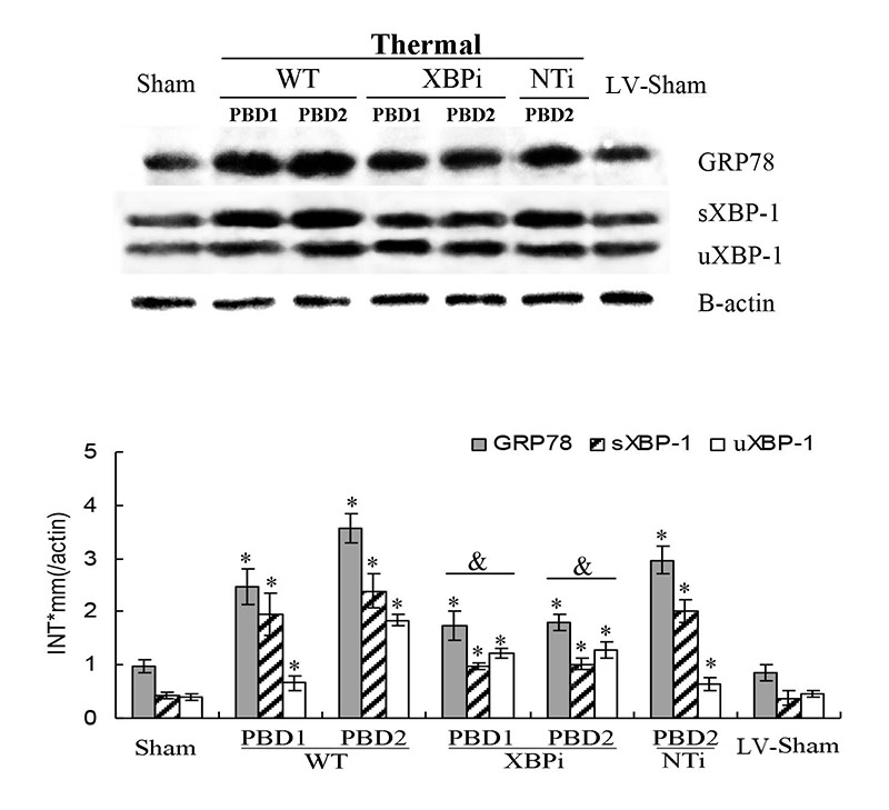
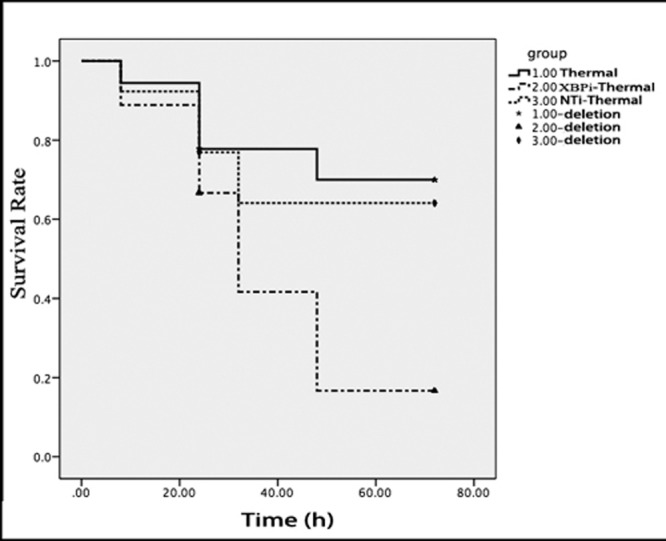
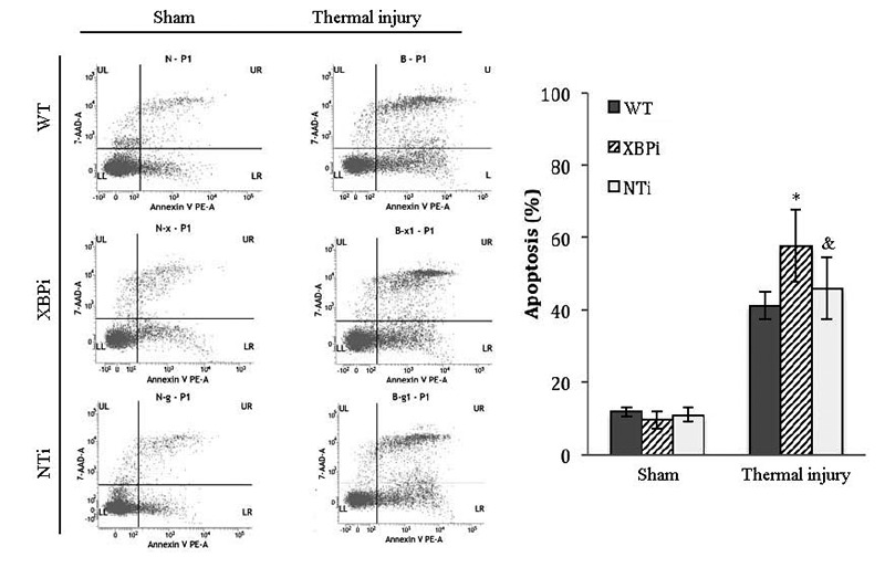
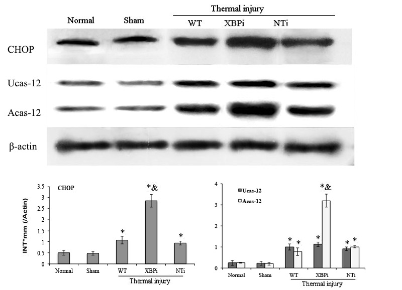
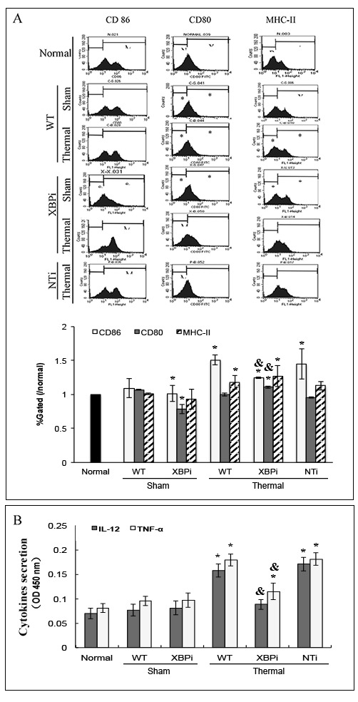
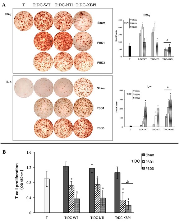
Suppressed adaptive immune function is one of the major concerns responsible for the development of opportunistic infections and subsequent sepsis with high mortality in severe burns. Endoplasmic reticulum stress (ERS) is the endogenous self-protective mechanism, and it plays an important role in almost every process of living by regulating the balance between homeostasis and apoptosis. The current study investigated the involvement of ERS in the pathogenesis of dysfunction of dendritic cells (DCs) in burn mice. Our results show a significant ERS response in splenic DC after burn injury. Treatment with salubrinal (Sal, reported to protect cells against ERS-induced apoptosis.) decrease the apoptotic rate of DC induced by burns, and promote maturation and activation of DC, as well as the ability to promote T cell proliferation and polarization towards Th1 immunity (all P<0.05). Gene silence of XBP-1 (key molecular in ERS response) results in the increased apoptosis and suppressed phenotypical maturation of splenic DC in burn mice. These results show that the excessive ERS is essential for immunosuppression during severe thermal injury. XBP-1 plays a pivotal role in DC functional immunomodulation in burn mice. Inhibition of apoptotic ERS response benefits mice from major burns.

Keywords: Pathology Section; burns; dendritic cell; endoplasmic reticulum stress; immune dysfunction; sepsis.


Conflict of interest statement

COMPETING INTERESTS

No authors have any financial interests to disclose.

Figures

Figure 1
Figure 1. Thermal injury induced dysfunction of splenic DC in mice
Splenic DCs were isolated with CD11c MicroBeads from burn mice. A. The expression of co-stimulatory molecules including CD80, CD86 and MHC-II on surface of DCs was determined by flow cytometry and analyzed by the ratios to normal value in the same batch. B. Apoptosis rate of splenic DCs was determined by flow cytometric analysis. The apoptosis rates were assessed using the sum of area UR (Annexin-V/7-AAD indicates late apoptotic cells) and area LR (Annexin-V+/7-AAD- indicates early apoptotic cells). Results were shown in mean ± SD (n = 6). * Statistically significant difference when compared with sham group at the same time point (p < 0.05).
Figure 2
Figure 2. Thermal injury induced ERS response in splenic DC in mice
Expressions of GRP78, PERK, p-PERK, XBP-1 (including full length and spliced form) in DC were assessed by Western blot analysis. Representative images from the same membrane were shown. Data were shown in mean ± SD (n = 4). * Statistically significant difference when compared with sham group at the same time point (p < 0.05). P.s uXBP-1: full length XBP-1 (unactivated). sXBP-1: spliced XBP-1(activated)
Figure 3
Figure 3. Protective effects of Sal on thermal injured mice
A. Survival rate of burn mice were monitored for over 7 d (= 48). Sal (1mg/kg) was administered at 1 h (Sal1) or 6 h (Sal6) after thermal injury. B.-D. Inflammatory mediators in serum of thermal mice model were assessed with ELISA (n = 6). Sal (1mg/kg) was administered at 6 h after thermal injury. * Statistically significant difference when compared with sham group at the same time point (p < 0.05). & Statistically significant difference when compared with thermal-injured group at the same time point (p < 0.05).
Figure 4
Figure 4. Sal-treatment decreased the apoptosis rate of splenic DCs in thermal injured mice
Splenic DCs were isolated with CD11c+ MicroBeads from mice model. Apoptosis rate of splenic DCs was determined by flow cytometric analysis. Sal (1mg/kg) was administered at 6 h after thermal injury. The apoptosis rates were assessed using the sum of area UR (Annexin-V+/7-AAD- indicates late apoptotic cells) and area LR (Annexin-V+/7-AAD- indicates early apoptotic cells). Results were shown in mean ± SD (n = 6). * Statistically significant difference when compared with sham group at the same time point (p < 0.05). & Statistically significant difference when compared with thermal-injured group (p < 0.05).
Figure 5
Figure 5. Sal-treatment relieved the ERS response in splenic DCs in thermal injured mice
Splenic DCs were isolated from mice model on PBD 1. The expression of ERS markers in splenic DCs was detected with Western blot analysis. Sal (1mg/kg) was administered at 6 h after thermal injury. Representative images from the same membrane were shown. Data were shown in mean ± SD (n = 4). * Statistically significant difference when compared with Sham group (p < 0.05). & Statistically significant difference when compared with thermal-injured group (p < 0.05). P.s uCas-12: unactivated caspase-12. aCas-12: activated caspase-12.
Figure 6
Figure 6. Sal-treatment promote the phenotypic maturation of splenic DC in thermal injured mice
Splenic DCs were isolated with CD11c+ MicroBeads from mice model. The expression of co-stimulatory molecules including CD80, CD86 and MHC-II on surface of DCs was determined by flow cytometry and analyzed by the ratios to normal value in the same batch. Sal (1mg/kg) was administered at 1 h (Sal1) or 6 h (Sal6) after thermal injury. Results were shown in mean ± SD (n = 6). * Statistically significant difference was found when compared with sham group at the same time point (p < 0.05). & Statistically significant difference when compared with thermal-injured group (p < 0.05).
Figure 7
Figure 7. Sal treatment improved the secretion ability of splenic DCs in thermal injured mice
Splenic DCs were isolated with CD11c+ MicroBeads from mice model and plated to the microplate of ELISpot Assay (2*105cell/well) and cultured overnight (18 hours). Spots were analyzed with ELISpot reader system. Sal (1mg/kg) was administered at 6 h after thermal injury. Results were shown in mean ± SD (n = 6). * Statistically significant difference when compared with sham group at the same time point (p < 0.05). & Statistically significant difference when compared with thermal-injured group (p < 0.05).
Figure 8
Figure 8. Sal-treatment ameliorated regulating ability of splenic DCs on T cell polarization and proliferation
T cells were isolated from normal mice, stimulated with soluble CD3 (1 μg/ml) and soluble CD28 (5 μg/ml) for 24 hours. Splenic DCs were isolated with CD11c+ MicroBeads from mice model and co-cultured with T mentioned above at a DC: T ratio of 1: 200 (the concentration of T was 2*105cell/well) and cultured for 3 days. Production of IFN-γ and IL-4 was assessed with ELISpot Assay A. T cell proliferation was detected with CCK-8 cell counting kit B. Sal (1mg/kg) was administered at 6 h after thermal injury. Results were shown in mean ± SD (n = 6). * Statistically significant difference when compared with sham group at the same time point (p < 0.05). & Statistically significant difference when compared with thermal-injured group (p < 0.05).
Figure 9
Figure 9. Effects of XBP-1 gene silence in splenic DC in thermal injured mice model
Mice transfected with XBPi-LV (3.5×10TU per mice) were subjected to thermal injury. Expressions of GRP78 and XBP-1 (including full length and spliced form) in splenic DCs were assessed by Western blot analysis. Representative images from the same membrane were shown. Data were shown in mean ± SD (n = 4). * Statistically significant difference when compared with sham group at the same time point (p < 0.05). Statistically significant difference when compared with thermal injured mice in WT or NTi group (p < 0.05).
Figure 10
Figure 10. Gene silence of XBP-1 lowered the survival rate of thermal-injured mice on PBD1
Totally18 normal mice, 18 mice injected with XBPi-LV and 13 mice injected with NTi-LV were subjected to thermal injury in this experiment. Four live mice on PBD1 and two on PBD3 were picked randomly from every group and killed for sample collection. Log-rank test was used to compare the survival times of the rest mice till 72 hours after thermal injury (p = 0.041). The survival rate of XBPi- group was markedly lower than that of normal mice (p = 0.017).
Figure 11
Figure 11. XBPi-transfection increased the apoptosis rate of splenic DCs in burn mice on PBD1
Splenic DCs were isolated with CD11c+ MicroBeads from mice model on PBD1. Apoptosis rate of splenic DCs was determined by flow cytometric analysis. The apoptosis rates were assessed using the sum of area UR (Annexin-V+/7-AAD- indicates late apoptotic cells) and area LR (Annexin-V+/7-AAD- indicates early apoptotic cells). Results were shown in mean ± SD (n = 4). * Statistically significant difference when compared with sham group at the same time point (< 0.05). & Statistically significant difference when compared with thermal-injured group (p < 0.05).
Figure 12
Figure 12. Gene silence of XBP-1 up-regulated the activation of ERS-related apoptosis pathway in splenic DCs in thermal injury
Splenic DCs were isolated from mice model on PBD 1. The expression of CHOP and activation of caspase-12 in splenic DCs was detected with Western blot analysis. Representative images from the same membrane were shown. Data were shown in mean ± SD (n = 4). * Statistically significant difference when compared with Sham group (n < 0.05). & Statistically significant difference when compared with WT or NTi group (p < 0.05).
Figure 13
Figure 13. XBPi-transfection affected the surface phenotype and cytokines secretion of splenic DC in mice
Splenic DCs were isolated with CD11c+ MicroBeads from mice model on PBD1 and cultured at 37°C in a humidified atmosphere with 5% CO2 overnight. A. The expression of co-stimulatory molecules including CD80, CD86 and MHC-II on surface of DCs was determined by flow cytometry and analyzed by the ratios to normal value in the same batch. B. Protein level of TNF-α and IL-12 in cell culture medium was determined with ELISA. Results were shown in mean ± SD (n = 4). * Statistically significant difference when compared with sham group (p < 0.05). & Statistically significant difference when compared XBPi with WT or NTi group (p < 0.05).
Figure 14
Figure 14. DCs from XBPi-burn mice could induce more notable hypo-responsiveness of CD4+ T
Normal CD4 T (2*105cell/well), after being stimulated with soluble CD3 (1 μg/ml) and soluble CD28 (5 μg/ml) for 24 hours, were co-cultured with splenic DCs isolated from XBPi-burn mice model for 3 days at a DC: T ratio of 1: 200 cultured. Production of IFN-γ and IL-4 were assessed with ELISpot Assay. T cell proliferation was detected with CCK-8 cell counting kit. Results were shown in mean ± SD (n = 6). * Statistically significant difference when compared with WT-sham group (p < 0.05). & Statistically significant difference when compared XBPi with WT or NTi group at the same time points (p < 0.05).

References

    1. Jeschke MG, Pinto R, Kraft R, Nathens AB, Finnerty CC, Gamelli RL, Gibran NS, Klein MB, Arnoldo BD, Tompkins RG, Herndon DN. Inflammation and the Host Response to Injury Collaborative Research Program. Morbidity and survival probability in burn patients in modern burn care. Crit Care Med. 2015;43(4):808–15. doi: 10.1097/CCM.0000000000000790. - DOI - PMC - PubMed
    1. Bahemia IA, Muganza A, Moore R, Sahid F, Menezes CN. Microbiology and antibiotic resistance in severe burns patients: A 5 year review in an adult burns unit. Burns. 2015;41(7):1536–42. doi: 10.1016/j.burns.2015.05.007. - DOI - PubMed
    1. O’Sullivan ST. O’Connor TP. Immunosuppression following thermal injury: the pathogenesis of immunodysfunction. Br J Plast Surg. 1997;50(8):615–23. - PubMed
    1. Lederer JA, Rodrick ML, Mannick JA. The effects of injury on the adaptive immune response. Shock. 1999;11(3):153–9. - PubMed
    1. Barlow Y. T lymphocytes and immunosuppression in the patient: a review. Burns. 1994;20(6):487–90. - PubMed

MeSH terms