Review
. 2016 Dec;11(4):489-501.
doi: 10.1016/j.jsmc.2016.07.003. Epub 2016 Oct 27.

Model-Derived Markers of Autonomic Cardiovascular Dysfunction in Sleep-Disordered Breathing

Affiliations
Review

Model-Derived Markers of Autonomic Cardiovascular Dysfunction in Sleep-Disordered Breathing

Michael C K Khoo et al. Sleep Med Clin. 2016 Dec.

Abstract

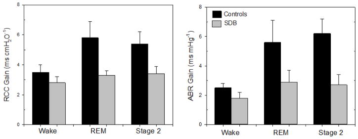
Evidence indicates that sleep-disordered breathing leads to elevated sympathetic tone and impaired vagal activity, promoting hypertension and cardiometabolic disease. Low-cost but accurate monitoring of autonomic function is useful for the aggressive management of sleep apnea. This article reviews the development and application of multivariate dynamic biophysical models that enable the causal dependencies among respiration, blood pressure, heart rate variability, and peripheral vascular resistance to be quantified. The markers derived from these models can be used in conjunction with heart rate variability to increase the sensitivity with which abnormalities in autonomic cardiovascular control are detected in subjects with sleep-disordered breathing.

Keywords: Cardiorespiratory control; Heart rate variability; Minimal model; Peripheral vascular resistance; Sleep apnea.


Figures

Figure 1
Figure 1
Schematic block diagram of the closed-loop minimal model of heart rate and blood pressure variability. Fluctuations in R-R interval (RRI) result from respiratory-cardiac coupling (RCC) and a component mediated through the arterial baroreflexes (ABR). Other factors are represented as “extraneous influences on RRI”. These components account for the “forward” part of the model that generates heart rate variability (rectangular block with broken lines). The “other half” of the closed-loop model consists of the components responsible for generating blood pressure variability: fluctuations in cardiac output (from heart rate and stroke volume changes), fluctuations in systemic vascular resistance, and respiration,
Figure 2
Figure 2
Effects of sleep-disordered breathing and sleep-wake state on the estimated impulse response magnitudes of the RCC (left panel) and ABR (right panel) components of the model. Adapted from: Jo et al., Am J Physiol Heart Circ Physiol 288: H1103–H1112, 2005.
Figure 3
Figure 3
(Left) Data collected from a subject during spontaneous breathing. “BP” represents beat-averaged arterial blood pressure, “ΔV” is change in lung volume (respiration), and “PATamp” is the beat-to-beat amplitude of the peripheral arterial tonometer signal. “BP” and “ΔV” are considered the inputs, and “PATamp”, taken to represent the change in peripheral vascular conductance (ΔGPV), is treated as the output. (Right) The linear and nonlinear components of the model (see Equation 4) are estimated from these data.
Figure 4
Figure 4
Autonomic reactivity to orthostatic stress in obese adolescents with sleep-disordered breathing (SDB) and insulin resistance versus obese adolescent controls. The model-derived marker shown here is the magnitude of the blood pressure to peripheral vascular conductance nonlinear kernel. BPC nonlinear gain decreased significantly more in SDB subjects compared to controls following change in posture from supine to standing. Lines connect the supine and standing values of each individual. Closed circles and error bars represent group means and standard errors of the parameter estimates.

Similar articles

Cited by

References

    1. Leung RS, Bradley TD. Sleep apnea and cardiovascular disease. Am J Respir Crit Care Med. 2001;164(12):2147–2165. doi: 10.1164/ajrccm.164.12.2107045. - DOI - PubMed
    1. Caples SM, Garcia-Touchard A, Somers VK. Sleep-disordered breathing and cardiovascular risk. [Accessed April 19, 2016];Sleep. 2007 30(3):291–303. http://www.ncbi.nlm.nih.gov/pubmed/17425225. - PubMed
    1. Peppard PE, Young T, Palta M, Skatrud J. Prospective study of the association between sleep-disordered breathing and hypertension. N Engl J Med. 2000;342(19):1378–1384. doi: 10.1056/NEJM200005113421901. - DOI - PubMed
    1. Shahar E, Whitney CW, Redline S, et al. Sleep-disordered breathing and cardiovascular disease: cross-sectional results of the Sleep Heart Health Study. Am J Respir Crit Care Med. 2001;163(1):19–25. doi: 10.1164/ajrccm.163.1.2001008. - DOI - PubMed
    1. Nieto FJ, Young TB, Lind BK, et al. Association of sleep-disordered breathing, sleep apnea, and hypertension in a large community-based study. Sleep Heart Health Study. [Accessed April 19, 2016];JAMA. 2000 283(14):1829–1836. http://www.ncbi.nlm.nih.gov/pubmed/10770144. - PubMed

MeSH terms