Review
. 2017 Jul 15:529:193-215.
doi: 10.1016/j.ab.2017.01.018. Epub 2017 Jan 21.

In-vivo31P-MRS of skeletal muscle and liver: A way for non-invasive assessment of their metabolism

Affiliations
Review

In-vivo31P-MRS of skeletal muscle and liver: A way for non-invasive assessment of their metabolism

Ladislav Valkovič et al. Anal Biochem. .

Abstract

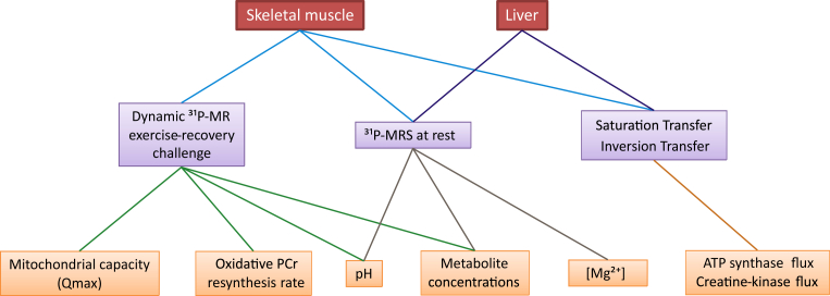
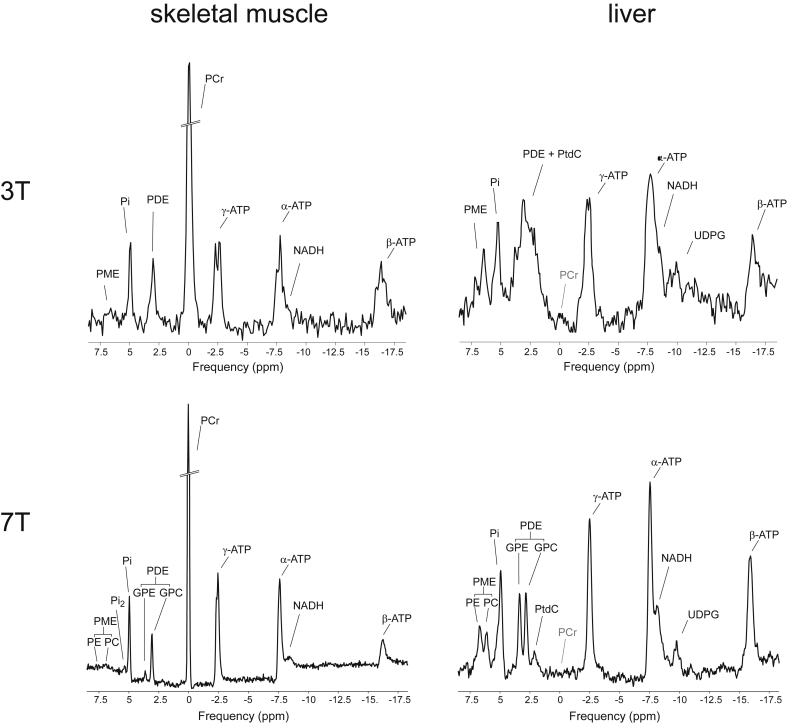
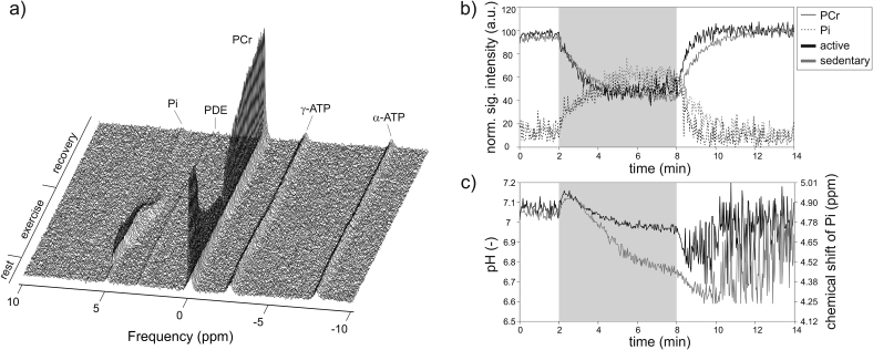
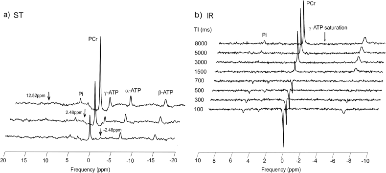
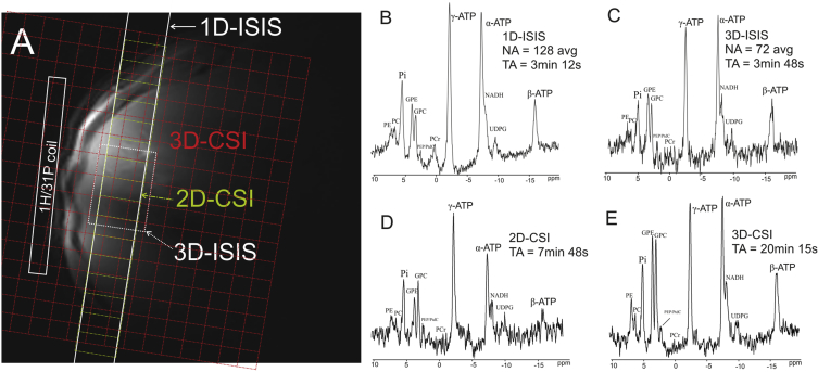
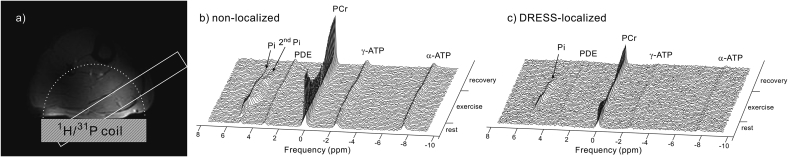
In addition to direct assessment of high energy phosphorus containing metabolite content within tissues, phosphorus magnetic resonance spectroscopy (31P-MRS) provides options to measure phospholipid metabolites and cellular pH, as well as the kinetics of chemical reactions of energy metabolism in vivo. Even though the great potential of 31P-MR was recognized over 30 years ago, modern MR systems, as well as new, dedicated hardware and measurement techniques provide further opportunities for research of human biochemistry. This paper presents a methodological overview of the 31P-MR techniques that can be used for basic, physiological, or clinical research of human skeletal muscle and liver in vivo. Practical issues of 31P-MRS experiments and examples of potential applications are also provided. As signal localization is essential for liver 31P-MRS and is important for dynamic muscle examinations as well, typical localization strategies for 31P-MR are also described.

Keywords: Energy metabolism; Exercise-recovery; Liver; Phosphorus magnetic resonance spectroscopy; Saturation transfer; Skeletal muscle.


Figures

Fig. 1
Fig. 1
An overview of the biochemical parameters (orange) assessed and quantified through in vivo 31P-MRS/MRSI experimental approaches reviewed and discussed in the text (violet) in the organs of interest, i.e. skeletal muscle and liver, (red).
Fig. 2
Fig. 2
Typical 31P-MR spectra acquired at 3 T (top) and 7 T (bottom), at rest, in skeletal muscle (left) and liver tissue (right). All spectra are depicted relative to the resonance frequency of phosphocreatine (PCr), although this is not present in healthy human liver tissue. Phosphorus metabolites common to both tissues include resonance lines of adenosine-triphosphate (ATP), nicotinamide adenine dinucleotide (NADH), phosphodiesters (PDEs) – glycerol-phoshocholine (GPC) and glycerol-phosphoethanolamine (GPE), inorganic phosphate (Pi) and phosphomonoesters (PMEs) – phosphocholine (PC) and phopshoethanolamine (PE). Note that PDEs and PMEs are readily resolved at 7 T. Another metabolite resolved in the muscle at 7 T is the recently described alkaline Pi (Pi2) pool. The liver spectrum, on the other hand, contains a resonance line of uridine diphosphate glucose (UDPG) and also a recently assigned spectral line of the bile component phosphatidylcholine (PtdC). Note that, due to the large frequency range at 7 T and the in vivo linewidth, the J-coupling of the ATP resonances is no longer resolved and the frequency limitations of the excitation pulse cause the β-ATP frequency line to be suppressed.
Fig. 3
Fig. 3
Bar plot showing a significantly higher Pi2/Pi (marked here as Pi2/Pi1) in the endurance trained athletes compared to the normal physical active group. Similarly, the Pi2/Pi was found lower in overweight-to-obese sedentary subjects than in lean, active individuals . Figure was reproduced from Ref. .
Fig. 4
Fig. 4
The 31P-MRS dynamic experiment with an isotonic aerobic exercise at a single workload (25% of maximal voluntary contraction force). A stack of dynamically acquired spectra is depicted in a). Note that while PCr depletes and Pi rises, ATP levels remain constant. The time-courses of the normalized PCr (full line) and Pi (dotted line) signal intensities are given in b). Panel c) shows the dynamic evolution of the calculated pH based on the chemical shift of Pi. In order to demonstrate the differences in training status observable by dynamic 31P-MRS, data from a regularly active (black lines) and sedentary (grey lines) volunteer are visualized in panels b) and c). The grey area indicates the 6-min long exercise period.
Fig. 5
Fig. 5
Standard continuous saturation transfer (cST) experiment performed in the gastrocnemius medialis muscle at 7 T. Saturation of the γ-ATP resonance and control spectra are depicted in a) and the inversion recovery (IR) experiment with continuous saturation of γ-ATP to determine the apparent T1app is shown in panel b). Arrows depict the saturation frequency in each experiment, i.e. in a) saturation of γ-ATP at −2.48 ppm (bottom); control saturation for the PCr-to-ATP reaction at 2.48 ppm (middle); and control saturation for the Pi-to-ATP reaction at 12.52 ppm (top). Figure was adapted from Ref. .
Fig. 6
Fig. 6
Typical 31P-MRS localization strategies for liver examinations. Localizer image of the human liver with indicated surface coil position overlaid with different localization volumes is given in a). Slab-selective 1D image-selected in vivo spectroscopy (ISIS) localization with the slab parallel to the coil, as shown by full white lines provides the spectrum depicted in b). The spectrum in c) was acquired by a single voxel 3D-ISIS technique visualized by the white dotted rectangle. 2D chemical shift imaging (CSI) localization, shown as the yellow dashed matrix, delivers the spectrum depicted in d). The spectrum in e) was acquired using a 3D-CSI technique – red dotted matrix. Figure was reproduced from Ref. .
Fig. 7
Fig. 7
A comparison of spectra acquired in a dynamic examination using simple topical surface-coil localization (non-localized) and a slice-selective, depth-resolved surface coil MRS (DRESS) localization. An in vivo localizer image with the depicted RF-coil position overlaid with localization volumes is given in a). The full line represents the DRESS selection placed over the gastrocnemius medialis, and the dotted line represents the approximate RF-coil sensitivity volume, containing several muscles. Stack plots of the 31P spectra acquired during rest, exercise and subsequent recovery are shown for the non-localized (b) and DRESS-localized (c) acquisitions. The spectra are scaled for equal noise to show the lower signal intensity of the localized experiment. On the other hand, the specificity to challenged muscle improves the dynamic range of PCr depletion. Note the Pi split in the non-localized data, which is lacking in the DRESS-localized MRS time course. Figure was reproduced from Ref. .

Similar articles

Cited by

References

    1. Ackerman J.J., Grove T.H., Wong G.G., Gadian D.G., Radda G.K. Mapping of metabolites in whole animals by 31P NMR using surface coils. Nature. 1980;283:167–170. - PubMed
    1. Chance B., Eleff S., Leigh J.S., Jr., Sokolow D., Sapega A. Mitochondrial regulation of phosphocreatine/inorganic phosphate ratios in exercising human muscle: a gated 31P NMR study. Proc. Natl. Acad. Sci. U. S. A. 1981;78:6714–6718. - PMC - PubMed
    1. Prompers J.J., Jeneson J.A., Drost M.R., Oomens C.C., Strijkers G.J., Nicolay K. Dynamic MRS and MRI of skeletal muscle function and biomechanics. NMR Biomed. 2006;19:927–953. - PubMed
    1. Brindle K.M., Blackledge M.J., Challiss R.A., Radda G.K. 31P NMR magnetization-transfer measurements of ATP turnover during steady-state isometric muscle contraction in the rat hind limb in vivo. Biochemistry. 1989;28:4887–4893. - PubMed
    1. Cadoux-Hudson T.A., Blackledge M.J., Radda G.K. Imaging of human brain creatine kinase activity in vivo. FASEB J. 1989;3:2660–2666. - PubMed

Publication types

Substances