

Synovial features of patients with rheumatoid arthritis and psoriatic arthritis in clinical and ultrasound remission differ under anti-TNF therapy: a clue to interpret different chances of relapse after clinical remission?
- PMID: 28119289
- PMCID: PMC5530352
- DOI: 10.1136/annrheumdis-2016-210424
Synovial features of patients with rheumatoid arthritis and psoriatic arthritis in clinical and ultrasound remission differ under anti-TNF therapy: a clue to interpret different chances of relapse after clinical remission?
Abstract
Objective: To define the synovial characteristics of patients with rheumatoid arthritis (RA) and psoriatic arthritis (PsA) in clinical and ultrasound remission achieved by combination therapy with methotrexate (MTX) and tumour necrosis factor (TNF) blockers.
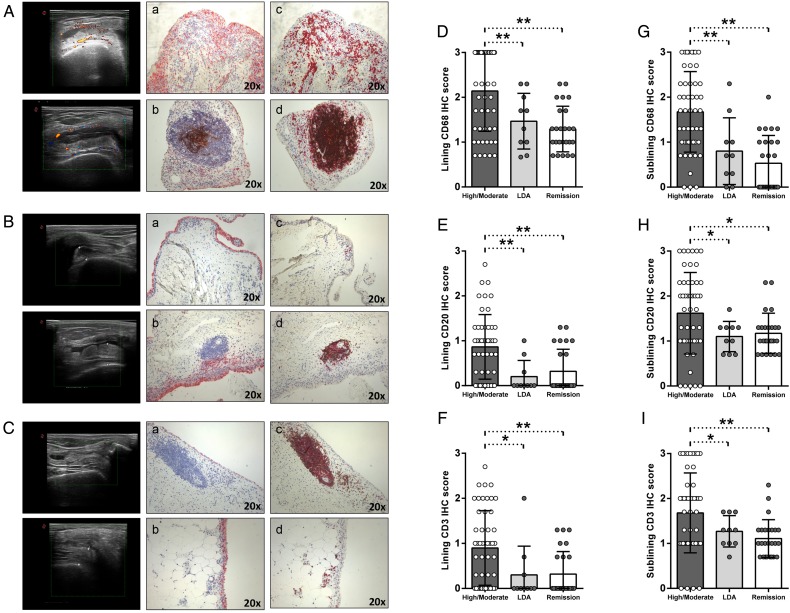
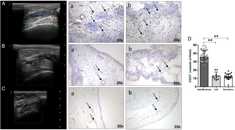
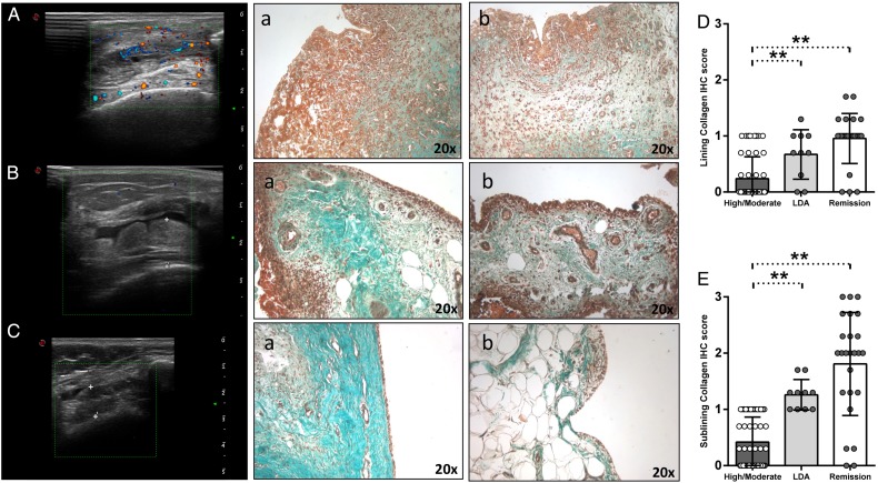
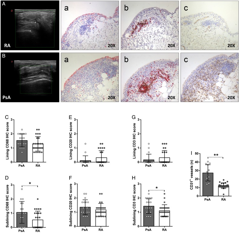
Methods: Patients with RA in remission (n=25) (disease activity score (DAS)<1.6 for at least 6 months), patients with RA in low disease activity (LDA) (n=10) (1.6<DAS<2.4 for at least 6 months) and patients with PsA in remission (n=18) (DAS<1.6 and Psoriasis Area Severity Index (PASI)=0 for at least 6 months) achieved by MTX+anti-TNF (adalimumab 40 mg or etanercept 50 mg) with power Doppler (PDUS)-negative synovial hypertrophy underwent synovial tissue biopsy. Patients with RA with high/moderate disease naïve to treatment (n=50) were included as a comparison group. Immunostaining for cluster designation (CD)68, CD21, CD20, CD3, CD31 and collagen was performed.
Results: PDUS-negative patients with RA in remission showed lower histological scores for synovial CD68+, CD20+, CD3+ cells and CD31+ vessels and collagen deposition (p<0.05 for both lining and sublining) compared with PDUS-positive patients with RA with high/moderate disease. In addition, there was no significant difference in terms of lining and sublining CD68+, CD20+, CD3+, CD31+ cells and collagen comparing PDUS-negative patients with RA in remission and in LDA, respectively. On the contrary, PDUS-negative patients with PsA in remission showed higher histological scores for sublining CD68+ (p=0.02) and CD3+ cells (p=0.04) as well as CD31+ vessels (p<0.001) than PDUS-negative patients with RA in remission.
Conclusions: PDUS-negative patients with RA in remission have comparable synovial histological features than PDUS-negative patients with RA in LDA. However, patients with PsA in remission are characterised by a higher degree of residual synovial inflammation than patients with RA in remission, despite PDUS negativity under TNF inhibition.
Keywords: Anti-TNF; Psoriatic Arthritis; Rheumatoid Arthritis; Synovitis; Ultrasonography.
Published by the BMJ Publishing Group Limited. For permission to use (where not already granted under a licence) please go to http://www.bmj.com/company/products-services/rights-and-licensing/.
Conflict of interest statement
Competing interests: None declared.
Figures
References
-
- Alivernini S, Peluso G, Fedele AL, et al. . Tapering and discontinuation of TNF-α blockers without disease relapse using ultrasonography as a tool to identify patients with rheumatoid arthritis in clinical and histological remission. Arthritis Res Ther 2016;18:39 10.1186/s13075-016-0927-z - DOI - PMC - PubMed
MeSH terms
Substances
LinkOut - more resources
Full Text Sources
Other Literature Sources
Medical
Research Materials
Miscellaneous
