. 2017 Apr;27(4):639-649.
doi: 10.1101/gr.202820.115. Epub 2017 Jan 24.

Large-scale analysis of branchpoint usage across species and cell lines

Affiliations

Large-scale analysis of branchpoint usage across species and cell lines

Allison J Taggart et al. Genome Res. 2017 Apr.

Abstract

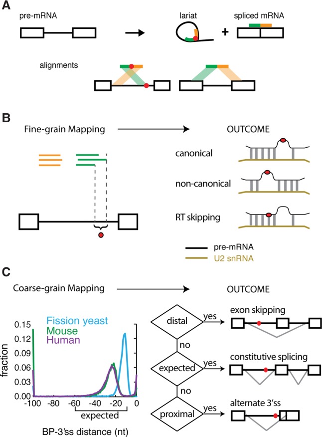
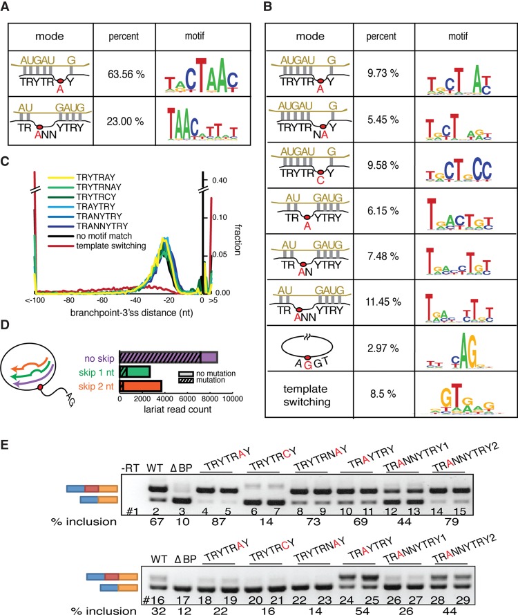
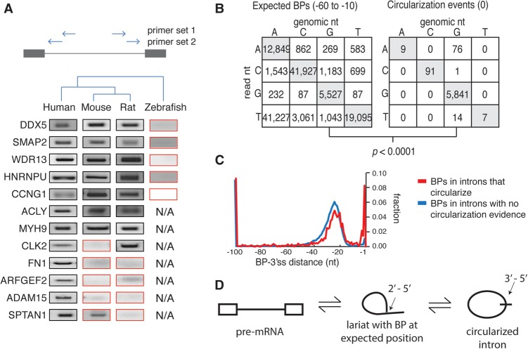
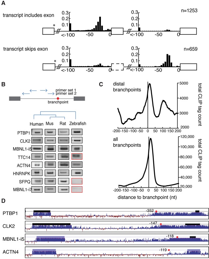
The coding sequence of each human pre-mRNA is interrupted, on average, by 11 introns that must be spliced out for proper gene expression. Each intron contains three obligate signals: a 5' splice site, a branch site, and a 3' splice site. Splice site usage has been mapped exhaustively across different species, cell types, and cellular states. In contrast, only a small fraction of branch sites have been identified even once. The few reported annotations of branch site are imprecise as reverse transcriptase skips several nucleotides while traversing a 2-5 linkage. Here, we report large-scale mapping of the branchpoints from deep sequencing data in three different species and in the SF3B1 K700E oncogenic mutant background. We have developed a novel method whereby raw lariat reads are refined by U2snRNP/pre-mRNA base-pairing models to return the largest current data set of branchpoint sequences with quality metrics. This analysis discovers novel modes of U2snRNA:pre-mRNA base-pairing conserved in yeast and provides insight into the biogenesis of intron circles. Finally, matching branch site usage with isoform selection across the extensive panel of ENCODE RNA-seq data sets offers insight into the mechanisms by which branchpoint usage drives alternative splicing.


Figures

Figure 1.
Figure 1.
The technique and purpose of mapping branchpoints. (A) Overview of branchpoint mapping (adapted by permission from Macmillan Publishers Ltd: [Nature Structural and Molecular Biology] (Taggart et al. 2012), © 2012). Inverted fragmental cDNA reads across the 2′-5′ linkage of the lariat RNAs were collected to map the branchpoint. (B) Fine-grain mapping. Discovering the precise locations of branch sites enables the discovery of canonical and noncanonical modes of U2snRNA pairing and the characterization of RT ‘skipping’ behavior around 2′-5′ linkages. (C) Coarse-grain mapping. General branchpoint location was analyzed in three species: fission yeast, mouse, and human. The human and mouse branchpoints were largely located within the expected region (between 10 and 60 nt upstream of the 3′ss AG), but the fission yeast branchpoints were more proximal. The ENCODE RNA-seq data sets were used to test the relationship between branchpoint location and alternative splicing outcomes.
Figure 2.
Figure 2.
Alternate modes of U2snRNA:pre-mRNA base-pairing emerged from lariat mapping. (A) U2snRNA:pre-mRNA base-pairing models found in Schizosaccharomyces pombe. Twenty-nucleotide windows around unprocessed branchpoint reads were iteratively aligned to U2snRNA in a manner that allowed branchpoint location, loop location, and the loop length to vary. Significantly enriched modes of base-pairing were identified and recorded. Left column: schematics of significantly enriched modes of base-pairing, middle column: percent of lariat reads that fit model, right column: motif of lariat reads that fit model. (B) U2snRNA:pre-mRNA base-pairing models found in human. Left column: schematics of significantly enriched modes of base-pairing, middle column: percent of lariat reads that fit model, right column: motif of lariat reads that fit model. (C) Branch site motif distribution relative to the 3′ss. (D) The skipping behavior and mutational profile of reverse transcriptase (RT) was inferred from the human canonical branchpoint (TRYTRAY) mapping. The histogram describes the frequency of each size of RT-induced skip (x-axis). The striped portion of bars represents the fraction of misincorporation events at the apparent branchpoint. (E) Exemplars of branch site motifs were validated in a strong (APRT, upper) and weak (SHMT2, lower) branch site reporter minigene. Branch sites of the first intron (between the blue and red exons) were first deleted (ΔBP, lanes 3,17) or replaced by branch site motifs identified in B to test for their ability to restore splicing. Inclusion level of the middle exon is indicated under the gel image.
Figure 3.
Figure 3.
Intron circularization via 3′-5′ linkage is conserved and arises in introns that also contain branchpoints in the expected region. (A) Conservation of the intron circles was assessed by inverted nested RT-PCR in multiple species (primer location indicated by arrows) and Sanger sequencing. The PCR results inconsistent with conserved distal branchpoints are boxed in red. ‘N/A’ indicates certain introns were not tested. (B) Mutational profile of branchpoints from expected region (left) compared with mutational profile from full intron circularization events (right). (C) Location of additional branchpoints in introns which circularize (red) was compared to introns with no observed evidence for circularization (blue). (D) Model for circles implicates a 3′-5′ circular linkage arising from a conventional lariat.
Figure 4.
Figure 4.
Distal branchpoints are conserved and associated with alternative exon usage. (A) The location of branchpoints was mapped in exon inclusion (top) and exon skipping (bottom) transcripts. Distal branchpoints more than 100 nt upstream of the 3′ss were binned at −100 nt. y-axes indicate the fraction of the bars. All branchpoints depicted use the same donor site (marked by *). (B) Distal branchpoints were validated by nested lariat PCR in human, mouse, rat, and zebrafish. Sequencing was used to distinguish conserved branch site usage (black box) from nonconserved (red). (C) The CLIP signal of U2AF2 binding was mapped relative to distal branchpoints (top) and all branchpoints (bottom). (D) Conservation of four introns that contain distal branchpoints. Thick bars indicate exons, and the red dots are distal branchpoints.
Figure 5.
Figure 5.
Distal branchpoints are functional elements that are associated with delayed splicing. (A) Both or either one of the distal (BP1) and expected (BP2) branchpoints was mutated in a splicing minigene as depicted at the top. Minigenes were transfected into HEK293T cells and inclusion of the middle exon was measured by RT-PCR as a readout of branchpoint activity (two replicates displayed for each construct). The splicing inclusion level was estimated by ImageQuant software across ≥3 replicates and recorded below each panel and in the histogram to the right. (B) Expected branch site of APRT (i.e., BP2) was replaced by the sequence of the distal branch site (BP2→BP1) and the functionality of branch sites was assayed by RT-PCR as in A. (C) Scheme for determining relative order of intron removal from paired-end reads, using all human introns. The bar plot indicates the degree to which the upstream (blue) and downstream (orange) introns splice first. (D) Introns that contain distal branch sites (orange) were analyzed for order of intron removal. “Upstream first” contains loci where the upstream intron reliably spliced first (i.e., >90%) and “downstream first” contains loci where the distal intron spliced before the upstream intron (left). Similar comparisons were performed on the downstream intron (right).
Figure 6.
Figure 6.
Branchpoint location predetermines the 3′ss choice in wild-type and SF3B1 mutant background. (A) Branchpoint location relative to the 3′ss analyzed in transcripts undergoing alternative 3′ss usage across ENCODE cell lines. Transcripts in cell lines using predominantly the upstream 3′ss (top) are compared to branchpoint usage in cases where the downstream 3′ss is used (bottom). (B) Branchpoint to 3′ss distance analyzed in wild type or the oncogenic mutant SF3B1 K700E NALM-6 cell line.

References

    1. Alioto TS. 2007. U12DB: a database of orthologous U12-type spliceosomal introns. Nucleic Acids Res 35: D110–D115. - PMC - PubMed
    1. Alsafadi S, Houy A, Battistella A, Popova T, Wassef M, Henry E, Tirode F, Constantinou A, Piperno-Neumann S, Roman-Roman S, et al. 2016. Cancer-associated SF3B1 mutations affect alternative splicing by promoting alternative branchpoint usage. Nat Commun 7: 10615. - PMC - PubMed
    1. Awan AR, Manfredo A, Pleiss JA. 2013. Lariat sequencing in a unicellular yeast identifies regulated alternative splicing of exons that are evolutionarily conserved with humans. Proc Natl Acad Sci 110: 12762–12767. - PMC - PubMed
    1. Bitton DA, Rallis C, Jeffares DC, Smith GC, Chen YY, Codlin S, Marguerat S, Bahler J. 2014. LaSSO, a strategy for genome-wide mapping of intronic lariats and branch points using RNA-seq. Genome Res 24: 1169–1179. - PMC - PubMed
    1. Bonnal S, Vigevani L, Valcarcel J. 2012. The spliceosome as a target of novel antitumour drugs. Nat Rev Drug Discov 11: 847–859. - PubMed

LinkOut - more resources