Review
. 2017 Jan 9:7:682.
doi: 10.3389/fimmu.2016.00682. eCollection 2016.

The Role of IL-33-Dependent Inflammation in the Tumor Microenvironment

Affiliations
Review

The Role of IL-33-Dependent Inflammation in the Tumor Microenvironment

Marie-Hélène Wasmer et al. Front Immunol. .

Abstract

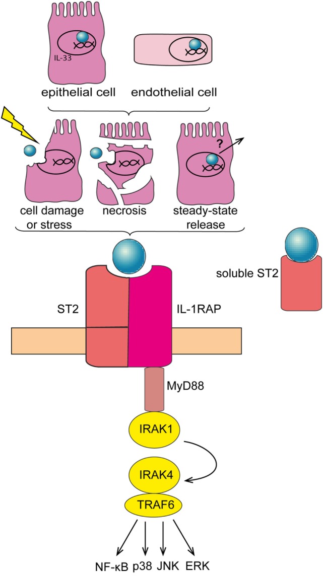
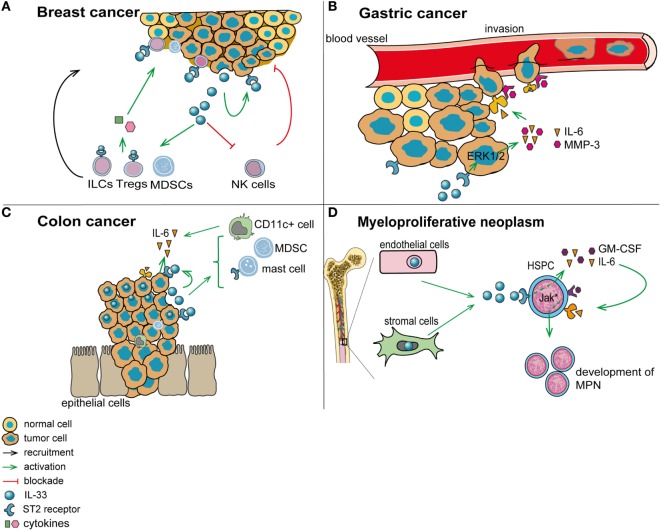
There is compelling evidence that inflammation contributes to tumorigenesis. Inflammatory mediators within the tumor microenvironment can either promote an antitumor immune response or support tumor pathogenesis. Therefore, it is critical to determine the relative contribution of tumor-associated inflammatory pathways to cancer development. Interleukin-33 (IL-33) is a member of the IL-1 family of cytokines that is released upon tissue stress or damage to operate as an alarmin. IL-33 has been primarily implicated in the induction of type-2 immune responses. However, recent findings have shown a role of IL-33 in several cancers where it may exert multiple functions. In this review, we will present the current knowledge on the role of IL-33 in the microenvironment of different tumors. We will highlight which cells produce and which cells are activated by IL-33 in cancer. Furthermore, we will explain how IL-33 modulates the tumor-associated inflammatory microenvironment to restrain or promote tumorigenesis. Finally, we will discuss the issues to be addressed first before potentially targeting the IL-33 pathway for cancer therapy.

Keywords: cancer; inflammation; interleukin-33; therapy; tumor microenvironment.


Figures

Figure 1
Figure 1
Interleukin-33 (IL-33)/ST2 signaling pathway. IL-33 is mainly expressed in the nucleus of epithelial and endothelial cells. IL-33 is released subsequent to cell damage, stress, or necrosis. Alternatively, IL-33 may be released under steady-state conditions—at low levels—via a so far unknown mechanism. Soluble IL-33 binds to its receptor, a heterodimeric complex comprising IL-1RL1/ST2 (ST2) and IL-1 receptor accessory protein (IL1RAP), thereby mobilizing downstream signaling molecules including MyD88 and TRAF-6, and eventually activating NF-κB, p38, JNK, and ERK pathways. Soluble ST2 acts as a decoy receptor for IL-33 to negatively regulate IL-33/ST2 signaling.
Figure 2
Figure 2
Role of interleukin-33 (IL-33) in the tumor microenvironment. (A) In breast cancer (mouse 4T1 breast cancer model), IL-33 is released by tumor cells and acts in an autocrine/paracrine manner. In addition, IL-33 promotes the recruitment of immunosuppressive cells and inhibits the function of antitumor cytotoxic natural killer cells (NK). (B) In gastric cancer, IL-33 may promote vessel invasion of tumor cells by stimulating the secretion of IL-6 and MMP-3 through activation of the ERK pathway. (C) In colorectal cancer, IL-33 supports the recruitment to the tumor environment of pro-tumorigenic immune cells, including mast cells and myeloid-derived suppressor cells (MDSCs). In addition, IL-33 promotes the secretion of pro-tumorigenic IL-6 by leukocytes in the tumor vicinity, which are possibly CD11c+ dendritic cells. (D) In patients with myeloproliferative neoplasms, IL-33 is released by bone marrow stromal and endothelial cells, and it engages ST2 on CD34+ hematopoietic stem/progenitor cells, thereby promoting their secretion of IL-6 and GM-CSF. These cytokines, in turn, stimulate in an auto/paracrine manner the uncontrolled proliferation of the malignant clone (due to its defect in JAK/STAT signaling).

References

    1. Hanahan D, Weinberg RA. The hallmarks of cancer. Cell (2000) 100(1):57–70.10.1016/S0092-8674(00)81683-9 - DOI - PubMed
    1. Hanahan D, Weinberg RA. Hallmarks of cancer: the next generation. Cell (2011) 144(5):646–74.10.1016/j.cell.2011.02.013 - DOI - PubMed
    1. Hanahan D, Coussens LM. Accessories to the crime: functions of cells recruited to the tumor microenvironment. Cancer Cell (2012) 21(3):309–22.10.1016/j.ccr.2012.02.022 - DOI - PubMed
    1. Grivennikov SI, Greten FR, Karin M. Immunity, inflammation, and cancer. Cell (2010) 140(6):883–99.10.1016/j.cell.2010.01.025 - DOI - PMC - PubMed
    1. Koontongkaew S. The tumor microenvironment contribution to development, growth, invasion and metastasis of head and neck squamous cell carcinomas. J Cancer (2013) 4(1):66–83.10.7150/jca.5112 - DOI - PMC - PubMed