. 2017 Jan;7(1):65-72.
doi: 10.1016/j.apsb.2016.04.003. Epub 2016 Jun 20.

Asiatic acid inhibits lung cancer cell growth in vitro and in vivo by destroying mitochondria

Affiliations

Asiatic acid inhibits lung cancer cell growth in vitro and in vivo by destroying mitochondria

Tiancong Wu et al. Acta Pharm Sin B. 2017 Jan.

Abstract

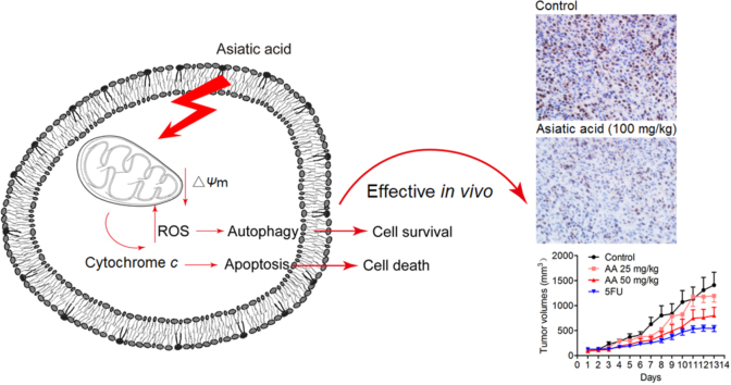
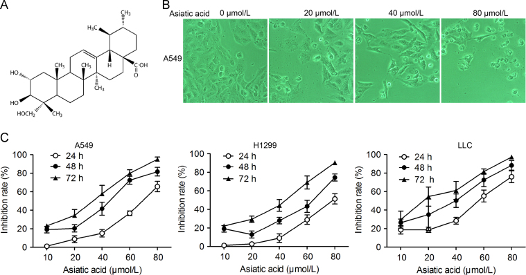
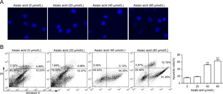
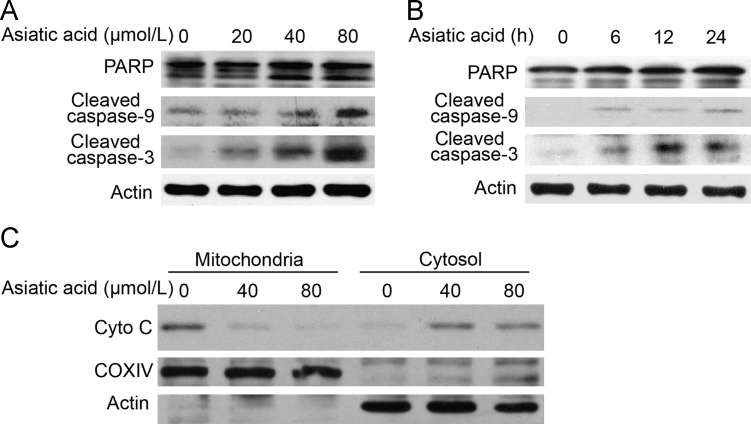
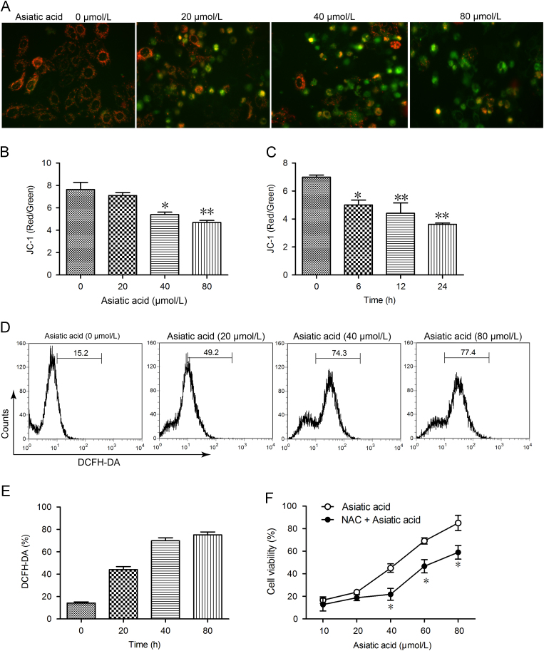
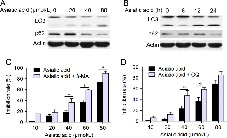
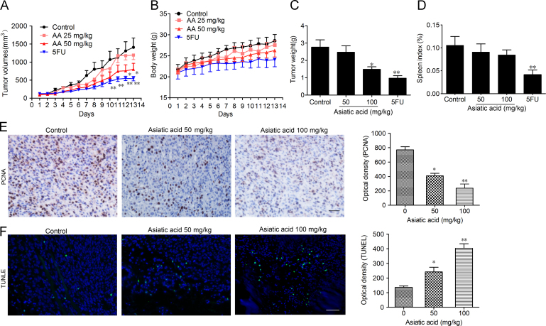
Asiatic acid (AA), a pentacyclic triterpene found in Centella asiatica, displays significant anti-proliferative effects on cancer cells in vitro although the underlying mechanism of this effect remains unknown. This study investigated the efficacy and mechanism of action of AA against lung cancer both in vivo and in vitro. Using the MTT assay, AA was found to induce apoptosis in a dose- and time-dependent manner, an effect enhanced by pretreatment with an autophagy inhibitor. It also elevated expression of microtubule-associated protein 1 light chain 3 (LC3) and decreased the expression of p62. Furthermore, exposure to AA resulted in collapse of the mitochondrial membrane potential and generation of reactive oxygen species (ROS), suggesting mitochondria are the target of AA. In the mouse lung cancer xenograft model, oral administration of AA significantly inhibited tumor volume and weight accompanied by significant apoptosis of lung cancer cells. In addition, it led to a significant decrease in the expression of proliferating cell nuclear antigen (PCNA). In summary, the results show that AA significantly reduces lung cancer cell growth both in vitro and in vivo and that the associated apoptosis is mediated through mitochondrial damage.

Keywords: Apoptosis; Asiatic acid; Lung cancer; Mitochondria; Reactive oxygen species.


Figures

fx1
Graphical abstract
Fig. 1
Figure 1
Asiatic acid inhibits lung cancer cell proliferation. (A) Structure of asiatic acid; (B) and (C) A549, H1299 and LLC cells exposed to various concentrations of asiatic acid. (B) Photographs showing morphological changes in cells; (C) Cell proliferation determined by the MTT assay every 24 h. Data represent mean±SD of three different experiments.
Fig. 2
Figure 2
Asiatic acid induces apoptotic cell death and cell cycle arrest in lung cancer cells. (A) A549 cells were treated with asiatic acid (20, 40 and 80 μmol/L) for 24 h, stained with DAPI and nuclei photographed. (B) A549 cells treated with asiatic acid were collected and subjected to annexin V/PI analysis (B). Data represent mean±SEM of three different experiments.
Fig. 3
Figure 3
Asiatic acid induces apoptotic cell death via a mitochondrial pathway. (A) and (B) A549 cells were exposed to asiatic acid (80 μmol/L for 3, 6, 12, 24 h or 20, 40, 80 μmol/L for 12 h) after which PARP, caspase-9 and caspase-3 activation was assessed by Western blot. (C) The release level of cytochrome c from mitochondria was examined by Western blotting.
Fig. 4
Figure 4
Asiatic acid treatment causes mitochondrial dysfunction. (A)–(C) A549 cells were exposed to asiatic acid (80 μmol/L for 3, 6, 12, 24 h or 20, 40, 80 μmol/L for 12 h) and the mitochondrial membrane potential (JC-1 staining, red/green fluorescence ratio) photographed by fluorescence microscopy and quantified by fluorescence spectrometry. (D) and (E) A549 cells were exposed to asiatic acid (20, 40, 80 μmol/L for 12 h) and the level of ROS detected by DCFH-DA staining. (F) A549 cells were exposed to asiatic acid (20, 40 and 80 μmol/L for 12 h) with or without NAC (2.5 mmol/L) for 48 h. The MTT assay was used to evaluate cell viability. Data represent mean±SD of three different experiments. *P<0.05, **P<0.01 vs. control.
Fig. 5
Figure 5
Asiatic acid induces cell protective autophagy. (A) and (B) A549 cells were treated with asiatic acid (20, 40, 80 μmol/L for 12 h or 80 μmol/L for 6, 12, 24 h) and protein expression of LC3 and p62 analyzed by Western blot. (C) and (D) A549 cells were treated with various concentrations of asiatic acid for 24 h with or without 3-MA and CQ pretreatment (2 h). The MTT assay was used to evaluate cell viability. Data are mean±SD of three different experiments. *P<0.05, **P<0.01 vs. indicated.
Fig. 6
Figure 6
Asiatic acid inhibits tumor growth in vivo. 1×10⁶ LLC cells were transplanted subcutaneously into the armpit of the C57 mice. Three days after transplantation, mice were randomly allocated to either control or treatment groups (n=6). Drugs were administered i.g. on days 1–13. (A) and (B) Tumor volume and mouse bodyweight were recorded daily. After sacrifice, solid tumors were separated and weighed. (C) and (D) The spleen index was calculated. (E) Paraffin sections of tumor tissues from mice were analyzed by H&E staining and TUNEL staining and (F) optical density was calculated. Scale bar, 50 μm. Data represent mean±SEM, n=8, *P<0.05, **P<0.01 vs. control.

References

    1. Siegel R.L., Miller K.D., Jemal A. Cancer statistics, 2015. CA Cancer J Clin. 2015;65:5–29. - PubMed
    1. Minguet J., Smith K.H., Bramlage P. Targeted therapies for treatment of non-small cell lung cancer—recent advances and future perspectives. Int J Cancer. 2016;138:2549–2561. - PubMed
    1. Landi L., Cappuzzo F. Experience with erlotinib in the treatment of non-small cell lung cancer. Ther Adv Respir Dis. 2015;9:146–163. - PubMed
    1. Schiller G.J., O׳Brien S.M., Pigneux A., Deangelo D.J., Vey N., Kell J. Single-agent laromustine, a novel alkylating agent, has significant activity in older patients with previously untreated poor-risk acute myeloid leukemia. J Clin Oncol. 2010;28:815–821. - PubMed
    1. Sharma S.V., Bell D.W., Settleman J., Haber D.A. Epidermal growth factor receptor mutations in lung cancer. Nat Rev Cancer. 2007;7:169–181. - PubMed