. 2017:3:5.
doi: 10.1051/sicotj/2016040. Epub 2017 Jan 24.

The Paley ulnarization of the carpus with ulnar shortening osteotomy for treatment of radial club hand

Affiliations

The Paley ulnarization of the carpus with ulnar shortening osteotomy for treatment of radial club hand

Dror Paley. SICOT J. 2017.

Abstract

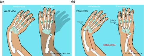
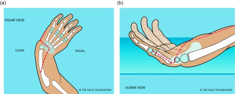
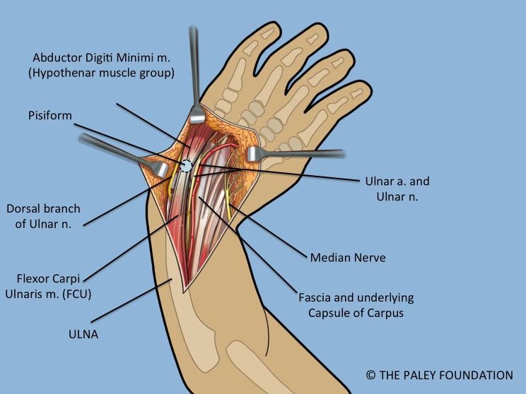
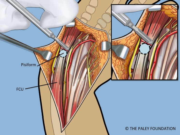
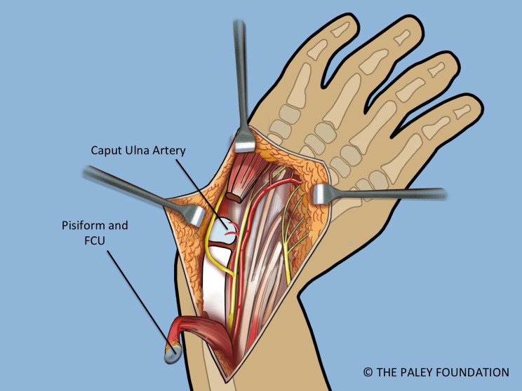
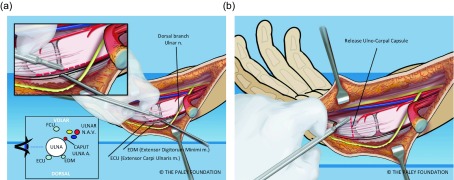
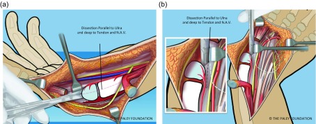
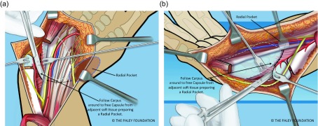
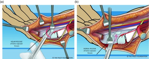
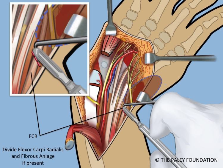
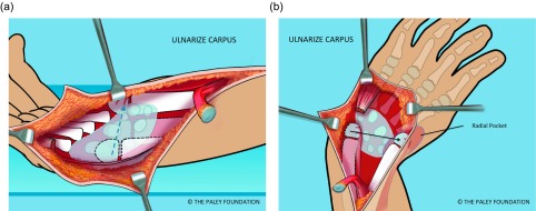
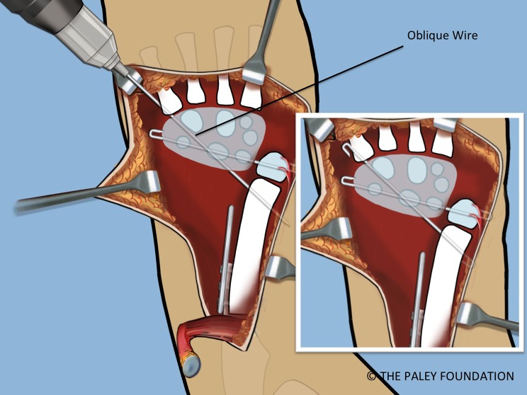
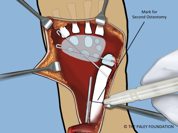
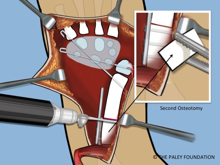
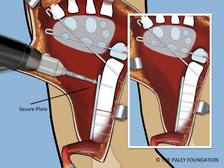
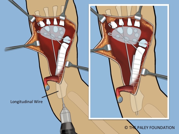
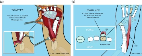
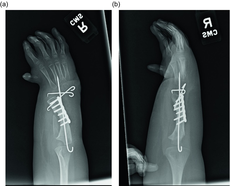
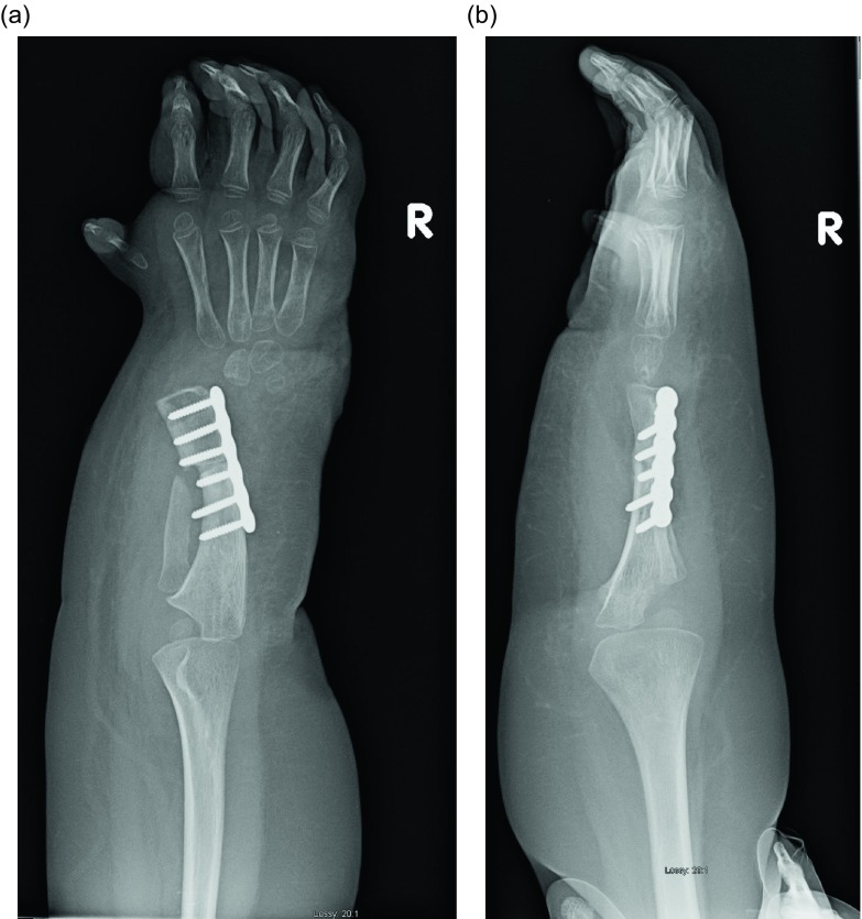
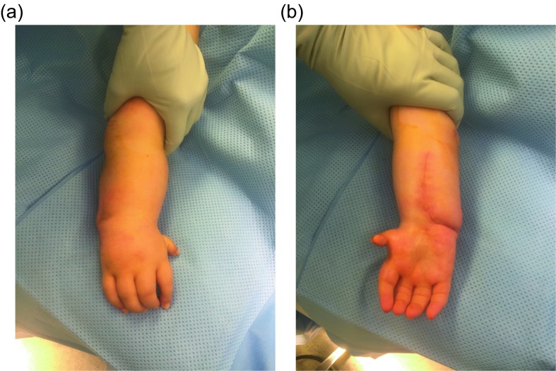
Recurrent deformity from centralization and radialization led to the development in 1999 of a new technique by the author called ulnarization. This method is performed through a volar approach in a vascular and physeal sparing fashion. It biomechanically balances the muscle forces on the wrist by dorsally transferring the flexor carpi ulnaris (FCU) from a deforming to a corrective force. The previous problems of a prominent bump from the ulnar head and ulnar deviation instability were solved by acutely shortening the diaphysis and by temporarily fixing the station of the carpus to the ulnar head at the level of the scaphoid. This is the first report of this modified Paley ulnarization method, which the author considers a significant improvement over his original procedure.


Figures

Figure 1.
Figure 1.
Pre- versus postoperative position of the hand following ulnarization with shortening (a). The muscle forces before surgery pull the hand into more radial deviation while after surgery there is a balance of forces with the ulnar head acting as a bony block to radial deviation (b).
Figure 2.
Figure 2.
Make a Z-shaped incision. Shown from the volar aspect (a) and from the ulnar side (b).
Figure 3.
Figure 3.
Volar forearm and wrist exposure after volar fasciotomy, showing the ulnar and median nerves, and the flexor tendons. Note the dorsal cutaneous branch of the ulnar nerve passes dorsal to the FCU.
Figure 4.
Figure 4.
The abductor digiti minimi is released off of the pisiform bone. The pisiform bone is separated from the triquetrum.
Figure 5.
Figure 5.
The FCU is reflected proximally. The circulation to the ulnar epiphysis comes from volar-radial. The ulnar and median nerves are decompressed into the hand.
Figure 6.
Figure 6.
The ulna is dissected free of the ECU and EDM on its ulno-dorsal aspect (a). The ulnar collateral ligament and capsule are cut (b). The caput ulnar vessels lie on the volar radial aspect of the ulna.
Figure 7.
Figure 7.
The capsule is released along the interval between the ulna and the carpus. The line of dissection is from distal to proximal, in line with the shaft of the ulna. Care must be taken to stay deep to the volar extra-articular soft tissues (a, b).
Figure 8.
Figure 8.
The capsular tissues are dissected by following around the proximal edge of the carpus towards the radial side (a, b).
Figure 9.
Figure 9.
The volar (a) and dorsal (b) extra-capsular soft tissues are separated from the carpal bones by dissection.
Figure 10.
Figure 10.
If the FCR is present it should be released. If a radial fibrous anlage is present it too should be released from the carpus.
Figure 11.
Figure 11.
A space is made radial to the carpus, called the radial pocket.
Figure 12.
Figure 12.
The wrist is ulnarized allowing the ulnar head to pass into the radial pocket (a, b).
Figure 13.
Figure 13.
The ulnar diaphysis is exposed by elevating the FCU proximally. The level of the planned osteotomy is marked.
Figure 14.
Figure 14.
A small plate is affixed to the proximal ulna with two or three screws.
Figure 15.
Figure 15.
The proximal ulnar osteotomy is made using a saw.
Figure 16.
Figure 16.
The distal ulna is stripped of some of its periosteum to allow the ulna to shorten and overlap the proximal end. Shortening of the ulna allows it to migrate proximally relative to the carpus. The ulnar head should sit opposite the scaphoid. This is referred to at station.
Figure 17.
Figure 17.
The carpus is pinned to the head of the ulna at station with a transverse 1.5 mm wire starting on the carpal side.
Figure 18.
Figure 18.
A second retrograde oblique wire is inserted from the fifth metatarsal base to the distal ulnar metaphysis. These wires should be cut and curled 180°.
Figure 19.
Figure 19.
With the bone ends overlapped and the wrist pinned, the level of the shortening osteotomy should be marked.
Figure 20.
Figure 20.
The shortening osteotomy is performed with a saw.
Figure 21.
Figure 21.
Fix the distal ulna to the plate. Adjust the rotation so that the plane of the hand and the plane of the elbow motion are the same. Stated differently, the lateral the profile view is a lateral of the elbow with an AP of the hand.
Figure 22.
Figure 22.
A third temporary wrist arthrodesis wire is inserted from the olecranon in a longitudinal antegrade fashion to exit the ulna and transfix the carpus. This wire is also cut and curled proximally.
Figure 23.
Figure 23.
The FCU should be transferred to the dorsum of the hand. This is shown from the volar (a) and dorsal (b) and cross-sectional views (b). The pisiform bone is sutured to the base of the 4th metacarpal. A non-absorbable suture is passed around the base of this metacarpal.
Figure 24.
Figure 24.
The proximal ulna is parallel and in line with the third metacarpal (a). The Z-shaped incision is closed over a drain (b). It is important to complete a volar fasciotomy before closing to allow for swelling without the risk of compartment syndrome.
Figure 25.
Figure 25.
AP and lateral radiographs of left hand of a four-year-old boy with radial club hand before surgery.
Figure 26.
Figure 26.
Photograph of the hand before surgery.
Figure 27.
Figure 27.
AP and lateral radiographs after ulnarization with shortening. Note the temporary arthrodesis wires in place and the plate on the ulna.
Figure 28.
Figure 28.
AP and lateral radiographs after removal of the wires. Note that the ulnar head remains at station.
Figure 29.
Figure 29.
Final photographs, dorsal view (a), volar view (b), showing the hand after ulnarization with shortening.

References

    1. Manske PR (1996) Longitudinal failure of upper-limb formation. Instructional Course Lectures, The American Academy of Orthopaedic Surgeons. J Bone Joint Surg 78A, 1600–1623. - PubMed
    1. Blauth W (1969) On the morphology and therapy of the radial club-hand [in German]. Arch Orthop Unfallchir 65, 97–123. - PubMed
    1. Lamb DW (1974) The treatment of radial club hand: absent radius, aplasia of the radius, hypoplasia of the radius, radial paraxial hemimelia. Hand 4, 22–30. - PubMed
    1. Lamb DW (1977) Radial club hand: a continuing study of sixty-eight patients with one hundred and seventeen club hands. J Bone Joint Surg 59A, 1–13. - PubMed
    1. Watson HK, Beebe RD, Cruz NI (1984) A centralization procedure for radial clubhand. J Hand Surg 9A, 541–547. - PubMed

LinkOut - more resources