. 2017 Jan 25:7:40685.
doi: 10.1038/srep40685.

Mitochondrial and Chromosomal Damage Induced by Oxidative Stress in Zn2+ Ions, ZnO-Bulk and ZnO-NPs treated Allium cepa roots

Affiliations

Mitochondrial and Chromosomal Damage Induced by Oxidative Stress in Zn2+ Ions, ZnO-Bulk and ZnO-NPs treated Allium cepa roots

Bilal Ahmed et al. Sci Rep. .

Abstract

Large-scale synthesis and release of nanomaterials in environment is a growing concern for human health and ecosystem. Therefore, we have investigated the cytotoxic and genotoxic potential of zinc oxide nanoparticles (ZnO-NPs), zinc oxide bulk (ZnO-Bulk), and zinc ions (Zn2+) in treated roots of Allium cepa, under hydroponic conditions. ZnO-NPs were characterized by UV-visible, XRD, FT-IR spectroscopy and TEM analyses. Bulbs of A. cepa exposed to ZnO-NPs (25.5 nm) for 12 h exhibited significant decrease (23 ± 8.7%) in % mitotic index and increase in chromosomal aberrations (18 ± 7.6%), in a dose-dependent manner. Transmission electron microcopy and FT-IR data suggested surface attachment, internalization and biomolecular intervention of ZnO-NPs in root cells, respectively. The levels of TBARS and antioxidant enzymes were found to be significantly greater in treated root cells vis-à-vis untreated control. Furthermore, dose-dependent increase in ROS production and alterations in ΔΨm were observed in treated roots. FT-IR analysis of root tissues demonstrated symmetric and asymmetric P=O stretching of >PO2- at 1240 cm-1 and stretching of C-O ribose at 1060 cm-1, suggestive of nuclear damage. Overall, the results elucidated A. cepa, as a good model for assessment of cytotoxicity and oxidative DNA damage with ZnO-NPs and Zn2+ in plants.


Figures

Figure 1
Figure 1. Characterization of ZnO-NPs.
(A) UV-Visible and fluorescence spectrum of ZnO-NPs, (B) FTIR spectrum of ZnO-NPs, (C) XRD pattern of ZnO-NPs, (D) SEM micrograph of ZnO-NPs, (E) TEM micrograph of ZnO-NPs, and (F) frequency size distribution of ZnO-NPs by TEM.
Figure 2
Figure 2. Optical and confocal microscopic analyses of chromosomal damage in A. cepa root meristem cells.
Chromosomal aberrations induced by ZnO-NPs, ZnO-Bulk, and Zn2+ ions in A. cepa root meristem cells at different cell division stages are shown as: normal metaphase (A1), sticky metaphase (A2–A5), normal metaphase (B1), sticky metaphase (B2), sticky metaphase with loop (B3), sticky metaphase with laggard chromosome(B4), and disoriented metaphase (B5), normal anaphase (C1), chromosome bridges with lag (C2,C3), broken chromosome bridge (C4), single chromosome bridge (C5), normal anaphase (D1), sticky multipolar anaphase (D2), multipolar anaphase with chromosome bridges (D3), multipolar anaphase (D4), multipolar anaphase with single chromosome bridge (D5). EMS (10 mM) was taken as a positive control. (All images were at 1000X magnification).
Figure 3
Figure 3. SEM micrographs showing ZnO-NPs deposition and tissue damage after exposure of A. cepa roots with ZnO-NPs.
Panel (A and B) shows untreated control root tip and surface. Panel C, and D depict the tip and surface of ZnO-NPs (1000 μg/ml) treated roots at 50 and 10 μm scale, respectively. The attachment of nanometer scale charged particles, fissures and fractured tissues at root surface are shown by arrows (D inset).
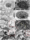
Figure 4
Figure 4. TEM micrographs showing internalization and sub-cellular damage in A. cepa root meristem cells upon exposure with ZnO-NPs.
Untreated cell showing normal architecture of nucleus (N), nucleolus (n), integrated mitochondria (M), intact nuclear membrane (Nm), vacuoles (V), plasmodesmata (P) (panel A and B). Triangular periplasmic space is clearly visible in untreated cell (panel B). Magnified view of nuclear matrix and nucleolus of untreated cell (panel C and D). Images of roots treated with ZnO-NPs (1000 μg/ml) showing the influx of ZnO-NPs in cytoplasm and attachment to the vacuoles (panel E). Yellow arrow heads showing degeneration of nuclear constituents and significant swelling of mitochondria while red arrows and arrow heads showing attachment of ZnO-NPs onto the nuclear membrane and infiltration of ZnO-NPs in intracellular junctions (panel F). Analysis was performed at 200 keV. Magnified view showing the sequesteration of ZnO-NPs on nuclear membrane (panel G). Distribution of ZnO-NPs in cytoplasmic matrix (panel H). Magnification for images (panel A) 10,000x, (panel B) 30,000x, (panel C) 12,000x, (panel D) 20,000x, (panel E) 15,000x, (panel E inset) 20,000x, (panel F) 15,000x, (panel G) 20,000x, (panel G inset) 30,000x, (panel H) 20,000x, and (panel H inset) 25,000x.
Figure 5
Figure 5. CLSM images of intracellular ROS generation and dissipation of mitochondrial membrane potential in A. cepa root after exposure with ZnO-NPs.
Panels show the untreated control (UC) and treated roots with varying concentrations of ZnO-Bulk (panel A), ZnO-NPs (panel B) and Zn2+ (panel C) stained with 25 μM DCFH-DA. Corresponding bar diagrams represent the % increase in DCF fluorescence of root meristem cells in UC and treated with ZnO-NPs, ZnO-Bulk, and Zn2+ ions. CLSM images show the changes in mitochondrial membrane potential (ΔΨm) in A. cepa UC roots and those treated with varying concentrations of ZnO-Bulk (panel D), ZnO-NPs (panel E) and Zinc ions (panel F) after staining with Rhodamine123 (1 μg/ml). Dotted, dashed, and solid white color rectangular outlines denote the root elongation zone, meristematic region, and root tip, respectively.
Figure 6
Figure 6. Assessment of in vivo interactions of ZnO-NPs, ZnO-Bulk and Zn2+ ions with A. cepa chemical constituents by FTIR analysis.
FTIR spectra of dry root tip powder obtained from untreated roots and those treated under hydroponic conditions in ddw for 12 h, are shown as (a) untreated control, and (b–d) as roots grown in presence of 100 μg/ml of ZnO-Bulk, ZnO-NPs and Zn2+ ions, respectively.
Figure 7
Figure 7. Schematic representation of plausible mechanism of ZnO-NPs interaction with cellular components and induced oxidative stress, cytotoxicity and genotoxicity in A. cepa roots.
Abbreviations are as follows: N: Nucleus, V: Vacuole, Nm: Nuclear membrane, Cy: Cytoplasm, M: Mitochondria, ER: Endoplasmic reticulum, P:Plasmodesmata, and PS: periplasmic space.

References

    1. Nakada T. et al.. Novel device structure for Cu (In,Ga) Se2 thin film solar cells using transparent conducting oxide back and front contacts. Solar Energy 77(6), 739–747 (2004).
    1. Gedamu D. et al.. Rapid fabrication technique for interpenetrated ZnO nanotetrapod networks for fast UV sensors. Adv. Mater. 26(10), 1541–1550 (2014). - PubMed
    1. Meulenkamp E. A. Synthesis and Growth of ZnO Nanoparticles J. Phys. Chem. B 102(29), 5566–5572 (1998).
    1. Lin D. & Xing B. Phytotoxicity of nanoparticles: inhibition of seed germination and root growth. Environ. Pollut. 150, 243–250 (2007). - PubMed
    1. Wahab R. et al.. Formation of ZnO micro flowers prepared via solution process and their antibacterial activity. Nanoscale Res. Lett. 5(10), 1675–1681 (2010). - PMC - PubMed

Publication types

MeSH terms

LinkOut - more resources