. 2017 Jan 25:7:41221.
doi: 10.1038/srep41221.

Phosphorylated Histone 3 at Serine 10 Identifies Activated Spinal Neurons and Contributes to the Development of Tissue Injury-Associated Pain

Affiliations

Phosphorylated Histone 3 at Serine 10 Identifies Activated Spinal Neurons and Contributes to the Development of Tissue Injury-Associated Pain

Jose Vicente Torres-Pérez et al. Sci Rep. .

Abstract

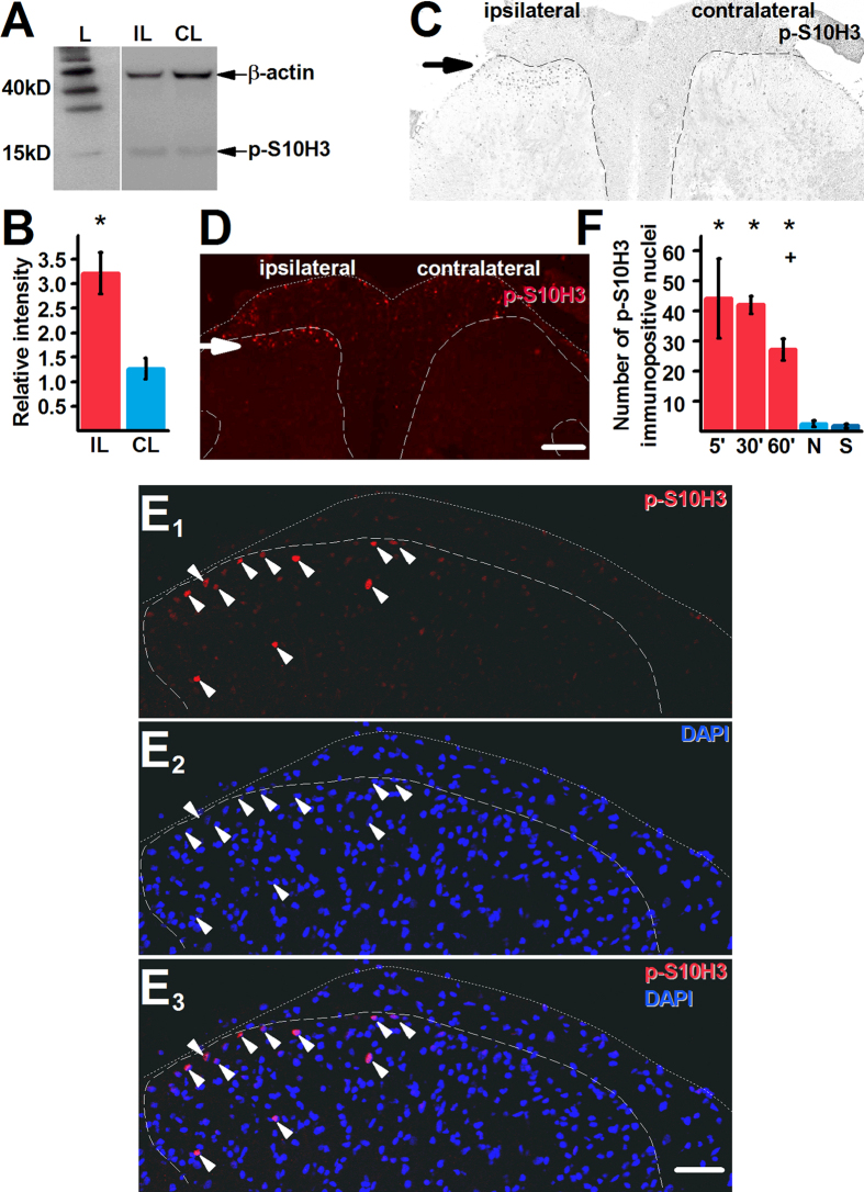
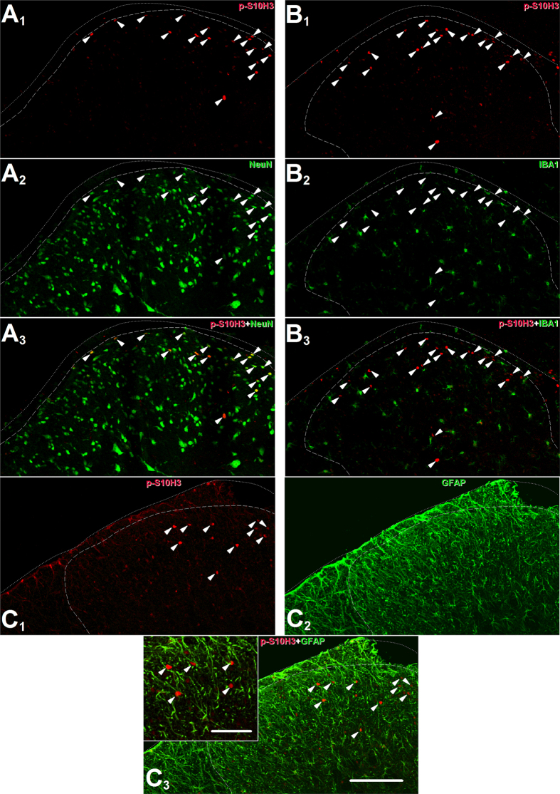
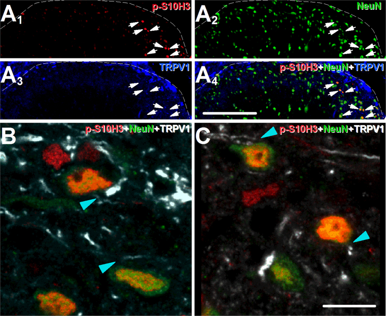
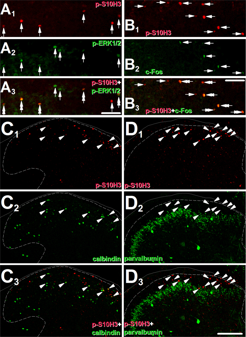
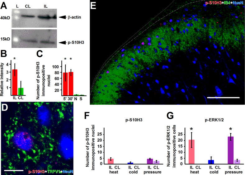
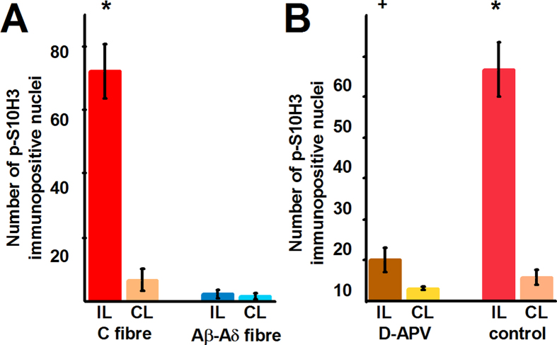
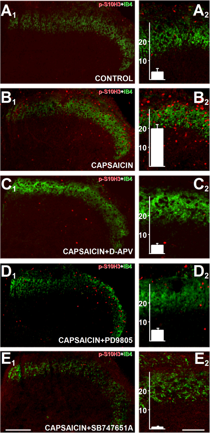
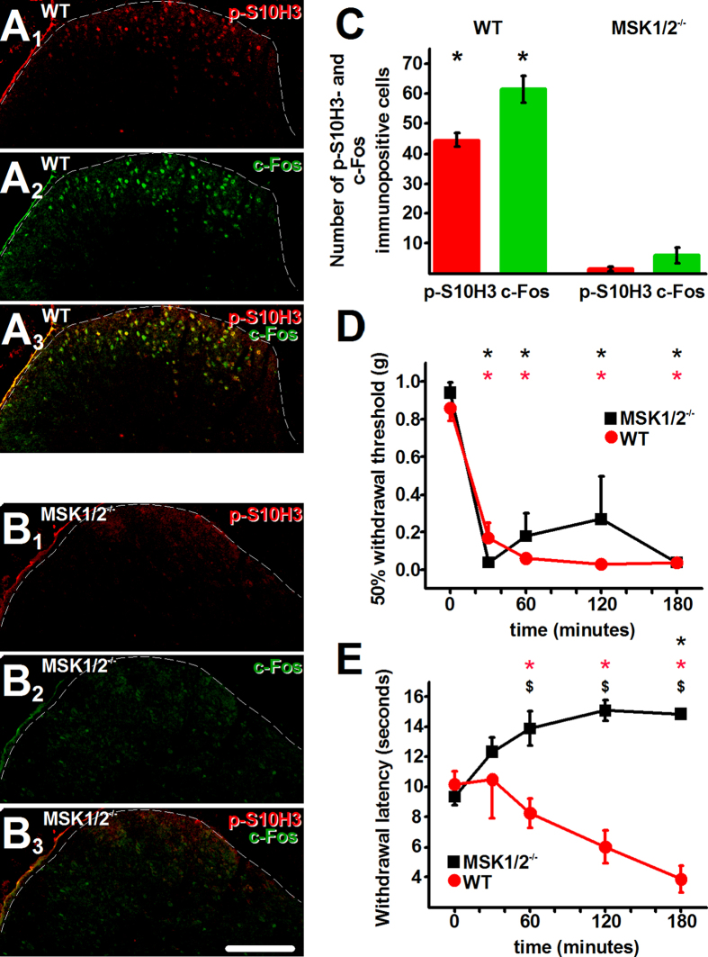
Transcriptional changes in superficial spinal dorsal horn neurons (SSDHN) are essential in the development and maintenance of prolonged pain. Epigenetic mechanisms including post-translational modifications in histones are pivotal in regulating transcription. Here, we report that phosphorylation of serine 10 (S10) in histone 3 (H3) specifically occurs in a group of rat SSDHN following the activation of nociceptive primary sensory neurons by burn injury, capsaicin application or sustained electrical activation of nociceptive primary sensory nerve fibres. In contrast, brief thermal or mechanical nociceptive stimuli, which fail to induce tissue injury or inflammation, do not produce the same effect. Blocking N-methyl-D-aspartate receptors or activation of extracellular signal-regulated kinases 1 and 2, or blocking or deleting the mitogen- and stress-activated kinases 1 and 2 (MSK1/2), which phosphorylate S10 in H3, inhibit up-regulation in phosphorylated S10 in H3 (p-S10H3) as well as fos transcription, a down-stream effect of p-S10H3. Deleting MSK1/2 also inhibits the development of carrageenan-induced inflammatory heat hyperalgesia in mice. We propose that p-S10H3 is a novel marker for nociceptive processing in SSDHN with high relevance to transcriptional changes and the development of prolonged pain.


Figures

Figure 1
Figure 1. Burn injury induces prolonged up-regulation in p-S10H3 expression in superficial spinal dorsal horn of the spinal cord.
(A) and (B) Burn injury increases p-S10H3 (expected MW: 17kD) expression within 5 minutes in the ipsilateral side (IL) compared to the contralateral side (CL). The expected molecular weight of β-actin is 42 kD. L indicates size marker. *p = 0.006; n = 4. The Western blot images shown in (A) are cropped. (C) and (D) Images of transverse sections of the L4-L5 spinal segments showing both the ipsilateral and contralateral sides. p-S10H3-immunopositive structures in the ipsilateral spinal cord (arrow; scale bar = 200 μm). (E1–E3) Images of the ipsilateral side of a transverse spinal cord section of the L4-L5 segments. Structures exhibiting p-S10H3-immunopositivity in the superficial spinal dorsal horn are invariably positive also to DAPI which binds to adenine-thymine regions of the DNA in the nucleus. Scale bar = 100 μm. (F) p-S10H3 expression lasts for at least 60 minutes (60′) after the injury. 5′, 30′ and 60′, 5, 30 and 60 minutes post-injury, respectively; N, naive; S, 5 minutes after a sham injury. *p < 0.001, + p = 0.001 (n = 4). Dotted and dashed lines indicate the surface of the spinal cord and the white-grey matter border, respectively on each image.
Figure 2
Figure 2. The burn injury-induced up-regulation in p-S10H3 expression occurs in neurons.
(A1–3) Images of the ipsilateral side of a section cut from the L4-L5 spinal segments. p-S10H3-expressing structures (arrows) 5 minutes after burn injury also express the neuronal nucleus marker NeuN. (B1–3) Images of the ipsilateral side of a section cut from the L4-L5 spinal segments. Microglia identified with an anti-ionised calcium-binding adaptor protein 1 (IBA1) antibody do not express p-S10H3 after burn injury. (C1–3) Images of the ipsilateral side of a section cut from the L4-L5 spinal segments. Astrocytes identified with an anti-glial fibrillary acidic protein (GFAP) antibody do not express p-S10H3 after burn injury. (Scale bar, 200 μm; scale bar in inset, 100 μm.) Dotted and dashed lines indicate the surface of the spinal cord and the white-grey matter border on each image.
Figure 3
Figure 3. p-S10H3 expression occurs in SSDHN, which are in close apposition to spinal terminals of nociceptive primary sensory neurons.
(A1A4) Images of the ipsilateral side of a section cut from the L4-L5 spinal segments. p-S10H3-expressing nuclei (arrows), 5 minutes after burn injury are distributed among primary sensory nerve fibres expressing TRPV1. (Scale bar, 400 μm). (B) and (C) Images of single optical sections of 2 μm thickness of the spinal dorsal horn 5 minutes following burn injury. Virtually, all neurons expressing p-S10H3 in the nucleus, 5 minutes after burn injury are in close apposition to TRPV1-expressing nerve fibres (arrow heads). (Scale bar, 10 μm). Dashed lines indicate the white-grey matter border on each image.
Figure 4
Figure 4. The majority of SSDHN expressing p-S10H3 following burn injury are excitatory neurons and also express p-ERK1/2 or c-Fos.
(A1A3) Images of the ipsilateral side of a section cut from the L4-L5 spinal segments. The great majority of p-S10H3-immunopositive nuclei (indicated by arrows in A1) also express p-ERK1/2 (double arrows in A3), 5 minutes after burn injury. Arrows in A2 indicate neurons expressing p-ERK1/2, arrows in A3 indicate neurons expressing either p-S10H3 or p-ERK1/2 alone. (Scale bar, 50 μm). (B1B3) Images of the ipsilateral side of a section cut from the L4-L5 spinal segments. The great majority of neurons with p-S10H3-immunopositive nuclei (indicated by arrows in B1) also express c-Fos (double arrows in B2 and B3) 30 minutes after burn injury. Arrows in B2 indicate neurons expressing c-Fos, arrows in B3 indicate neurons expressing either c-Fos or p-ERK1/2 alone. (Scale bar, 50 μm). (C1C3) Images of the ipsilateral side of a section cut from the L4-L5 spinal segments. A major proportion of neurons with p-S10H3-immunopositive nucleus also express calbindin, a marker of a sub-population of excitatory SSDHN. Arrowheads indicate double-labelled neurons in all images. (Scale bar, 200 μm). (D1D3) Images of the ipsilateral side of a section cut from the L4-L5 spinal segments. Neurons with p-S10H3-immunopositive nucleus were never seen to express parvalbumin, a marker of a sub-population of inhibitory SSDHN. Arrowheads indicate neurons expressing p-S10H3 or the location of neurons in D2. (Scale bar, 200 μm). Dotted and dashed lines indicate the surface of the spinal cord and the white-grey matter border, respectively on each image.
Figure 5
Figure 5. Capsaicin application does, whereas brief painful stimuli which do not damage tissues do not, induce p-S10H3 expression in SSDHN.
(A) and (B) Capsaicin injection increases p-S10H3 expression (expected MW: 17 kD) in the ipsilateral (IL) spinal cord within 5 minutes. CL, contralateral; L indicates size marker, *p < 0.04, n = 6. The expected molecular weight of β-actin is 42 kD. The Western blot images shown in (A) are cropped. (C) The ipsilateral L4-L5 spinal cord exhibits a significant increase in the number of neurons with p-S10H3-expressing nuclei 5 (5’) and 30 (30’) minutes after capsaicin injection. N, “ipsilateral” side of naive; S, “ipsilateral” side of saline-injection. *p < 0.001, n = 4. (D) p-S10H3-immunopositive nucleus-exhibiting neurons, following capsaicin injection are often seen in close apposition to TRPV1-expressing primary sensory neuron terminal-like profiles. Single optical section of 2.2 μm. (Scale bar, 10 μm). (E) An image of the ipsilateral side of a section cut from the L4-L5 spinal segments. Topical application of capsaicin onto the paw also induces p-S10H3 expression in the superficial laminae of the spinal cord. IB4-staining was used to indicate the ventral border of lamina IIi. The dotted and dashed lines indicate the surface of the spinal cord and the white-grey matter border, respectively. (Scale bar, 100 μm). (F) Quantification of p-S10H3 immunolabelled nuclei in the ipsilateral (IL) and contralateral (CL) superficial spinal dorsal horn 5 minutes after repeated noxious heat (44 °C), noxious cold (4 °C) or noxious pressure (6 N/cm2) stimulation. n = 3. (H) Quantification of p-ERK1/2-immunolabelled nuclei in the ipsilateral (IL) and contralateral (CL) superficial spinal dorsal horn 5 minutes after repeated noxious heat (44 °C), noxious cold (4 °C) or noxious pressure (6 N/cm2) stimulation. *p < 0.001, n = 3.
Figure 6
Figure 6. Repetitive electrical stimulation of nociceptive primary sensory neurons up-regulates p-S10H3 expression, in an NMDA receptor-dependent manner, in a group of SSDHN.
(A) Bar chart showing the number of p-S10H3-immunopositive nuclei in the ipsilateral (IL) and contralateral (CL) dorsal horn of the spinal cord after repetitive, C fibre strength (10 trains of 200 pulses of 30 V amplitude and 2 ms width at 10 Hz frequency in every 30 second for 5 minutes) and Aβ and Aδ fibre strength (10 trains of 2 pulses of 15 V amplitude and 100 μs width with 0.1 Hz frequency in every 30 second for 5 minutes) electrical stimulation of the femoral nerve. *p < 0.001, n = 3. (B) Bar chart showing the number of p-S10H3-immunopositive nuclei in the ipsilateral (IL) and contralateral (CL) dorsal horn of in vitro spinal cord preparations after the L4 and L5 dorsal roots were stimulated with electrical pulses with C fibre strength (500 μA in amplitude and 1 ms in width were delivered at 2 Hz for 15 seconds, followed by 15 seconds without stimulation) either in control bath solution (control) or in the presence of the NMDA receptor antagonist D-APV (50 μM; D-APV). *p < 0.001, n = 4; + p < 0.001, n = 4.
Figure 7
Figure 7. Phosphorylation of S10H3 by capsaicin involves the activation of the NMDA receptor, ERK1/2 and MSK1/2.
(A1–A2)–(E1–E2) Images of sections cut from slices prepared from the L4-L5 spinal segments and exposed to various drugs followed by immunostaining with an anti-p-S10H3 antibody. p-S10H3-immunolabelled nuclei in spinal cord slice preparations exposed to vehicle (A1 and A2), 10 μM capsaicin (B1 and B2), 50 μM D-APV (NMDA receptor blocker) + 10 μM capsaicin (C1 and C2), 50 μM PD98052 (MEK1/2 inhibitor) + 10 μM capsaicin (D1 and D2) or 10 μM SB 747651 A (MSK1/2 inhibitor + 10 μM capsaicin (E1 and E2). IB4 labelling was done to label the ventral border of lamina IIi. Chart inserts in (A2E2) show results of quantification (n = 9). (Scale bars on (A1-E1), 200 μm; on (A2E2), 100 μm.)
Figure 8
Figure 8. Carrageenan injection fails to up-regulate p-S10H3 and c-Fos expression and to induce heat hyperalgesia in MSK1/2−/− mice.
(A1A3) An image of the ipsilateral side of a section cut from the L4-L5 spinal segments. p-S10H3- and c-Fos-expressing nuclei in the L4-L5 spinal cord of WT mice 3 hours after intraplantar carrageenan injection. (B1B3) An image of the ipsilateral side of a section cut from the L4-L5 spinal segments. p-S10H3- and c-Fos-expressing nuclei in the L4-L5 spinal cord of MSK1/2−/− mice 3 hours after intraplantar carrageenan injection. (Scale bar, 100 μm). (C) Quantification of p-S10H3- and c-Fos-immunopositive cells in WT mice and MSK1/2−/− mice 3 hours after carrageenan injection. *p < 0.001, n = 4. (D) 50% mechanical withdrawal threshold for WT and MSK1/2−/− mice after carrageenan injection. *p < 0.001 from baseline value, n = 4. (E) Withdrawal latency of WT and MSK1/2−/− mice following injection carrageenan into the paw. *p < 0.003 from baseline value, $p < 0.001 between WT and MSK1/2−/−, n = 4. Dashed lines indicate the white-grey matter border on each image. (Scale bar, 100 μm).

References

    1. Breivik H., Collett B., Ventafridda V., Cohen R. & Gallacher D. Survey of chronic pain in Europe: prevalence, impact on daily life, and treatment. Eur. J. Pain 10, 287–333 (2006). - PubMed
    1. Ji R. R., Kohno T., Moore K. A. & Woolf C. J. Central sensitization and LTP: do pain and memory share similar mechanisms? Trends Neurosci. 26, 696–705 (2003). - PubMed
    1. Ganguly K. & Poo M. M. Activity-dependent neural plasticity from bench to bedside. Neuron 80, 729–741 (2013). - PubMed
    1. Edelmayer R. M., Brederson J. D., Jarvis M. F. & Bitner R. S. Biochemical and pharmacological assessment of MAP-kinase signaling along pain pathways in experimental rodent models: a potential tool for the discovery of novel antinociceptive therapeutics. Biochem. Pharmacol. 87, 390–398 (2014). - PubMed
    1. Todd A. J. Neuronal circuitry for pain processing in the dorsal horn. Nat. Rev. Neurosci. 11, 823–836 (2010). - PMC - PubMed

Publication types

MeSH terms

Substances