. 2017 Jul;37(7):2639-2643.
doi: 10.1177/0271678X16689801. Epub 2017 Jan 1.

Heterogeneous propagation of spreading depolarizations in the lissencephalic and gyrencephalic brain

Affiliations

Heterogeneous propagation of spreading depolarizations in the lissencephalic and gyrencephalic brain

Edgar Santos et al. J Cereb Blood Flow Metab. 2017 Jul.

Abstract

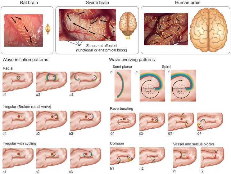
In the recently published article, "Heterogeneous incidence and propagation of spreading depolarizations," it is shown, in vivo and in vitro, how KCl-induced spreading depolarizations in mouse and rat brains can be highly variable, and that they are not limited, as once thought, to a concentric, isotropic, or homogenous depolarization wave in space or in time. The reported results serve as a link between the different species, and this paper contributes to changing the way in which SD expansion is viewed in the lissencephalic brain. Here, we discuss their results with our previous observations made in the gyrencephalic swine brain, in computer simulations, and in the human brain.

Keywords: Spreading depolarization; animal models; gyrencephalic brain; intrinsic optical signal imaging; lissencephalic brain.


Figures

Figure 1.
Figure 1.
Anatomical differences between gyrencephalic brains and a lissencephalic rat brain (upper). Typical propagation patters reported by Kaufmann et al. in rats; example of irregular propagation pattern seen in the swine model and a hypothetical irregular propagation pattern in a human brain. In the lower section, wave initiation and evolving SD patterns in a gyrencephalic brain are presented by type, presented by letters over time (represented by numbers). Initiation patterns a, b and c correspond to KCl stimulations. The typical initiation is a radial wave, but early breaking can originate irregular patterns. Evolving patterns (d to i) are described according to findings in swine experiments. The spirals (e and f) were predicted even before my mathematical models.
Figure 2.
Figure 2.
Personalized computer simulation of SD causing a propagating visual field defect in support of SD as a localized small wave segment. (a) Propagation pattern of a self-reported visual field defect during migraine with aura in the lower quadrant of the right visual hemifield. The position of the propagating visual field defect is marked by a line every minute for 8 min resulting in eight lines starting near the center of the visual field and passing roughly parallel to the right of the vertical meridian. (b) 3D form (blue) of the left primary visual cortex obtained by magnetic resonance imaging scanner readings from a migraine sufferer who draw his visual field defect in (a). The occipital pole is facing backwards. The computer simulation of SD (red wave segment) reproduced the path causing the visual field defect shown in (a). The wave segment was initiated near the occipital pole in such a way that the direction of SD propagation points toward the cunes. (c) Same as (b) showing the end of the computer simulation where the SD wave segment propagated for about 8 min a distance of about 24 mm on a particular path (white line) on the gyral crown on the cuneus. (d) Cortical areas successively affected reported by other patient with complex aura, which provides further evidence of a specified cortical path of a wave segment rather than an all-engulfing circular SD that spreads concentrically in all directions (modified from Vincent et al.).

References

    1. Ayata C, Lauritzen M. spreading depression, spreading depolarizations, and the cerebral vasculature. Physiol Rev 2015; 95: 953–993. - PMC - PubMed
    1. Dreier JP, Reiffurth C. The stroke-migraine depolarization continuum. Neuron 2015; 86: 902–922. - PubMed
    1. Dreier JP. The role of spreading depression, spreading depolarization and spreading ischemia in neurological disease. Nat Med 2011; 17: 439–447. - PubMed
    1. Hartings JA, Shuttleworth CW, Kirov SA, et al. The continuum of spreading depolarizations in acute cortical lesion development: examining Leão’s legacy. J Cereb Blood Flow Metab. Metab 2017; 37(5): 1571–1594. - PMC - PubMed
    1. Ayata C. Pearls and pitfalls in experimental models of spreading depression. Cephalalgia Int J Headache 2013; 33: 604–613. - PubMed

LinkOut - more resources