

Compromised Neurotrophic and Angiogenic Regenerative Capability during Tendon Healing in a Rat Model of Type-II Diabetes
- PMID: 28122008
- PMCID: PMC5266316
- DOI: 10.1371/journal.pone.0170748
Compromised Neurotrophic and Angiogenic Regenerative Capability during Tendon Healing in a Rat Model of Type-II Diabetes
Abstract
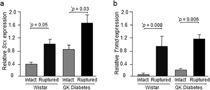
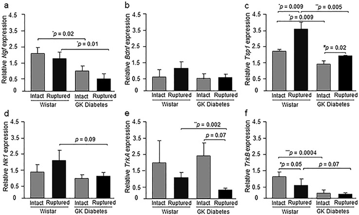
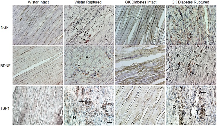
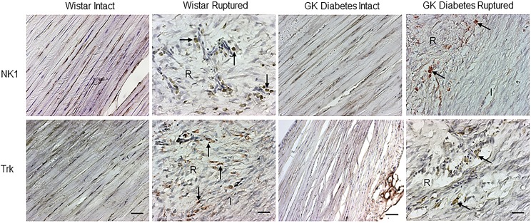
Metabolic diseases such as diabetes mellitus type-II (DM-II) may increase the risk of suffering painful connective tissue disorders and tendon ruptures. The pathomechanisms, however, by which diabetes adversely affects connective tissue matrix metabolism and regeneration, still need better definition. Our aim was to study the effect of DM-II on expressional changes of neuro- and angiotrophic mediators and receptors in intact and healing Achilles tendon. The right Achilles tendon was transected in 5 male DM-II Goto-Kakizaki (GK) and 4 age-matched Wistar control rats. The left Achilles tendons were left intact. At week 2 post-injury, NGF, BDNF, TSP, and receptors TrkA, TrkB and Nk1 gene expression was studied by quantitative RT-PCR (qRT-PCR) and their protein distribution by immunohistochemistry in intact and injured tendons. The expression of tendon-related markers, Scleraxis (SCX) and Tenomodulin (TNMD), was evaluated by qRT-PCR in intact and injured tendons. Injured tendons of diabetic GK rats exhibited significantly down-regulated Ngf and Tsp1 mRNA and corresponding protein levels, and down-regulated Trka gene expression compared to injured Wistar controls. Intact tendons of DM-II GK rats displayed reduced mRNA levels for Ngf, Tsp1 and Trkb compared to corresponding intact non-diabetic tendons. Up-regulated Scx and Tnmd gene expression was observed in injured tendons of normal and diabetic GK rats compared to intact Wistar controls. However, these molecules were not up-regulated in injured DM-II GK rats compared to their corresponding controls. Our results suggest that DM-II has detrimental effects on neuro- and angiotrophic pathways, and such effects may reflect the compromised repair seen in diabetic Achilles tendon. Thus, novel approaches for regeneration of injured, including tendinopathic, and surgically repaired diabetic tendons may include therapeutic molecular modulation of neurotrophic pathways such as NGF and its receptors.
Conflict of interest statement
The authors have declared that no competing interests exist.
Figures
Similar articles
-
Expressional changes in growth and inflammatory mediators during Achilles tendon repair in diabetic rats: new insights into a possible basis for compromised healing.Cell Tissue Res. 2014 Jul;357(1):109-17. doi: 10.1007/s00441-014-1871-3. Epub 2014 May 6. Cell Tissue Res. 2014. PMID: 24797839
-
Type 2 diabetes impairs tendon repair after injury in a rat model.J Appl Physiol (1985). 2012 Dec 1;113(11):1784-91. doi: 10.1152/japplphysiol.00767.2012. Epub 2012 Oct 4. J Appl Physiol (1985). 2012. PMID: 23042903
-
Dynamic patterns of BDNF expression in injured sensory neurons: differential modulation by NGF and NT-3.Eur J Neurosci. 2002 Oct;16(8):1449-62. doi: 10.1046/j.1460-9568.2002.02205.x. Eur J Neurosci. 2002. PMID: 12405958
-
Does Diabetes Mellitus Affect Tendon Healing?Adv Exp Med Biol. 2016;920:179-84. doi: 10.1007/978-3-319-33943-6_16. Adv Exp Med Biol. 2016. PMID: 27535259 Review.
-
Effects of Type II Diabetes Mellitus on Tendon Homeostasis and Healing.J Orthop Res. 2020 Jan;38(1):13-22. doi: 10.1002/jor.24388. Epub 2019 Jun 24. J Orthop Res. 2020. PMID: 31166037 Free PMC article. Review.
Cited by
-
Scaffold-Mediated Immunoengineering as Innovative Strategy for Tendon Regeneration.Cells. 2022 Jan 13;11(2):266. doi: 10.3390/cells11020266. Cells. 2022. PMID: 35053383 Free PMC article. Review.
-
Mammal comparative tendon biology: advances in regulatory mechanisms through a computational modeling.Front Vet Sci. 2023 Apr 27;10:1175346. doi: 10.3389/fvets.2023.1175346. eCollection 2023. Front Vet Sci. 2023. PMID: 37180059 Free PMC article.
-
Effect of photobiomodulation and exercise on early remodeling of the Achilles tendon in streptozotocin-induced diabetic rats.PLoS One. 2019 Feb 4;14(2):e0211643. doi: 10.1371/journal.pone.0211643. eCollection 2019. PLoS One. 2019. PMID: 30716140 Free PMC article.
-
The Implication of Substance P in the Development of Tendinopathy: A Case Control Study.Int J Mol Sci. 2017 Jun 9;18(6):1241. doi: 10.3390/ijms18061241. Int J Mol Sci. 2017. PMID: 28598390 Free PMC article.
-
Metabolic Regulation of Tendon Inflammation and Healing Following Injury.Curr Rheumatol Rep. 2021 Feb 10;23(3):15. doi: 10.1007/s11926-021-00981-4. Curr Rheumatol Rep. 2021. PMID: 33569739 Free PMC article. Review.
References
-
- Cagliero E, Apruzzese W, Perlmutter GS, Nathan DM. Musculoskeletal disorders of the hand and shoulder in patients with diabetes mellitus. Am J Med. 2002;112(6):487–90. - PubMed
-
- Rosenbloom AL, Silverstein JH. Connective tissue and joint disease in diabetes mellitus. Endocrinol Metab Clin North Am. 1996;25(2):473–83. - PubMed
-
- Ackermann PW, Salo PT, Hart DA. Neuronal pathways in tendon healing. Front Biosci. 2009;14:5165–87. - PubMed
MeSH terms
Substances
LinkOut - more resources
Full Text Sources
Other Literature Sources
Medical
Miscellaneous
