. 2017 Apr;65(4):207-221.
doi: 10.1369/0022155416689670. Epub 2017 Jan 25.

Histochemical Examination on Periodontal Tissues of Klotho-Deficient Mice Fed With Phosphate-Insufficient Diet

Affiliations

Histochemical Examination on Periodontal Tissues of Klotho-Deficient Mice Fed With Phosphate-Insufficient Diet

Kumiko Hikone et al. J Histochem Cytochem. 2017 Apr.

Abstract

To elucidate which of elevated serum concentration of inorganic phosphate (Pi) or disrupted signaling linked to αklotho/fibroblast growth factor 23 (FGF23) is a predominant regulator for senescence-related degeneration seen in αKlotho-deficient mice, we have examined histological alteration of the periodontal tissues in the mandibular interalveolar septum of αKlotho-deficient mice fed with Pi-insufficient diet. We prepared six groups of mice: wild-type, kl/kl, and αKlotho-/- mice with normal diet or low-Pi diet. As a consequence, kl/klnorPi and αKlotho-/-norPi mice showed the same abnormalities in periodontal tissues: intensely stained areas with hematoxylin in the interalveolar septum, dispersed localization of alkaline phosphatase-positive osteoblasts and tartrate-resistant acid phosphatase-reactive osteoclasts, and accumulation of dentin matrix protein 1 in the osteocytic lacunae. Although kl/kllowPi mice improved these histological abnormalities, αKlotho-/- lowPi mice failed to normalize those. Gene expression of αKlotho was shown to be increased in kl/kl lowPi specimens. It seems likely that histological abnormalities of kl/kl mice have been improved by the rescued expression of αKlotho, rather than low concentration of serum Pi. Thus, the histological malformation in periodontal tissues in αKlotho-deficient mice appears to be due to not only increased concentration of Pi but also disrupted αklotho/FGF23 signaling.

Keywords: DMP-1; osteopontin; periodontal tissue; phosphate; αKlotho-deficient mice.


Conflict of interest statement

Competing Interests: The author(s) declared no potential conflicts of interest with respect to the research, authorship, and/or publication of this article.

Figures

Figure 1.
Figure 1.
Body weights and the serum concentration of Pi in wild-type and kl/kl mice fed with normal and Pi-insufficient diet. (A) Appearance of whole body size at the 7 weeks of age in wild-type norPi, kl/kl norPi, wild-type lowPi, and kl/kl lowPi mice. The average body weight of wild-type lowPi mice significantly decreases compared with wild-type norPi mice. In contrast, the body weight of kl/kl lowPi mice increases compared with kl/kl norPi mice (B; **p<0.01). Serum concentration of Pi shows no significant difference in kl/kl lowPi mice and kl/kl lowPi mice (C). The values are expressed as mean ± SE. Abbreviation: Pi, phosphate.
Figure 2.
Figure 2.
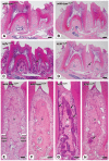
Histological analysis of interalveolar septum in wild-type and kl/kl mice fed with normal and Pi-insufficient diet. Panels (E)–(H) are highly magnified images of panels (A)–(D). Hematoxylin–eosin staining demonstrates the similar histology of alveolar bone and PDL in the interalveolar septum between the first and second molars of wild-type norPi and wild-type lowPi mice (A, B, E, F). The regions intensely stained with hematoxylin (arrows in C and G) are observed in the alveolar bone and narrow PDL of kl/kl norPi mice (C, G). However, these histological alterations are rescued by feeding low-Pi diet as shown in kl/kl lowPi mice (D, H). Note only a few regions of intensely stained with hematoxylin in alveolar bones of kl/kl lowPi mice (arrows in D and H). Abbreviations: Pi, phosphate; PDL, periodontal ligament; is, interalveolar septum. Bars: A–D = 150 µm; E–H = 50 µm.
Figure 3.
Figure 3.
Histology of mesial and distal regions in the interalveolar septum of wild-type and kl/kl mice fed with normal and Pi-insufficient diet. In wild-type norPi and wild-type lowPi mice, the mesial regions of the interalveolar septum demonstrate scalloped bone surfaces (A, C), whereas the distal regions demonstrate smooth bone surfaces (B, D). Kl/kl norPi mice shows the abnormal features at both mesial and distal regions—amorphous materials intensely stained with hematoxylin (arrows) in the superficial layer of alveolar bone and in the cementum (E, F). There are several empty osteocytic lacunae in the alveolar bone. Note the disturbed orientation of PDL cells in the narrow periodontal space. However, kl/kl lowPi mice show the intact histology of alveolar bone and PDL as can be seen in the wild-type mice (G, H). Abbreviations: Pi, phosphate; PDL: periodontal ligament; is, interalveolar septum. Bars: A–H = 20 µm.
Figure 4.
Figure 4.
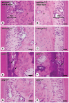
Immunolocalization of ALPase in the interalveolar septum of wild-type and kl/kl mice fed with normal and Pi-insufficient diet. Panels (E)–(H) are the highly magnified images of the distal walls of interalveolar septum (A–D). Distal walls of interalveolar septum of wild-type norPi mice (arrows, E), wild-typelowPi mice (arrows, F), and kl/kl lowPi mice (arrows, H) display intense ALPase immunoreactivity (brown color, A, B, D, E, F, H) and smooth bone surfaces. However, in kl/kl norPi interalveolar bone, weak ALPase positivity can be seen (C, G). Abbreviations: ALPase, alkaline phosphatase; Pi, phosphate; is, interalveolar septum. Bars: A–D = 50 µm; E–H = 20 µm.
Figure 5.
Figure 5.
Distribution of TRAPase-positive osteoclasts in the interalveolar septum of wild-type and kl/kl mice fed with normal and Pi-insufficient diet. Panels (E)–(H) are the highly magnified images of the mesial walls of interalveolar septum (A–D). TRAPase-reactive osteoclasts (red color) are located on the mesial surfaces of wild-type norPi, wild-type lowPi, and kl/kl lowPi interalveolar septum (arrows, A, B, D, E, F, H). However, TRAPase-positive osteoclasts are hardly seen in kl/kl norPi mice (C, G), but instead, some TRAPase-positive cement lines (arrows, G) are discernible in the central regions of the interalveolar septum. Abbreviations: TRAPase, tartrate-resistant acid phosphatase; Pi, phosphate; is, interalveolar septum. Bars: A–D = 50 µm; E–H = 20 µm.
Figure 6.
Figure 6.
Immunolocalization of DMP-1 in the interalveolar septum of wild-type and kl/kl mice fed with normal and Pi-insufficient diet. DMP-1 positivity (brown color) is found in osteocytes and their canaliculi located in the interalveolar septum of wild-type norPi and wild-type lowPi (A, B, E, F). In contrast, kl/kl norPi interalveolar septum displays many patchy materials with DMP-1 immunoreactivity in osteocytic lacunae and on cementum (arrows, C, G). When being fed with low-Pi diet, DMP-1 immunoreactivity of kl/kl lowPi looks similar to that seen in the wild-type mice, although some regions show DMP-1 accumulation in the osteocytic lacunae in kl/kl lowPi mice (arrows, D, H). Abbreviations: DMP-1, dentin matrix protein 1; Pi, phosphate; is, interalveolar septum. Bars: A–D = 50 µm; E–H = 20 µm.
Figure 7.
Figure 7.
Immunolocalization of osteopontin in the interalveolar septum of wild-type and kl/kl mice fed with normal and Pi-insufficient diet. Panels (E)–(H) are the highly magnified images of the mesial regions of interalveolar septum (A–D). The mesial surfaces of interalveolar septum are intensely stained with osteopontin in wild-type norPi and wild-type lowPi mice (brown color, arrows, E, F). Alternatively, amorphous materials bearing an intense osteopontin reactivity (arrows) are present in kl/kl norPi alveolar bone (C, G). In kl/kl lowPi mice, however, immunolocalization of osteopontin (arrows) shows the same distribution as wild-type mice in the mesial region of interalveolar septum (D, H). Abbreviations: Pi, phosphate; is, interalveolar septum. Bars: A–D = 50 µm; E–H = 20 µm.
Figure 8.
Figure 8.
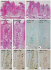
Histochemical analyses of interalveolar septum in αKlotho−/− mice fed with normal and Pi-insufficient diet. Panels (C)–(J) are the magnified images of the region indicated by arrows in panels A and B. Both of αKlotho−/−norPi and αKlotho−/−lowPi mice exhibit the areas intensely stained with hematoxylin in the interalveolar septum (arrows, C, D). TRAPase-positive osteoclasts are not obvious in the mesial region (E, F). Note that DMP-1 accumulates in the osteocytic lacunae (brown color, arrows, G, H), and abundant osteopontin is discernible in the amorphous material (brown color, arrows, I, J). Note the distribution of amorphous materials strongly stained with hematoxylin (C, D), DMP-1 (G, H), and osteopontin (I, J) is similar between αKlotho−/−norPi and αKlotho−/−lowPi interalveolar septum. Abbreviations: Pi, phosphate; TRAPase, tartrate-resistant acid phosphatase; DMP-1, dentin matrix protein 1; is, interalveolar septum. Bars: A, B = 150 µm; C–F = 50 µm; G–J = 30 µm.
Figure 9.
Figure 9.
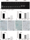
Gene expression of Fgfr1c and αKlotho in kidney and mandibles of kl/kl mice fed with normal and phosphate (Pi)-insufficient diet. RT-PCR shows relatively intense band representing αKlotho gene in the kidney of kl/kl lowPi mice compared with that of kl/kl norPi mice (A). In real-time PCR analysis, αKlotho gene is elevated in kl/kl lowPi mice compared with kl/kl norPi mice, whereas it does not reach the levels of wild-type (WT) specimens (B). No obvious difference can be seen in the expression level of Fgfr1c mRNA among the specimens from WT norPi, WT lowPi, kl/kl norPi, and kl/kl lowPi mice (C). Panels (D)–(G) show FGF23 immunolocalization in the interalveolar septum. FGF23 immunoreactivity is observed in osteocytes (arrows in D, E, and G). Real-time PCR shows an elevated expression of αKlotho gene in kl/kl lowPi specimens compared with kl/kl norPi mice (H). Notice no obvious differences concerning Fgfr1c mRNA among all groups (I). Bars: D–G = 50 µm.

Similar articles

Cited by

References

    1. Skougaard MR, Frandsen A, Baker DG. Collagen metabolism of skin and periodontal membrane in the squirrel monkey. Scand J Dent Res. 1970;78:374–7. - PubMed
    1. Fullmer HM, Sheetz JH, Narkates AJ. Oxytalan connective tissue fibers: a review. J Oral Pathol. 1974;3:291–316. - PubMed
    1. Rippin JW. Collagen turnover in the periodontal ligament under normal and altered functional forces. I. Young rat molars. J Periodontal Res. 1976;11:101–7. - PubMed
    1. Sodek J. A comparison of the rates of synthesis and turnover of collagen and non-collagen proteins in adult rat periodontal tissues and skin using a microassay. Arch Oral Biol. 1977;22:655–65. - PubMed
    1. Rippin JW. Collagen turnover in the periodontal ligament under normal and altered functional forces. II. Adult rat molars. J Periodontal Res. 1978;13:149–54. - PubMed