Review
. 2017 Jan 24;112(2):207-214.
doi: 10.1016/j.bpj.2016.12.015.

Mechanochemical Signaling Directs Cell-Shape Change

Affiliations
Review

Mechanochemical Signaling Directs Cell-Shape Change

Eric S Schiffhauer et al. Biophys J. .

Abstract

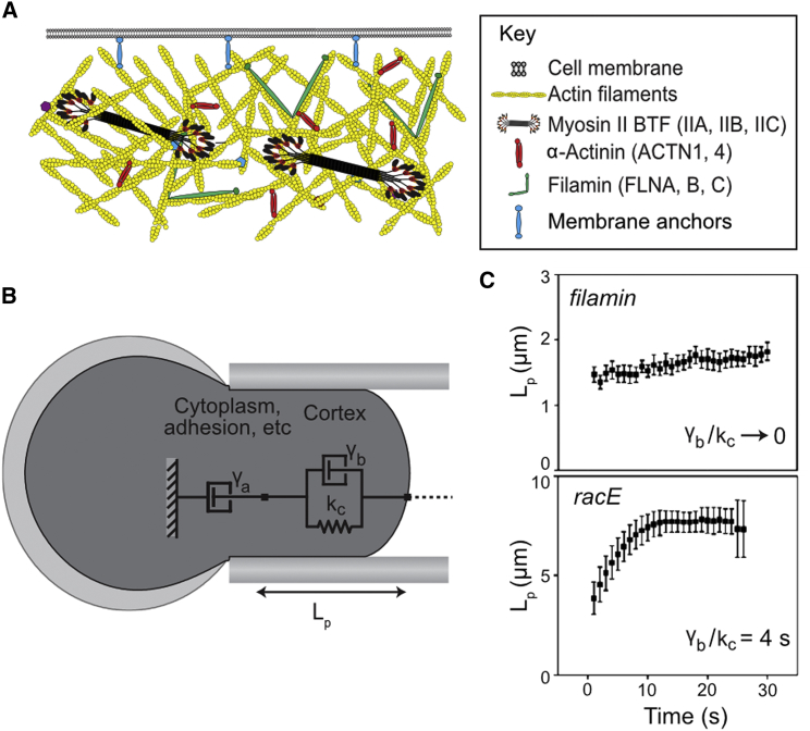
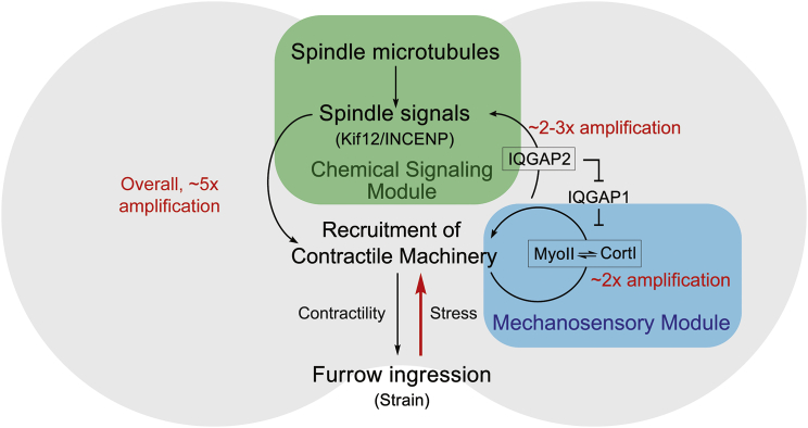
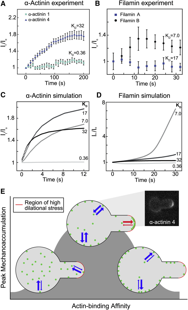
For specialized cell function, as well as active cell behaviors such as division, migration, and tissue development, cells must undergo dynamic changes in shape. To complete these processes, cells integrate chemical and mechanical signals to direct force production. This mechanochemical integration allows for the rapid production and adaptation of leading-edge machinery in migrating cells, the invasion of one cell into another during cell-cell fusion, and the force-feedback loops that ensure robust cytokinesis. A quantitative understanding of cell mechanics coupled with protein dynamics has allowed us to account for furrow ingression during cytokinesis, a model cell-shape-change process. At the core of cell-shape changes is the ability of the cell's machinery to sense mechanical forces and tune the force-generating machinery as needed. Force-sensitive cytoskeletal proteins, including myosin II motors and actin cross-linkers such as α-actinin and filamin, accumulate in response to internally generated and externally imposed mechanical stresses, endowing the cell with the ability to discern and respond to mechanical cues. The physical theory behind how these proteins display mechanosensitive accumulation has allowed us to predict paralog-specific behaviors of different cross-linking proteins and identify a zone of optimal actin-binding affinity that allows for mechanical stress-induced protein accumulation. These molecular mechanisms coupled with the mechanical feedback systems ensure robust shape changes, but if they go awry, they are poised to promote disease states such as cancer cell metastasis and loss of tissue integrity.


Figures

Figure 1
Figure 1
The structure of the cortex determines its physical properties. (A) Major components of the actin cytoskeleton in the cell cortex, shown roughly to scale for mammalian cells. (B) A simple model of cell mechanics, where the spring kc and the viscous damper γb respectively describe the elastic and viscous contributions from the cortex. The damper γa primarily describes the viscous contribution from the cytoplasm. Upon aspiration of Dictyostelium cells into a micropipette using a fixed pressure, the length of the cell protruding into the pipette (Lp) is observed over time. The model in (B) can be accurately used to describe the creep of the cells into the pipette in (C). The slope of the first and second phases of deformation can be used to compute the value of the viscous dampers and γa, respectively. The amplitude of the initial length deformation can be used to determine the elastic parameter kc. In the filamin-null, the contribution of the initial damper γb is much smaller than that of kc, so the initial deformation happens in <1 s. The continued flow can be described by γa. In the racE-null, the contribution of γb is quite large, causing a slow initial deformation, whereas γa is quite small, displaying no creep in the second phase of deformation. To see this figure in color, go online.
Figure 2
Figure 2
Integrated chemical and mechanical feedback loops drive cleavage furrow ingression. At the cleavage furrow of dividing Dictyostelium, the chemical signaling module, including INCENP and Kif12 (the kinesin 6 family protein), can activate the recruitment of contractile machinery, including cortexillin I (CortI) and myosin II (MyoII). Simultaneously, the contractile machinery, which comprises the mechanosensory module, can accumulate in response to the forces created by furrow ingression and drive the activation of the chemical signaling module through IQGAP2. The overall system allows for an ∼5-fold amplification of myosin II accumulation at the cleavage furrow in response to mechanical stress. To see this figure in color, go online.
Figure 3
Figure 3
Mechanoaccumulation by actin-binding proteins is determined by an optimal zone of actin-binding affinity. (A) The low-affinity α-actinin 4, transfected into HeLa cells, accumulates over time in response to micropipette aspiration at a region of high network dilation, the tip of the cell (calculated by fluorescence intensity at the tip, It, normalized to the fluorescence intensity at the opposite side of the cell, Io). The high-affinity α-actinin 1 does not accumulate. All KD values in panels (A)–(D) have the units of μM. (B) The high-affinity filamin B accumulates to a region of high shear deformation, the neck of the cell, during micropipette aspiration (calculated by the fluorescence intensity at the neck, In, normalized to the fluorescence intensity at the opposite side of the cell, Io). The lower-affinity filamin A does not accumulate. For filamin B, a second phase of myosin II-mediated flow carries the protein to the tip, which accounts for the decrease beginning at ∼12 s. (C) The accumulation of a force-dependent actin-binding protein (e.g., α-actinin) is modeled with four different actin-binding affinities, using a reaction-diffusion model of force-dependent actin binding with physiological G- and F-actin concentrations, cross-linker concentrations, and published actin-binding affinities. (D) The accumulation of filamin is modeled using the same four actin-binding affinities, but considering cooperativity to account for the accelerating rate of accumulation. The results show that an optimal dissociation equilibrium constant (KD) to actin exists for both noncooperative (α-actinin) and cooperative (filamin) actin-binding proteins where mechanoaccumulation is maximized. Please note that for (C) and (D), the affinities represent the affinities of single actin-binding heads, and not an overall apparent affinity from the complete cross-linking reaction. (A)–(D) are reproduced here from Schiffhauer et al. 2016 (49). (E) At a high actin-binding affinity, actin cross-linking proteins do not have a large enough unbound pool to dynamically respond to force applied during micropipette aspiration. At a very low actin-binding affinity, actin-binding proteins do not bind the cortex with enough affinity to remain locked on at sites of mechanical stress. Thus, there is an optimal zone for actin-binding affinity where mechanoaccumulation is maximal. This is demonstrated by the inset, which shows accumulation of α-actinin 4 during micropipette aspiration. To see this figure in color, go online.

References

    1. Xu K., Zhong G., Zhuang X. Actin, spectrin, and associated proteins form a periodic cytoskeletal structure in axons. Science. 2013;339:452–456. - PMC - PubMed
    1. Nussenzveig R.H., Christensen R.D., Agarwal A.M. Novel α-spectrin mutation in trans with α-spectrin causing severe neonatal jaundice from hereditary spherocytosis. Neonatology. 2014;106:355–357. - PubMed
    1. Clark A.G., Dierkes K., Paluch E.K. Monitoring actin cortex thickness in live cells. Biophys. J. 2013;105:570–580. - PMC - PubMed
    1. Reichl E.M., Ren Y., Robinson D.N. Interactions between myosin and actin crosslinkers control cytokinesis contractility dynamics and mechanics. Curr. Biol. 2008;18:471–480. - PMC - PubMed
    1. Biro M., Romeo Y., Paluch E.K. Cell cortex composition and homeostasis resolved by integrating proteomics and quantitative imaging. Cytoskeleton. 2013;70:741–754. - PubMed

LinkOut - more resources