. 2017 Jan 24;112(2):313-324.
doi: 10.1016/j.bpj.2016.12.025.

Three-Phase Coexistence in Lipid Membranes

Affiliations

Three-Phase Coexistence in Lipid Membranes

Anders Aufderhorst-Roberts et al. Biophys J. .

Abstract

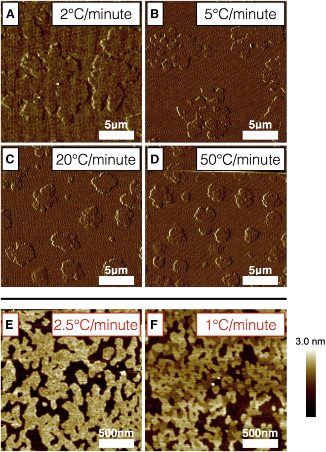
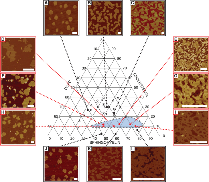
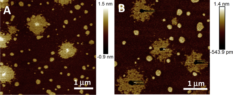
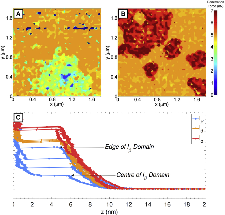
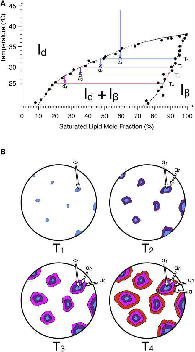
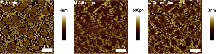
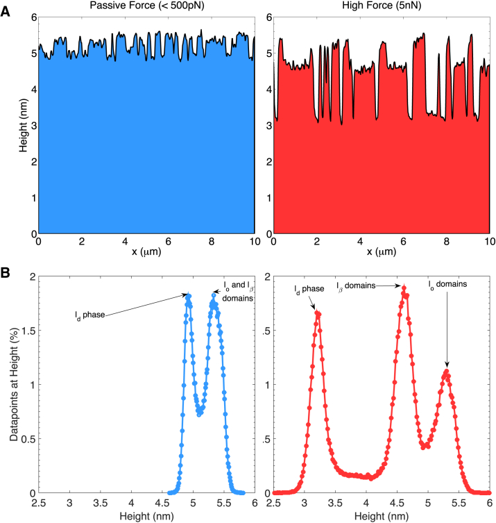
Phospholipid ternary systems are useful model systems for understanding lipid-lipid interactions and their influence on biological properties such as cell signaling and protein translocation. Despite extensive studies, there are still open questions relating to membrane phase behavior, particularly relating to a proposed state of three-phase coexistence, due to the difficulty in clearly distinguishing the three phases. We look in and around the region of the phase diagram where three phases are expected and use a combination of different atomic force microscopy (AFM) modes to present the first images of three coexisting lipid phases in biomimetic cell lipid membranes. Domains form through either nucleation or spinodal decomposition dependent upon composition, with some exhibiting both mechanisms in different domains simultaneously. Slow cooling rates are necessary to sufficiently separate mixtures with high proportions of lo and lβ phase. We probe domain heights and mechanical properties and demonstrate that the gel (lβ) domains have unusually low structural integrity in the three-phase region. This finding supports the hypothesis of a "disordered gel" state that has been proposed from NMR studies of similar systems, where the addition of small amounts of cholesterol was shown to disrupt the regular packing of the lβ state. We use NMR data from the literature on chain disorder in different mixtures and estimate an expected step height that is in excellent agreement with the AFM data. Alternatively, the disordered solid phase observed here and in the wider literature could be explained by the lβ phase being out of equilibrium, in a surface kinetically trapped state. This view is supported by the observation of unusual growth of nucleated domains, which we term "tree-ring growth," reflecting compositional heterogeneity in large disordered lβ phase domains.


Figures

Figure 1
Figure 1
Two-phase and three phase bilayers prepared at variable cooling rates. (AD) Contact-mode AFM images (deflection signal) of a lipid bilayer (40% egg sphingomyelin and 60% DOPC) formed at different linear cooling rates, as labeled. Slowing the cooling rate has no effect on height mismatch (1.5 ± 0.1 nm) or domain area fraction (area lβ = 24 ± 3%), only on the number and size of domains. Cooling more slowly than 1°C/min leads to domains that are too large to observe clearly by AFM at this composition. (E and F) Three-phase bilayers are imaged with peak-force QNM AFM (composition, 68% egg sphingomyelin, 20% DOPC, and 12% cholesterol). Here, faster cooling causes the bilayer to appear as a two-phase system, although a fine structure is apparent in the higher of the two phases. For slower cooling, the three phases can be clearly seen, suggesting that under increased cooling rates very small domains become kinetically trapped and unable to effectively separate. To see this figure in color, go online.
Figure 2
Figure 2
Selected AFM images of bilayers exhibiting two-phase (AC and JL) and three-phase (DI) behavior. Image labels correspond to the compositions listed in Table 1, and black circles are previously published two-phase compositions (25). In the two-phase regions, lateral structure varies gradually and uniformly with composition. By contrast, in the three-phase region (approximated by the blue shaded region), lateral structure of the different domains varies significantly between samples, suggesting that nucleation pathways are very sensitive to sample composition. Image sizes are chosen to show fine structure. Scale bars, 1 μm. To see this figure in color, go online.
Figure 3
Figure 3
Tapping-mode AFM images of composition F formed at a slower cooling rate of 0.4°C/min. At a high-amplitude setpoint, the core of the lβ phase appears slightly higher than the surrounding lβ phase (A), whereas at a lower-amplitude setpoint (higher force), the core collapses (B), demonstrating variable domain compressibility. Domains are approximately round, suggesting binodal formation. lo domains appear homogeneous, whereas lβ domains are laterally heterogeneous, suggesting a noncontinuous composition. An image of a larger area of the sample shown in (B) can be found in Fig. S1.
Figure 4
Figure 4
Heat map of the penetration force of the lβ and ld domains (A) and the lo and ld domains (B). The lo domains collapse at ∼5.7–6.7 nN (B), whereas the ld phase collapses at ∼4.7–5 nN. (A) The lβ domains collapse gradually over a much broader range of forces, 1.4–4.6 nN, with the core of the domains collapsing at low force and the edge at high force; hence, the compressiblity of the lβ domains varies radially. The penetration force is shown as a discontinuity in the force curves (C) and is shown to be approximately constant for different positions in the lo and ld domains but variable across radially different positions in the lβ domains, further evidence of radial variation in lβ domain mechanical properties.
Figure 5
Figure 5
Schematic describing the process of “tree-ring growth.” As a saturated lipid/unsaturated lipid binary-mixture bilayer cools, the temperature drops below the phase transition temperature (A; figure for a simple two-phase system as taken from a DOPC/DPPC phase diagram in the literature (39)). With cooling, nucleated lβ domains begin to appear (B (38)). Subsequent drops in temperature result in further phase separation, but only between lipids in the ld phase, as lipids in the lβ domains are kinetically trapped due to the domain immiscibility. Thus, each incremental layer of the lβ domain has a subtly different composition and therefore different mechanical properties. Experimentally, the temperature ramp is smooth and continuous; hence, the composition of the domains will follow the solidus curve, and the composition of the melt will follow the liquidus curve.
Figure 6
Figure 6
Peak-force QNM AFM image at high force (5 nN) of a phospholipid bilayer formed from composition E. The step heights between the three phases are highly pronounced, indicating that the different domains are compressed to different extents (A). The adhesion channel shows high adhesion in the ld phase but negligible adhesion in the other two phases, suggesting substantial compression of the ld domains (B). The deformation channel shows that the ld phase is the most deformable, followed by the lβ phase, whereas the lo phase shows negligible deformability (C).
Figure 7
Figure 7
Representative height cross section and image-averaged height histogram of bilayers prepared using composition E at two different forces. (A) The step-height differences between phases is shown to increase when the force is increased from ∼200 pN (left) to 5 nN (right). (B) Similarly, at lower forces (left), the height histogram shows two distinct peaks, as the lo and lβ phases are indistinguishable, whereas at higher forces (right), differential domain compression results in three clearly distinguishable peaks.

References

    1. Lingwood D., Simons K. Lipid rafts as a membrane-organizing principle. Science. 2010;327:46–50. - PubMed
    1. Chini B., Parenti M. G-protein coupled receptors in lipid rafts and caveolae: how, when and why do they go there? J. Mol. Endocrinol. 2004;32:325–338. - PubMed
    1. Batada N.N., Shepp L.A., Siegmund D.O. Stochastic model of protein-protein interaction: why signaling proteins need to be colocalized. Proc. Natl. Acad. Sci. USA. 2004;101:6445–6449. - PMC - PubMed
    1. Klaasse E.C., Ijzerman A.P., Beukers M.W. Internalization and desensitization of adenosine receptors. Purinergic Signal. 2008;4:21–37. - PMC - PubMed
    1. Uppamoochikkal P., Tristram-Nagle S., Nagle J.F. Orientation of tie-lines in the phase diagram of DOPC/DPPC/cholesterol model biomembranes. Langmuir. 2010;26:17363–17368. - PMC - PubMed

MeSH terms

Substances