. 2017 Jan 24;18(4):831-839.
doi: 10.1016/j.celrep.2016.12.074.

Germ Cell-less Promotes Centrosome Segregation to Induce Germ Cell Formation

Affiliations

Germ Cell-less Promotes Centrosome Segregation to Induce Germ Cell Formation

Dorothy A Lerit et al. Cell Rep. .

Abstract

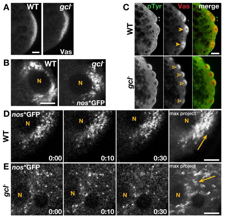
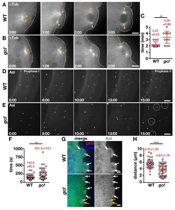
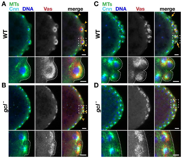
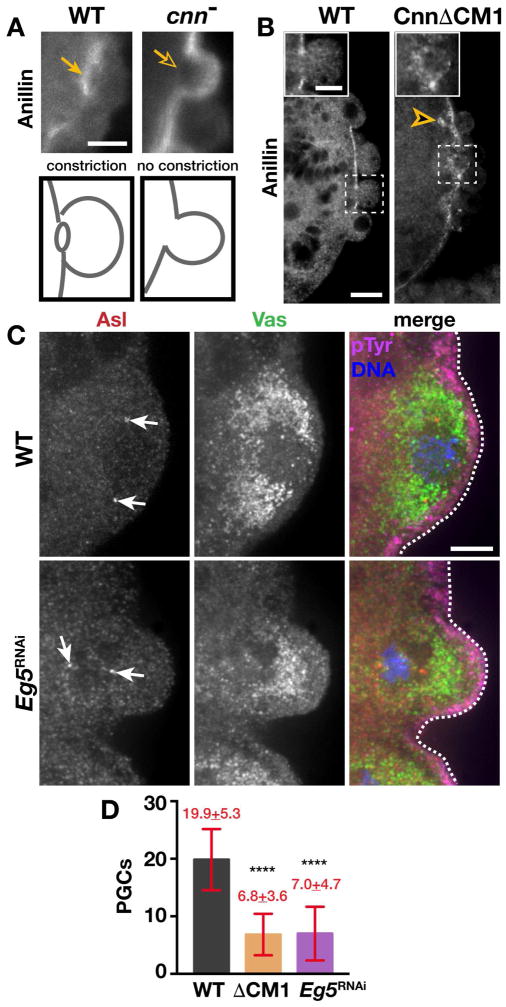
The primordial germ cells (PGCs) specified during embryogenesis serve as progenitors to the adult germline stem cells. In Drosophila, the proper specification and formation of PGCs require both centrosomes and germ plasm, which contains the germline determinants. Centrosomes are microtubule (MT)-organizing centers that ensure the faithful segregation of germ plasm into PGCs. To date, mechanisms that modulate centrosome behavior to engineer PGC development have remained elusive. Only one germ plasm component, Germ cell-less (Gcl), is known to play a role in PGC formation. Here, we show that Gcl engineers PGC formation by regulating centrosome dynamics. Loss of gcl leads to aberrant centrosome separation and elaboration of the astral MT network, resulting in inefficient germ plasm segregation and aborted PGC cellularization. Importantly, compromising centrosome separation alone is sufficient to mimic the gcl loss-of-function phenotypes. We conclude Gcl functions as a key regulator of centrosome separation required for proper PGC development.

Keywords: cell fate; centrosome; germ cell-less; germ cells; germ plasm; stem cells.


Figures

Figure 1
Figure 1. Germ plasm distribution is aberrant in gcl embryos
In all images, posterior is to the right. (A) Immunofluorescence for Vas in 0–1 hr WT and gcl embryos. (B) Maximum intensity projections show 8.3s of elapsed time from live 1–2 hr embryos expressing nos*GFP. (C) 1–2 hr embryos stained for the indicated proteins; closed arrowheads, symmetric Vas; open arrowheads, uneven/reduced Vas. (D and E) Time-lapse imaging of nos*GFP in live 1–2 hr embryos. Time is shown min:s; “N”, PB nuclei; arrows mark trajectory of nos*GFP. Bars: (A and C) 20 μm; (B, D, and E) 5 μm.
Figure 2
Figure 2. Gcl is required for MT organization
(A and B) Time-lapse imaging of αTub-GFP in 1–2 hr embryos; arrowheads, MTOCs; dashed lines, cortex. (C) Quantification of MTOC separation timing. Each data point indicates the time it takes for a pair of MTOCs to separate in WT (N=14 events in 12 embryos) and gcl (N=13 events in 9 embryos). (D and E) Maximum intensity projections from live imaging of Asl-YFP. Time is relative to prophase onset (0:00) and shown min:s. Centrosome separation delays are highlighted (dashed circles). (F) Quantification of centrosome segregation timing in WT (N=53 events in 9 embryos) and gcl (N=112 events in 17 embryos). (G) Prophase-stage NC 10 embryos stained for the indicated proteins; arrows mark complete (white) or incomplete (orange) centrosome separation. (H) Quantification of centrosome distance during prophase NC 10 (N=42 pairs from 10 WT embryos and N=49 pairs from 9 gcl embryos). Statistical significance was determined using a two-tailed Mann-Whitney test, **** p<0.0001, ** p<0.01. Bars: 5 μm. See also Figures S1 and S2.
Figure 3
Figure 3. Centrosome segregation instructs germ plasm distribution
Immunofluorescence for the indicated proteins during (A and B) PB formation and (C and D) PGC formation. Boxes show inset enlarged below; dashed lines mark the posterior cortex. Arrowheads show PBs, and arrows delineate the PGC cluster. Bars: 10 μm; insets 5 μm. See also Figure S3.
Figure 4
Figure 4. Functional centrosomes promote PGC formation
(A and B) Immunofluorescence for Anillin in 1–2 hr embryos. (A) Arrows note the presence (closed) or absence (open) of the Anillin ring (PB furrow). (B) Insets show Anillin distribution; arrowhead, Anillin puncta. (C) Immunofluorescence for the indicated proteins in NC 10 embryos; arrows, centrosomes; dashed lines, cortex. (D) Quantification of PGCs in NC 13–14 embryos. Statistical significance was determined by Student’s t-test, **** p<0.0001. Bars: (A and B) 10 μm; insets in (B), 5 μm; (C) 5 μm. See also Figure S4.

References

    1. BRECHBIEL JL, GAVIS ER. Spatial regulation of nanos is required for its function in dendrite morphogenesis. Curr Biol. 2008;18:745–750. - PMC - PubMed
    1. CAO J, et al. Cortical actin dynamics facilitate early-stage centrosome separation. Curr Biol. 2010;20:770–776. - PMC - PubMed
    1. CINALLI RM, LEHMANN R. A spindle-independent cleavage pathway controls germ cell formation in Drosophila. Nat Cell Biol. 2013;15:839–845. - PMC - PubMed
    1. EWEN-CAMPEN B, SCHWAGER EE, EXTAVOUR CG. The molecular machinery of germ line specification. Mol Reprod Dev. 2010;77:3–18. - PubMed
    1. FIELD CM, COUGHLIN M, DOBERSTEIN S, MARTY T, SULLIVAN W. Characterization of anillin mutants reveals essential roles in septin localization and plasma membrane integrity. Development. 2005;132:2849–2860. - PubMed

MeSH terms

LinkOut - more resources