. 2017 Feb 28;8(9):14680-14692.
doi: 10.18632/oncotarget.14747.

Cytoprotective effect of chlorogenic acid against hydrogen peroxide-induced oxidative stress in MC3T3-E1 cells through PI3K/Akt-mediated Nrf2/HO-1 signaling pathway

Affiliations

Cytoprotective effect of chlorogenic acid against hydrogen peroxide-induced oxidative stress in MC3T3-E1 cells through PI3K/Akt-mediated Nrf2/HO-1 signaling pathway

Dandan Han et al. Oncotarget. .

Abstract

Osteoporosis is a disorder of bone and its development is closely associated with oxidative stress and reactive oxygen species (ROS). Chlorogenic acid (CGA) has potential antioxidant effects and its pharmacological action in osteoblasts is not clearly understood. The present study aimed to clarify the protective effects and mechanisms of CGA on hydrogen peroxide (H2O2)-induced oxidative stress in osteoblast cells. MC3T3-E1 cells were treated with H2O2 to induce oxidative stress model in vitro. Cells were treated with CGA prior to H2O2 exposure, the intracellular ROS production, malondialdehyde content, nitric oxide release and glutathione level were measured. We also investigated the protein levels of heme oxygenase-1 (HO-1), the nuclear translocation of transcription factor NF-erythroid 2-related factor (Nrf2) and the phosphorylation levels of Akt in CGA-treated cells. The results showed that pretreatment of CGA could reverse the inhibition of cell viability and suppress the induced apoptosis and caspase-3 activity. Additionally, it significantly reduced H2O2-induced oxidative damage in a dose-dependent manner. Furthermore, it induced the protein expression of HO-1 together with its upstream mediator Nrf2, and activated the phosphorylation of Akt in MC3T3-E1 cells. LY294002, a PI3K/Akt inhibitor, significantly suppressed the CGA-induced Nrf2 nuclear translocation and HO-1 expression. Reduction of cell death mediated by CGA in presence of H2O2 was significantly inhibited by Zinc protoporphyrin IX (a HO-1 inhibitor) and LY294002. These data demonstrated that CGA protected MC3T3-E1 cells against oxidative damage via PI3K/Akt-mediated activation of Nrf2/HO-1 pathway, which may be an effective drug in treatment of osteoporosis.

Keywords: MC3T3-E1 cells; Nrf2/HO-1 pathway; chlorogenic acid; cytoprotection; oxidative stress.


Conflict of interest statement

CONFLICTS OF INTEREST

The author declares that there is no conflict of interest regarding the publication of this paper.

Figures

Figure 1
Figure 1. Effects of H2O2 on the apoptosis of MC3T3-E1 cells
(A) MC3T3-E1 cells were treated with various concentrations of H2O2 (0~1000 μM) for 1, 2, 4 and 6 h, and the cell viability was analyzed by MTT assay. (B) Cells were treated with 400 μM H2O2 for the indicated times (0, 1, 4, 6, and 12 h), and the cells were stained with DCFH-DA to detect the intracellular ROS production in different times by flow cytometry. (C) Cells were treated with 0, 400 and 800 μM H2O2 for 4 h, and apoptosis was determined by flow cytometry followed by Annexin V–PI double staining. (D) Cells were treated with 400 μM H2O2 for 4 h, and the cell morphology was observed using an inverted/phase-contrast microscope. Data represent means ± S.E.M of three independent experiments and differences between mean values were assessed by one-way ANOVA. *p < 0.01 indicates the significant difference compared with control group.
Figure 2
Figure 2. Effects of CGA on cell viability
Effect of CGA on the viability of MC3T3-E1 cells was measured using MTT assay. Cells were treated with various concentrations (0, 5, 25, 50, 100, 200, 400 μM) of CGA for 24, 48 h. Data represent means ± S.E.M of six separate experiments and differences between mean values were assessed by one-way ANOVA. *p < 0.05 and *p < 0.01 indicate the significant difference compared with control group.
Figure 3
Figure 3. Protective effect of CGA on H2O2-induced cytotoxicity and inhibitory effect of CGA on H2O2-induced apoptosis in MC3T3-E1 cells
(A) Cells were pretreated with or without CGA at the indicated concentrations for 1, 3, 6 h and then incubated in the presence of H2O2 (400 μM). The cell viability was determined by MTT assay. (B) Cells were pretreated with or without CGA at the indicated concentrations for 3 h before treatment with H2O2 (400 μM). Apoptosis was measured by flow cytometry, followed by Annexin V-EGFP (FL 1 channels) and PI (FL 2 channels) double staining. (C) The percentage of apoptosis was counted including early apoptosis (in the lower-right quadrants) and late apoptosis (in the upper-right quadrant). (D) The activity of caspase-3 in cell lysates was measured using respective substrate peptide Ac-DEVD-ρNA. Data represent means ± S.E.M of three independent experiments and differences between mean values were assessed by one-way ANOVA. *p < 0.05 and *p < 0.01 indicate the significant difference compared with control group; #p < 0.05 and ##p < 0.01 indicate the significant difference compared with H2O2-treated group.
Figure 4
Figure 4. Effects of CGA on intracellular ROS, MDA, GSH and NO levels after H2O2 treatment
(A) and (B) MC3T3-E1 cells were pretreated with or without CGA at the indicated concentrations in the presence or absence of 50 μM NAC for 1 h before treatment with 400 μM H2O2, ROS generation was observed using fluorescence microscopy and the fluorescence intensity of ROS was measured by a fluorescence microplate reader. (C), (D) and (E) Cells were pretreated with or without CGA at the indicated concentrations for 3 h and incubated in the presence of H2O2 (400 μM). Data represent means ± S.E.M of three independent experiments and differences between mean values were assessed by one-way ANOVA. *p < 0.05 and *p < 0.01 indicate the significant difference compared with control group; #p < 0.05 and ##p < 0.01 indicate the significant difference compared with H2O2-treated group.
Figure 5
Figure 5. Effects of CGA on HO-1 protein induction
(A) Cells were treated with CGA (100 μM) for indicated time periods (0, 1, 3, 6 and 12 h). (B) Cells were treated with indicated concentrations (0, 25, 50 and 100 μM) of CGA for 3 h. Cell lysates were subjected to western blot analysis with anti-HO-1 antibody. (C) Cells were pretreated with ZnPP-IX (5 μM) for 1 h prior to incubation with CGA (100 μM) for 3 h and then proteins were extracted for western blot analysis using anti-HO-1 antibody. (D) Cells were pretreated with ZnPP-IX (5 μM) for 1 h, and then incubated with or without CGA (100 μM) for 3 h, followed by exposure of H2O2. Cell viability was measured using the MTT assay. *p < 0.01 indicates the significant difference compared with control group; ##p < 0.01 indicates the significant difference compared with H2O2-treated group; &&p < 0.01 indicates the significant difference compared with CGA-treated group.
Figure 6
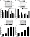
Figure 6. Effects of CGA on Nrf2 nuclear translocation and PI3K/Akt phosphorylation in MC3T3-E1 cells
(A) and (C) Cells were treated with CGA (100 μM) for indicated time points (0, 1, 3, 6 and 12 h). (B) and (D) Cells were treated with indicated concentrations (0, 25, 50 and 100 μM) of CGA for 3 h. (A) and (B) The nuclear and cytosolic levels of Nrf2 were determined by western blot analysis. PCNA was used as nuclear loading control. (C) and (D) Cell lysates were subjected to western blot analysis with anti-Akt or p-Akt antibodies. β-actin was used as loading control. *p < 0.01 indicates the significant difference compared with control group.
Figure 7
Figure 7. Role of the PI3K/Akt pathway in CGA-induced Nrf2 transcription and HO-1 expression
(A), (B) and (C) Cells were pretreated with 20 μM LY294002 for 1 h, and then treated with 100 μM CGA for 3 h. Nrf2, p-Akt and HO-1 levels were determined by western blot analysis. *p < 0.01 indicates the significant difference compared with control group; ##p < 0.01 indicates the significant difference compared with H2O2-treated group.
Figure 8
Figure 8. Effects of CGA and selective inhibitor LY294002 on H2O2-induced cytotoxicity and apoptosis in MC3T3-E1 cells
Cells were pretreated with 20 μM LY294002 for 1 h, and then incubated with or without CGA (100 μM) for 3 h, followed by exposure with H2O2. (A) Cell viability was measured using the MTT assay. (B) and (C) Cell apoptosis was determined by the Annexin V-EGFP/PI staining assay. *p < 0.01 indicates the significant difference compared with control group; ##p < 0.01 indicates the significant difference compared with H2O2-treated group; &p < 0.05 and &&p < 0.01 indicate the significant difference compared with CGA-treated group.
Figure 9
Figure 9. A proposed signaling pathway involved in CGA against H2O2-induced oxidative damage
Schematic diagram shows that CGA induces Nrf2 –mediated cytoprotective protein via activation of PI3K/Akt signaling, which protects against oxidative stress of osteoblast cells. H2O2 induced ROS generation resulting in cell apoptosis. Meanwhile, CGA activates Nrf2/HO-1 through the PI3K/Akt pathway. Activation of Nrf2/HO-1 protects against H2O2 –induced osteoblast apoptosis. Green arrow indicates stimulation and red bar indicates inhibition.

References

    1. Baek KH, Oh KW, Lee WY, Lee SS, Kim MK, Kwon HS, Rhee EJ, Han JH, Song KH, Cha BY, Lee KW, Kang MI. Association of oxidative stress with postmenopausal osteoporosis and the effects of hydrogen peroxide on osteoclast formation in human bone marrow cell cultures. Calcif Tissue Int. 2010;87:226–235. - PubMed
    1. Cervellati C, Bergamini CM. Oxidative damage and the pathogenesis of menopause related disturbances and diseases. Clin Chem Lab Med. 2015;54:739–753. - PubMed
    1. Liu YJ, Zhou CY, Qiu CH, Lu XM, Wang YT. Chlorogenic acid induced apoptosis and inhibition of proliferation in human acute promyelocytic leukemia HL60 cells. Mol Med Rep. 2013;8:1106–1110. - PubMed
    1. Arai M, Shibata Y, Pugdee K, Abiko Y, Ogata K. Effects of reactive oxygen species (ROS) on antioxidant system and osteoblastic differentiation in MC3T3-E1 cells. IUBMB Life. 2007;59:27–33. - PubMed
    1. Pisoschi AM, Pop A. The role of antioxidants in the chemistry of oxidative stress: A review. Eur J Med Chem. 2015;97:55–74. - PubMed

MeSH terms