Review
. 2017 Jan;10(1):1-17.
doi: 10.14802/jmd.16062. Epub 2017 Jan 18.

Functional Neuroanatomy for Posture and Gait Control

Affiliations
Review

Functional Neuroanatomy for Posture and Gait Control

Kaoru Takakusaki. J Mov Disord. 2017 Jan.

Abstract

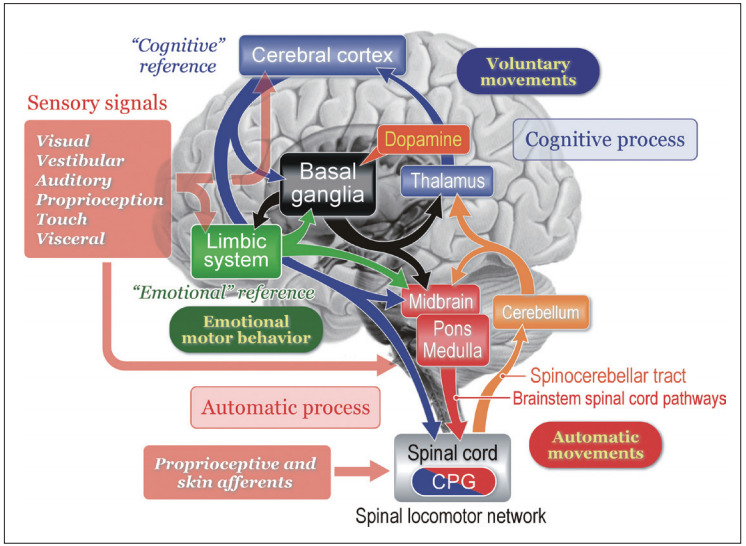
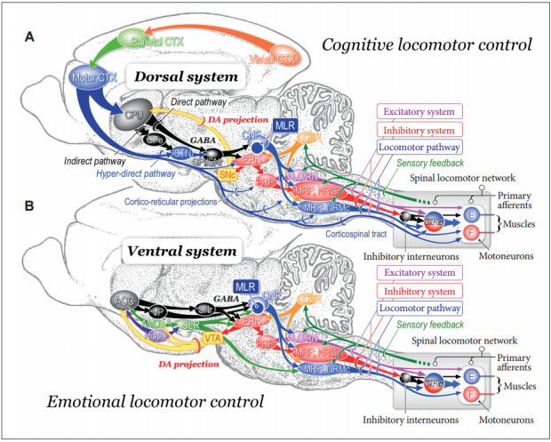
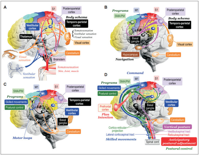
Here we argue functional neuroanatomy for posture-gait control. Multi-sensory information such as somatosensory, visual and vestibular sensation act on various areas of the brain so that adaptable posture-gait control can be achieved. Automatic process of gait, which is steady-state stepping movements associating with postural reflexes including headeye coordination accompanied by appropriate alignment of body segments and optimal level of postural muscle tone, is mediated by the descending pathways from the brainstem to the spinal cord. Particularly, reticulospinal pathways arising from the lateral part of the mesopontine tegmentum and spinal locomotor network contribute to this process. On the other hand, walking in unfamiliar circumstance requires cognitive process of postural control, which depends on knowledges of self-body, such as body schema and body motion in space. The cognitive information is produced at the temporoparietal association cortex, and is fundamental to sustention of vertical posture and construction of motor programs. The programs in the motor cortical areas run to execute anticipatory postural adjustment that is optimal for achievement of goal-directed movements. The basal ganglia and cerebellum may affect both the automatic and cognitive processes of posturegait control through reciprocal connections with the brainstem and cerebral cortex, respectively. Consequently, impairments in cognitive function by damages in the cerebral cortex, basal ganglia and cerebellum may disturb posture-gait control, resulting in falling.

Keywords: Multisensory information; Parkinson’s disease.; body schema; midbrain locomotor region; motor programs; reticulospinal system.


Conflict of interest statement

The author has no financial conflicts of interest.

Figures

Figure 1.
Figure 1.
Basic signal flow involved in postural control. Multisensory signals from the visual, vestibular, auditory, somatosensory (proprioceptive), and visceral receptors act on various sites in the central nervous system. These signals may provide cognitive and emotional references to the cerebral cortex and limbic system, respectively, so that the subject may elicit either voluntary movements or emotional motor behavior depending on the context. In each case, automatic process of postural control, such regulation of postural muscle tone and basic postural reflexes, by the brainstem and spinal cord is required. On the other hand, cognitive postural control is particularly important when the subject learns motor skills and behaves in unfamiliar circumstance. See text for detail explanation. Modified from Takakusaki. Mov Disord 2013;28:1483-1491, with permission of Wiley [6].
Figure 2.
Figure 2.
Effects of midbrain stimulation on posture and locomotion in decerebrate cat preparation. A: Stimulation sites in the right mesopontine tegmentum. Stimulation consists of 30 μA in intensity and 50 Hz in frequency with a duration of 10 seconds. B: Effects of stimulation applied to each site in (A) on left and right soleus muscle electromyograms. Stimulation of the dorsal part of the CNF induced muscle tone augmentation. While stimulation of the ventral CNF and the dorsal PPN induced locomotor rhythm, the latter was accompanied by a decrease in muscle tone. Stimulation of the PPN and PRF corresponding to the nucleus reticularis pontis oralis (NRPo) immediately suppressed soleus muscle activities. C: Topography of stimulus effects in the mesopontine tegmentum. Locomotion was evoked by stimulating the CNF (blue). Stimulation of the locus coeruleus (LC) and dorsolateral CNF induced hypertonia (violet; muscle tone augmentation). Ventrolateral part of the PPN and NRPo, induced muscular atonia (red) and hypotonia (orange). Stimuli applied to the locomotion-evoking sites and atonia-evoking sites elicited a mixture of rhythmic limb movements and muscle tone suppression (green). Modified from Takakusaki et al. J Neural Transm (Vienna) 2016;123:695-729, with permission of Springer [15]. CNF: cuneiform nucleus, PPN: pedunculopontine tegmental nucleus, IC: inferior colliculus, SCP: superior cerebellar peduncle.
Figure 3.
Figure 3.
Functional organization of medullary reticulospinal systems in decerebrate cats. A: Locations of the medullary reticulospinal neurons relating to muscle tone suppression (a), muscle tone augmentation (hypertonus) (b), and locomotion (c). During reflex standing of the decerebrate cats, reticulospinal neurons with a firing frequency more than 10 Hz during reflex standing of decerebrate cats are judged as hypertonus-related reticulospinal neurons (b; n = 76). When carbachol (long-acting cholinomimetic agents) was injected into the pontine reticular formation muscle tone of decerebrate cats was abolished. Reticulospinal neurons of which firing frequency was increased to more than 10 Hz during carbachol-induced atonia are judged as atonia-related reticulospinal neurons (a; n = 75). During reflex standing (decerebrate rigidity) these cells usually had no spontaneous firing. Locomotion-related neurons (n = 59) were judged as those displaying rhythmic firing relating to step cycles of locomotion. Recording was made in both high decerebrated cats which displayed spontaneous locomotion and normal decerebrated cats with stimulation of the MLR. B: Results obtained from five animals are superimposed on representative coronal planes of the caudal pons and medulla. Sites from which either suppression (red), augmentation (blue), or tegmental reflexes (green) was elicited in more than three out of five animals are marked. Sites from which the stimulation induced postural changes in more than four animals are indicated by darker colored squares; conversely, light colored squares indicate that the postural changes were induced in three animals. Modified from Takakusaki et al. J Neural Transm (Vienna) 2016;123:695-729, with permission of Springer [15]. P: pyramidal tract, MLF: medial longitudinal fasciculus, 5ST: spinal trigeminal tract, NRPc: nucleus reticularis pontis caudalis, TB: trapezoid body, RM: nucleus raphe magnus, SO: superior olive, NRGc: nucleus reticularis gigantocellularis, NRMc: nucleus reticularis magnocellularis, RPa: nucleus raphe pallidus, NRPv: nucleus reticulars parvocellularis.
Figure 4.
Figure 4.
Neuronal mechanisms of cognitive (A) and emotional (B) control of locomotion in the cat. A: Dorsal system for cognitive locomotor control. A visuo-motor pathway from the visual cortex to motor cortex via the parietal cortex contributes to this control. Corticofugal projections act on to the basal ganglia nuclei, brainstem and spinal cord. Dopaminergic projection from the substantia nigra pars compacta (SNc) to the caudate-putamen (CPu) may be involved in learning the locomotor behaviors. GABAergic output from the basal ganglia nuclei (internal segment of the globus pallidus and substantia nigra pars reticulata; GPi/SNr) acts on MLR/PPN area may control locomotion and posture. Efferents from the midbrain locomotor region (MLR) recruit excitatory system, inhibitory system and locomotor pathway. The excitatory system arises from the LC and the raphe nuclei. The inhibitory system which arises from cholinergic neurons in the PPN sequentially activates PRF neurons, medullary reticulospinal neurons in the nucleus reticularis gigantocellularis (NRGc) and spinal inhibitory interneurons. The inhibitory interneurons may inhibit both motoneurons and interneurons. The locomotor pathway consists of reticulospinal neurons arising from the ventromedial medulla corresponding to the nucleus reticularis magnocellularis (NRMc). Cholinergic and glutamatergic projections from the PPN excite SNc-DA neurons. These descending tracts act on CPGs in spinal cord so that muscle tone and locomotion are regulated. Efferents from the (CLR may excite locomotor pathway. B: Ventral system for emotional locomotor control. Efferents from the amygdala (AMD) and hippocampus (Hipp) project to the nucleus accumbens (NAc). GABAergic NAc neurons project to ventral pallidum (VP) and the SNr, which control activity of the MLR/PPN neurons. Efferents from the AMD and the Hipp also act on lateral hypothalamic area, which corresponds to the SLR. DA projections from the ventral tegmental area (VTA) may contribute to the rewardoriented locomotor behaviors. Modified from Snijders et al. Ann Neurol 2016;80:644-659, with permission of Wiley [164]. E: extensor motoneurons, F: flexor motoneurons, PRF: pontine reticular formation, PPN: pedunculopontine tegmental nucleus, LC: locus coeruleus, RN: raphe nuclei, DA: dopamine, CLR: cerebellar locomotor region, SLR: subthalamic locomotor region, CNF: cuneiform nucleus, CTX: cortex, GPe: external segment of the globus pallidus.
Figure 5.
Figure 5.
Hypotheses of cognitive process of posture-gait control. A: Cognition of bodily information. Sensory signals flowing into the central nervous system converge to the brainstem, cerebellum, thalamus, and cerebral cortex. At the level of cerebral cortex, signals from the visual cortex, vestibular cortex and primary sensory cortex (S1) is integrated and internal model of self-body, such as body schema and verticality can be constructed at the temporoparietal cortex including the vestibular cortex and posteroparietal cortex. Reciprocal connection between the temporoparietal cortex and cerebellum may contribute to this process. B: Transmission of the bodily information. The bodil y information is then transmitted to the supplementary motor area (SMA) and premotor area (PM) where the information can be utilized as materials to produce motor programs. Similarly, the information is transferred to hippocampus and is used to navigate further behaviors. C: Motor programming. The motor cortical areas closely cooperate with the basal ganglia and cerebellum so that appropriate motor programs are constructed. D: Postural control by corticofugal projections to the brainstem and spinal cord. The bodily information generated at the vestibular cortex may be utilized for sustention of vertical posture via cortico-vestibular and vestibulospinal tract. Signals from the prefrontal cortex including plans and intentions may trigger to run motor programs in the SMA/PM, which may include those for purposeful movements and associating postural control. The postural control program may be utilized to generate anticipatory postural adjustment via cortico-reticular and reticulospinal tract. Then motor programs are sent to the M1 so that goal-directed purposeful skilled movements can be achieved. Modified from http://dx.doi.org/10.1080/01691864.2016.1252690, with permisson of Taylor & Francis [165].

References

    1. Grillner S. In: Handbook of physiology. The nervous system II. Brookhart JM, Mountcastle VB, editors. Bethesda, MD: American Physiological Society; 1981. Control of locomotion in bipeds, tetrapods, and fish; pp. 1179–1236.
    1. Takakusaki K. Forebrain control of locomotor behaviors. Brain Res Rev. 2008;57:192–198. - PubMed
    1. Sinnamon HM. Preoptic and hypothalamic neurons and the initiation of locomotion in the anesthetized rat. Prog Neurobiol. 1993;41:323–344. - PubMed
    1. Takakusaki K, Takahashi K, Saitoh K, Harada H, Okumura T, Kayama Y, et al. Orexinergic projections to the cat midbrain mediate alternation of emotional behavioural states from locomotion to cataplexy. J Physiol. 2005;568(Pt 3):1003–1020. - PMC - PubMed
    1. Takakusaki K, Tomita N, Yano M. Substrates for normal gait and pathophysiology of gait disturbances with respect to the basal ganglia dysfunction. J Neurol. 2008;255 Suppl 4:19–29. - PubMed

LinkOut - more resources