. 2017 Jan 25;18(1):39.
doi: 10.1186/s12891-017-1410-y.

Pigment Epithelium-Derived Factor (PEDF) mediates cartilage matrix loss in an age-dependent manner under inflammatory conditions

Affiliations

Pigment Epithelium-Derived Factor (PEDF) mediates cartilage matrix loss in an age-dependent manner under inflammatory conditions

Daisy S Nakamura et al. BMC Musculoskelet Disord. .

Abstract

Background: Inflammation is a major cause of cartilage destruction and leads to the imbalance of metabolic activities in the arthritic joint. Pigment epithelium-derived factor (PEDF) has been reported to have both pro- and anti-inflammatory activities in various cell types and to be upregulated in the arthritic joint, but its role in joint destruction is unclear. Our aim was to investigate the role of PEDF in cartilage degeneration under inflammatory conditions.

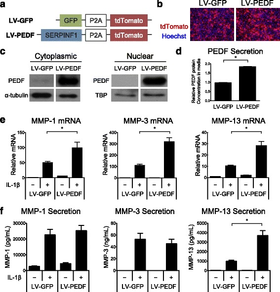
Methods: PEDF was ectopically expressed in primary human articular chondrocytes, and catabolic gene expression and protein secretion in response to the pro-inflammatory cytokine interleukin 1 beta (IL-1β) were evaluated. Metatarsal bones from PEDF-deficient and wild type mice were cultured in the presence or absence of IL-1β. Cartilage matrix integrity and matrix metalloproteinases MMP-1, MMP-3, and MMP-13 were evaluated. PEDF-deficient and wild type mice were evaluated in the monosodium iodoacetate (MIA) inflammatory joint destruction animal model to determine the role of PEDF in inflammatory arthritis in vivo. Student's t-tests and Mann-Whitney tests were employed where appropriate, for parametric and non-parametric data, respectively.

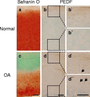
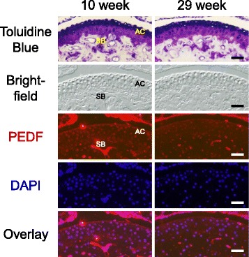
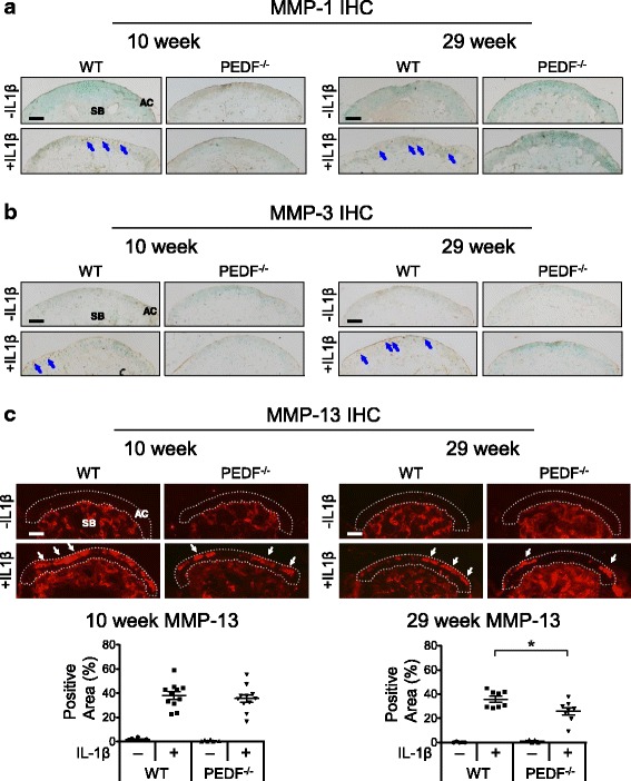
Results: We showed that PEDF protein levels were higher in human osteoarthritis samples compared to normal samples. We demonstrated that ectopic PEDF expression in primary human articular chondrocytes exacerbated catabolic gene expression in the presence of IL-1β. In whole bone organ cultures, IL-1β induced MMP-1, MMP-3 and MMP-13 protein production, and caused significant cartilage matrix loss. Interestingly, Toluidine Blue staining showed that PEDF-deficient bones from 29 week old animals, but not 10 week old animals, had reduced matrix loss in response to IL-1β compared to their wild type counterparts. In addition, PEDF-deficiency in 29 week old animals preserved matrix integrity and protected against cell loss in the MIA joint destruction model in vivo.

Conclusion: We conclude that PEDF exacerbates cartilage degeneration in an age-dependent manner under an inflammatory setting. This is the first study identifying a specific role for PEDF in joint inflammation and highlights the multi-faceted activities of PEDF.

Keywords: Age-related; Arthritis; IL-1β; Inflammation; MMPs; Matrix, cartilage; Pigment epithelium-derived factor; SERPINF1.


Figures

Fig. 1
Fig. 1
PEDF expression is elevated in human osteoarthritis (OA) cartilage specimens. Normal samples were obtained from the National Disease Research Interchange and OA samples were obtained from patients undergoing total knee replacement surgery. a Normal sample stained with Safranin O and counterstained with Hematoxylin and Fast Green. Mankin score = 1. b Immunohistochemistry (IHC) analysis on a normal cartilage sample using a mouse anti-PEDF antibody and counterstained with Methyl Green. Magnified superficial (b') and deeper (b'') zones are shown from insets. c OA sample stained with Safranin O and counterstained with Hematoxylin and Fast Green. Mankin score = 5. d IHC analysis on an OA sample using a mouse anti-PEDF antibody and counterstained with Methyl Green. Magnified superficial (d') and deeper (d'') zones are shown from insets. Arrows indicate positive PEDF staining. A negative control using a biotinylated horse anti-mouse IgG secondary antibody alone is shown in the Additional file 2: Figure S1. Scale bar = 200 μm
Fig. 2
Fig. 2
PEDF ectopic expression upregulates catabolic gene expression in normal human articular chondrocytes (NHACs). a Lentiviral constructs were generated that contained the human SERPINF1 gene, which encodes for the PEDF protein, and a tdTomato moiety that was separated from the SERPINF1 gene by a P2A self-cleaving peptide (LV-PEDF). A lentiviral construct encoding the gene for green fluorescent protein (GFP) and a tdTomato moiety (LV-GFP) was also constructed. b tdTomato expression is comparable between the two expression vectors. c Western Blot analysis confirmed elevated PEDF protein production in the cytoplasm and nucleus of NHACs transduced with LV-PEDF. α-tubulin was used as a loading control for cytoplasmic proteins. TATA binding protein (TBP) was used as a loading control for nuclear proteins. LV-GFP: GFP-encoding lentivirus; LV-PEDF: PEDF-encoding lentivirus. d ELISA analysis of PEDF protein secretion into the media of NHACs transduced with LV-GFP or LV-PEDF following 4 days in culture. Data are represented as fold change relative to LV-GFP samples. e Quantitative RT-PCR analysis of MMP-1, MMP-3 and MMP-13 on NHACs transduced with LV-GFP or LV-PEDF and cultured in the presence or absence of 1 ng/mL IL-1β for 4 days. Data are represented as fold change relative to LV-GFP-transduced cells in the absence of IL-1β within each gene. Gene expression was normalized to TATA-binding protein mRNA expression levels. f ELISA analysis of MMP-1, MMP-3 and MMP-13 protein secreted into the media of NHACs transduced with LV-GFP or LV-PEDF and cultured in the presence or absence of 1 ng/mL IL-1β for 4 days. Data are presented as fold change relative to that found in the media of LV-GFP-transduced cells cultured in the absence of IL-1β. All data are plotted as mean ± SEM. * p < 0.05 (Mann–Whitney test)
Fig. 3
Fig. 3
Detection of PEDF in the articular surface of adult mouse metatarsal bones. Metatarsal bones from 10 and 29 week old mice were subjected to histological analysis by Toluidine Blue staining. AC: articular cartilage. SB: subchondral bone. Immunohistochemistry (IHC) analysis of PEDF protein was performed on sections adjacent to those used for Toluidine Blue staining. Nuclei were stained with DAPI. Bright field, PEDF staining (red), DAPI and PEDF/DAPI overlay images of the same section are shown. A negative control using goat anti-mouse IgG secondary antibody alone is shown in the Additional file 2: Figure S1. Scale bar = 200 μm
Fig. 4
Fig. 4
PEDF-deficiency protects against IL-1β-induced matrix loss in organ cultures of metatarsal bones. a Metatarsal bones from 10 week old wild type or PEDF-deficient mice were harvested and cultured in the presence or absence of 10 ng/mL IL-1β for 7 days. Samples were cryosectioned and stained with Toluidine Blue. Scale bar = 100 μm. b Percent loss of Toluidine Blue staining along the articular surface in 10 week old samples with respect to the total surface area was calculated. c Metatarsal bones from 29 week old wild type or PEDF-deficient mice were harvested and cultured in the presence or absence of 10 ng/mL IL-1β for 7 days. Samples were cryosectioned and stained with Toluidine Blue. Scale bar = 100 μm. d Percent loss of Toluidine Blue staining along the articular surface in 29 week old samples with respect to the total surface area was calculated. For experiments involving either 10 or 29 week old mice, the middle three metatarsal bones isolated from three animals were used. Each data point in the graphs represents an individual metatarsal bone. Each treatment was repeated 5–11 times. Data are plotted as mean ± SEM. ** p = 0.0021 (Mann–Whitney test)
Fig. 5
Fig. 5
PEDF-deficiency protects against IL-1β-induced MMP-1, MMP-3 and MMP-13 protein production in metatarsal bone cultures. Metatarsal bones from 10 and 29 week old wild type or PEDF-deficient mice were harvested and cultured in the presence or absence of 10 ng/mL IL-1β for 7 days. Cryosectioned samples were analyzed for MMP protein levels by immunohistochemistry (IHC). AC: articular cartilage. SB: subchondral bone. Scale bar = 100 μm. a Samples were stained with rabbit anti-MMP-1 and counterstained with 0.5% Methyl Green in 0.1 M Sodium Acetate. Arrows indicate positive MMP-1 staining. b Samples were stained with rabbit anti-MMP-3 and counterstained with 0.5% Methyl Green in 0.1 M Sodium Acetate. Arrows indicate positive MMP-3 staining. c Samples were stained with mouse anti-MMP-13 and nuclei were stained with DAPI. Arrows indicate positive MMP-13 staining. The percent area staining positive for MMP-13 in the 10 and 29 week old samples was calculated as the area with MMP-13 staining along the articular cartilage area against the total articular cartilage area. For experiments involving either 10 or 29 week old mice, the middle three metatarsal bones isolated from three animals were used. Each data point in the graphs represents an individual metatarsal bone. Each treatment was repeated 5–11 times. Negative controls using goat anti-rabbit IgG or biotinylated horse anti-mouse IgG followed by streptavidin DyLight 594 alone are shown in the Additional file 2 Figure S1. Data are plotted as mean ± SEM. * p = 0.0281 (Mann–Whitney test)
Fig. 6
Fig. 6
PEDF-deficiency protects against monosodium iodoacetate (MIA)-induced joint cartilage damage. a Immunofluorescence staining was used to determine PEDF protein levels in knee joints from 29 week old wild type animals using a mouse anti-PEDF antibody. Nuclei were stained with DAPI. A negative control using goat anti-mouse IgG secondary antibody alone is shown in the Additional file 2: Figure S1. M: meniscus; F: femur; T: tibia. Scale bar = 200 μm. b MIA or PBS was intraarticularly injected into 29 week old wild type or PEDF-deficient knees, which were harvested after 10 days. Samples were stained with Safranin O and counterstained with Hematoxylin and Fast Green. Arrows indicate areas exhibiting a loss of Safranin O staining. Scale bar = 100 μm. c The percent area loss of Safranin O staining along the articular surface was calculated for the femur and tibia independently, as well as for the whole joint (femur + tibia). Each point represents the averaged score of an individual animal. Data are plotted as mean ± SEM. d OARSI scores were calculated from Safranin O stained joints. Each point represents an individual animal. The bar represents the median. e Quantification of percent cell loss based on DAPI cell counts of the articular surface and empty lacunae. ** p < 0.01 (Mann–Whitney test) * p < 0.05 (Student’s t-test)

References

    1. Hootman JM, et al. Updated projected prevalence of self-reported doctor-diagnosed arthritis and arthritis-attributable activity limitation among US adults, 2015–2040. Arthritis Rheumatol. 2016;68(7):1582–7. doi: 10.1002/art.39692. - DOI - PMC - PubMed
    1. Wong R, et al. Prevalence of Arthritis and Rheumatic Diseases Around the World: A Growing Burden and Implications for Health Care Needs. Models of Care in Arthritis (MOCA). 2010. Working Paper 10-02. http://modelsofcare.ca/resources.html#workingpaper.
    1. Joosten LA, et al. IL-1 alpha beta blockade prevents cartilage and bone destruction in murine type II collagen-induced arthritis, whereas TNF-alpha blockade only ameliorates joint inflammation. J Immunol. 1999;163(9):5049–55. - PubMed
    1. Joosten LA, et al. Anticytokine treatment of established type II collagen-induced arthritis in DBA/1 mice. A comparative study using anti-TNF alpha, anti-IL-1 alpha/beta, and IL-1Ra. Arthritis Rheum. 1996;39(5):797–809. doi: 10.1002/art.1780390513. - DOI - PubMed
    1. Palmer G, et al. Mice transgenic for intracellular interleukin-1 receptor antagonist type 1 are protected from collagen-induced arthritis. Eur J Immunol. 2003;33(2):434–40. doi: 10.1002/immu.200310018. - DOI - PubMed

Publication types