Review
. 2017 Apr 1;312(4):F732-F743.
doi: 10.1152/ajprenal.00337.2016. Epub 2017 Jan 25.

Sleeping beauty: awakening urothelium from its slumber

Affiliations
Review

Sleeping beauty: awakening urothelium from its slumber

Zarine R Balsara et al. Am J Physiol Renal Physiol. .

Abstract

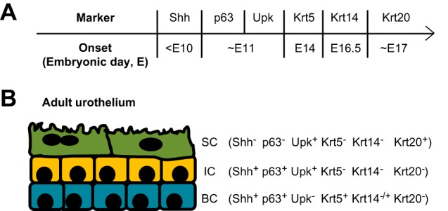
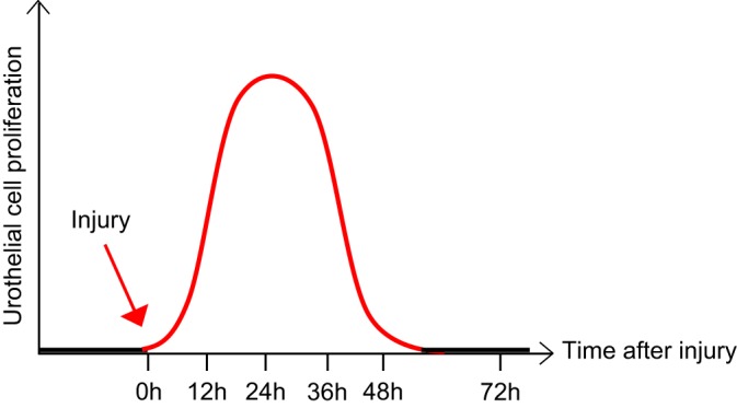
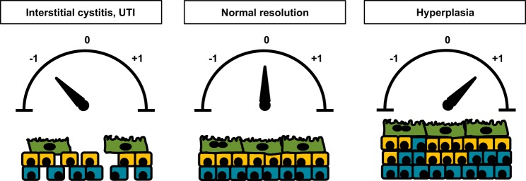
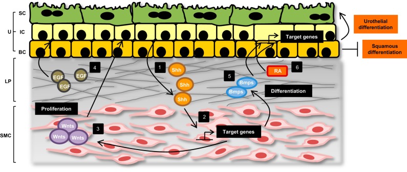
The bladder urothelium is essentially quiescent but regenerates readily upon injury. The process of urothelial regeneration harkens back to the process of urothelial development whereby urothelial stem/progenitor cells must proliferate and terminally differentiate to establish all three urothelial layers. How the urothelium regulates the level of proliferation and the timing of differentiation to ensure the precise degree of regeneration is of significant interest in the field. Without a carefully-orchestrated process, urothelial regeneration may be inadequate, thereby exposing the host to toxins or pathogens. Alternatively, regeneration may be excessive, thereby setting the stage for tumor development. This review describes our current understanding of urothelial regeneration. The current controversies surrounding the identity and location of urothelial progenitor cells that mediate urothelial regeneration are discussed and evidence for each model is provided. We emphasize the factors that have been shown to be crucial for urothelial regeneration, including local growth factors that stimulate repair, and epithelial-mesenchymal cross talk, which ensures feedback regulation. Also highlighted is the emerging concept of epigenetic regulation of urothelial regeneration, which additionally fine tunes the process through transcriptional regulation of cell cycle genes and growth and differentiation factors. Finally, we emphasize how several of these pathways and/or programs are often dysregulated during malignant transformation, further corroborating their importance in directing normal urothelial regeneration. Together, evidence in the field suggests that any attempt to exploit regenerative programs for the purposes of enhanced urothelial repair or replacement must take into account this delicate balance.

Keywords: epigenetics; epithelial-mesenchymal cross talk; label retention; lineage tracing; progenitor cells; regeneration; superficial cells; urothelium.


Figures

Fig. 1.
Fig. 1.
Urothelium is stratified into three major cell types. A: temporal expression of several key urothelial cell markers is depicted. Shh is the earliest of these markers to be expressed in the urothelium, while the cytokeratins are expressed much later in embryogenesis. B: adult urothelium comprises basal cells (BC), intermediate cells (IC), and superficial cells (SC), each of which have discrete patterns of cell marker expression.
Fig. 2.
Fig. 2.
Adult urothelium is normally quiescent but rapidly responds and proliferates upon urothelial injury. At baseline, mature urothelium remains in a quiescent state, with extremely slow turnover. However, in response to injury, the urothelium rapidly awakens and undergoes proliferation and differentiation to restore the damaged epithelium. Maximal proliferation occurs within 12–36 h, depending on the stimulus, followed by differentiation and a return to the dormant state.
Fig. 3.
Fig. 3.
A fine balance is necessary to ensure normal urothelial regeneration after injury. Following injury, several outcomes are possible. Most commonly, regeneration results in restoration of the urothelium to its original state (designated as “0”). However, failure to fully regenerate the urothelium (designated as “−1”) results in potential breaches in barrier function that may increase susceptibility to infection or increase sensory fiber stimulation and set the stage for interstitial cystitis. Alternatively, unrestrained regeneration (designated as “+1”) can lead to urothelial hyperplasia that may ultimately lead to bladder tumor formation.
Fig. 4.
Fig. 4.
Proposed models of urothelial formation and regeneration. A: the linear model of urothelial formation and regeneration suggests that the urothelial stem/progenitor cells resides within the basal cell population, likely the Krt5+/Krt14+ basal cells. These cells are capable of self renewal but also give rise to intermediate cells, which subsequently give rise to superficial cells. B: in the nonlinear model, the transient population of P cells, distinguished by Foxa2 expression, gives rise to intermediate and superficial cells during embryogenesis. In contrast to the linear model, the nonlinear model argues that a separate progenitor cell (unidentified at present) initially gives rise to basal cells during development. In adults, stem cells are thought to reside within both the intermediate and the basal cell populations and are capable of self-renewal and/or differentiation into superficial cells. It is unclear, however, whether stem cells in the basal layer need to go through an intermediate step, such as transient amplifying cells, before they differentiate into superficial cells.
Fig. 5.
Fig. 5.
Urothelial regeneration is achieved through multiple layers of regulation. The urothelium comprises three main cell types: superficial cells (SC), intermediate cells (IC), and basal cells (BC). Below the urothelium lies the lamina propria (LP) and smooth muscle cell (SMC) layers. Significant epithelial-mesenchymal cross talk facilitates urothelial regeneration after injury (1). In response to damage, urothelial cells upregulate and secrete the soluble molecule Shh. Shh acts on the underlying mesenchymal cells and promotes SMC proliferation and differentiation (2). Shh signaling also leads to activation of target genes, including the Gli family of transcription factors, and (3) increased stromal expression of Wnt molecules. Wnt signals subsequently promote urothelial proliferation and differentiation (4). Injured urothelium also secretes local growth factors like EGF, which function in an autocrine manner to stimulate urothelial cell proliferation (5). Likewise, BMP signaling through BMP receptors on the urothelium is important in ensuring terminal differentiation of regenerating superficial cells (6). Finally, RA produced by stromal cells acts on RA-responsive urothelial cells to promote superficial cell regeneration and inhibit squamous differentiation, in part through activation of transcription factors like FOXA1.

References

    1. Acharya P, Beckel J, Ruiz WG, Wang E, Rojas R, Birder L, Apodaca G. Distribution of the tight junction proteins ZO-1, occludin, and claudin-4, -8, and -12 in bladder epithelium. Am J Physiol Renal Physiol 287: F305–F318, 2004. doi:10.1152/ajprenal.00341.2003. - DOI - PubMed
    1. Adam RM, Danciu T, McLellan DL, Borer JG, Lin J, Zurakowski D, Weinstein MH, Rajjayabun PH, Mellon JK, Freeman MR. A nuclear form of the heparin-binding epidermal growth factor-like growth factor precursor is a feature of aggressive transitional cell carcinoma. Cancer Res 63: 484–490, 2003. - PubMed
    1. Baskin LS, Sutherland RS, Thomson AA, Nguyen HT, Morgan DM, Hayward SW, Hom YK, DiSandro M, Cunha GR. Growth factors in bladder wound healing. J Urol 157: 2388–2395, 1997. doi:10.1016/S0022-5347(01)64786-4. - DOI - PubMed
    1. Bellmunt J, Hussain M, Dinney CP. Novel approaches with targeted therapies in bladder cancer. Crit Rev Oncol Hematol 46, Suppl: 85–104, 2003. doi:10.1016/S1040-8428(03)00067-2. - DOI - PubMed
    1. Berger MS, Greenfield C, Gullick WJ, Haley J, Downward J, Neal DE, Harris AL, Waterfield MD. Evaluation of epidermal growth factor receptors in bladder tumours. Br J Cancer 56: 533–537, 1987. doi:10.1038/bjc.1987.238. - DOI - PMC - PubMed

Publication types

MeSH terms

Substances

LinkOut - more resources