Controlled Clinical Trial
. 2017 Apr 1;312(4):R529-R538.
doi: 10.1152/ajpregu.00348.2016. Epub 2017 Jan 25.

Acute high-intensity interval exercise reduces human monocyte Toll-like receptor 2 expression in type 2 diabetes

Affiliations
Controlled Clinical Trial

Acute high-intensity interval exercise reduces human monocyte Toll-like receptor 2 expression in type 2 diabetes

Cody Durrer et al. Am J Physiol Regul Integr Comp Physiol. .

Abstract

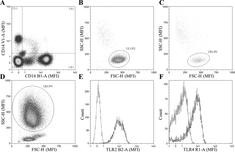
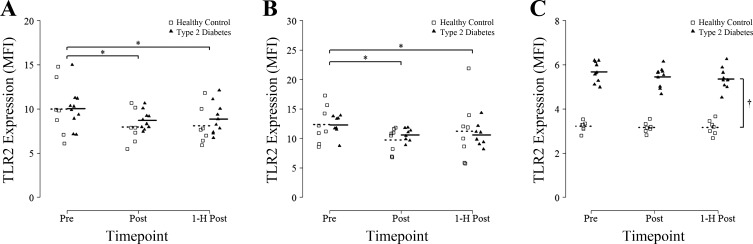
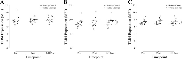
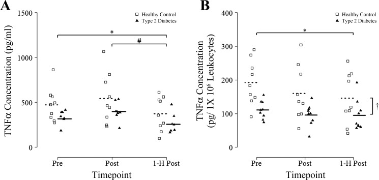
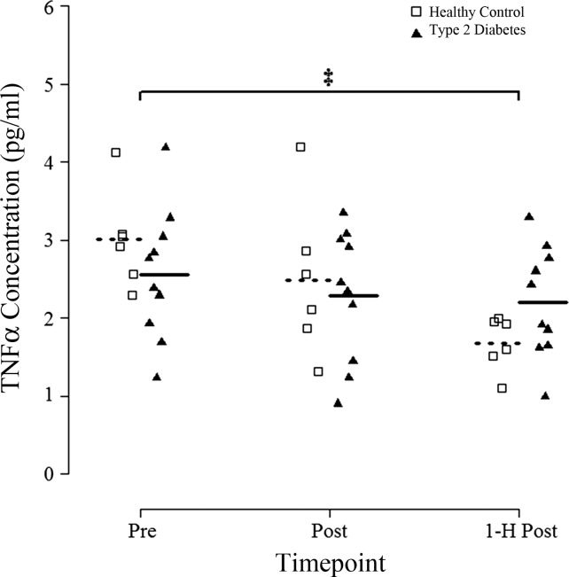
Type 2 diabetes (T2D) is characterized by chronic low-grade inflammation that contributes to disease pathophysiology. Exercise has anti-inflammatory effects, but the impact of high-intensity interval training (HIIT) is not known. The purpose of this study was to determine the impact of a single session of HIIT on cellular, molecular, and circulating markers of inflammation in individuals with T2D. Participants with T2D (n = 10) and healthy age-matched controls (HC; n = 9) completed an acute bout of HIIT (7 × 1 min at ~85% maximal aerobic power output, separated by 1 min of recovery) on a cycle ergometer with blood samples obtained before (Pre), immediately after (Post), and at 1 h of recovery (1-h Post). Inflammatory markers on leukocytes were measured by flow cytometry, and TNF-α was assessed in both LPS-stimulated whole blood cultures and plasma. A single session of HIIT had an overall anti-inflammatory effect, as evidenced by 1) significantly lower levels of Toll-like receptor (TLR) 2 surface protein expression on both classical and CD16+ monocytes assessed at Post and 1-h Post compared with Pre (P < 0.05 for all); 2) significantly lower LPS-stimulated TNF-α release in whole blood cultures at 1-h Post (P < 0.05 vs. Pre); and 3) significantly lower levels of plasma TNF-α at 1-h Post (P < 0.05 vs. Pre). There were no differences between T2D and HC, except for a larger decrease in plasma TNF-α in HC vs. T2D (group × time interaction, P < 0.05). One session of low-volume HIIT has immunomodulatory effects and provides potential anti-inflammatory benefits to people with, and without, T2D.

Keywords: CD14+ monocyte; inflammation; innate immunity; leukocyte; tumor necrosis factor-α.


Figures

Fig. 1.
Fig. 1.
Gating strategy for analysis of surface Toll-like receptor (TLR) 2 and TLR4 on monocytes and neutrophils. Cells that stained positive for propidium idodide were first excluded from analysis. Cells are first gated on expression of CD14+/CD16− (classical monocytes), CD14−/CD16+ (CD16+ neutrophils), or both CD14+/CD16+ (CD16+ monocytes) (A). Cell type is then confirmed via characteristic forward and side-scatter profile for classical monocytes, CD16+ monocytes, and neutrophils (B, C, and D, respectively). Cell surface TLR2 and TLR4 expression were then measured on each cell type (i.e., classical monocytes, CD16+ monocytes, and CD16+ neutrophils), Fluorescence minus one controls are displayed in gray (E and F). MFI, median fluorescence intensity; SSC-H, side scatter height; FSC-H, forward scatter height.
Fig. 2.
Fig. 2.
Toll-like receptor 2 expression on CD14+/CD16− classic monocytes, CD16+ monocytes, and CD16+ neutrophils in response to an acute bout of high-intensity interval training (HIIT). Blood samples were obtained before (Pre), immediately after (Post), and 1 h after (1-h Post) a single session of HIIT involving 7 × 1-min at 85% peak power output and TLR2 MFI was measured by flow cytometry on CD14+/CD16− monocytes (A), CD16+ monocytes (B), and CD16+ neutrophils (C). Group means are denoted by dotted (Healthy Controls) or solid (Type 2 Diabetes) horizontal lines. Repeated-measures ANOVA revealed a significant main effect of time for classical monocytes and CD16+ monocytes (all P < 0.05). *P < 0.05 vs. Pre [Fisher least significant difference (LSD) post hoc]. †A main effect of group was also detected for CD16+ neutrophils (P < 0.05).
Fig. 3.
Fig. 3.
TLR4 expression on CD14+/CD16− classic monocytes, CD16+ monocytes, and CD16+ neutrophils in response to an acute bout of HIIT. Blood samples were obtained before (Pre), immediately after (Post), and 1 h after (1-H Post) a single session of HIIT involving 7 × 1 min at 85% peak power output and TLR4 MFI was measured by flow cytometry on CD14+/CD16− monocytes (A), CD16+ monocytes (B), and CD16+ neutrophils (C). Group means are denoted by dotted (Healthy Controls) or solid (Type 2 Diabetes) horizontal lines. †Repeated-measures ANOVA revealed a significant main effect of group for CD16+ monocytes and CD16+ neutrophils (both P < 0.05).
Fig. 4.
Fig. 4.
Whole blood culture TNF-α concentration from 4-h supernatants stimulated with 10 ng/ml LPS in response to an acute bout of HIIT. Blood samples were obtained before (Pre), immediately after (Post), and 1 h after (1-H Post) a single session of HIIT involving 7 × 1-min at 85% peak power output and absolute TNF-α (A) and leukocyte concentration corrected TNF-α (B) secretion in whole blood cultured in the presence of 10 ng/ml LPS was measured. Supernatants were collected after 4 h in culture, and TNF-α was measured by Magpix ELISA. Group means are denoted by dotted (Healthy Controls) or solid (Type 2 Diabetes) horizontal lines. Repeated-measures ANOVA revealed a significant main effect of time for absolute TNF-α concentration (P < 0.05) and a significant main effect of group for leukocyte-corrected TNF-α concentration (†P < 0.05). *P < 0.05 vs. Pre (Fisher LSD post hoc). #P < 0.05 vs. Post (Fisher LSD post hoc).
Fig. 5.
Fig. 5.
Circulating plasma TNF-α concentration in response to an acute bout of HIIT. Blood samples were obtained before (Pre), immediately after (Post), and 1 h after (1-H Post) a single session of HIIT involving 7 × 1-min at 85% peak power output and TNF-α in plasma samples was measured by Magpix ELISA. Groups means are denoted by dotted (Healthy Controls) or solid (Type 2 Diabetes) horizontal lines. Repeated-measures ANOVA revealed a significant group × time interaction for TNF-α concentration (P < 0.05). ‡P < 0.05 vs. Pre within each group (Fisher LSD post hoc).

References

    1. Abbasi A, Hauth M, Walter M, Hudemann J, Wank V, Niess AM, Northoff H. Exhaustive exercise modifies different gene expression profiles and pathways in LPS-stimulated and un-stimulated whole blood cultures. Brain Behav Immun 39: 130–141, 2014. doi:10.1016/j.bbi.2013.10.023. - DOI - PubMed
    1. American College of Sports Medicine; American Diabetes Association; Colberg SR, Sigal RJ, Fernhall B, Regensteiner JG, Blissmer BJ, Rubin RR, Chasan-Taber L, Albright AL, Braun B. Exercise and type 2 diabetes: the American College of Sports Medicine and the American Diabetes Association: joint position statement. Diabetes Care 33: e147–e167, 2010. doi:10.2337/dc10-9990. - DOI - PMC - PubMed
    1. Asea A, Rehli M, Kabingu E, Boch JA, Bare O, Auron PE, Stevenson MA, Calderwood SK. Novel signal transduction pathway utilized by extracellular HSP70: role of toll-like receptor (TLR) 2 and TLR4. J Biol Chem 277: 15,028–15,034, 2002. doi:10.1074/jbc.M200497200. - DOI - PubMed
    1. Balducci S, Zanuso S, Nicolucci A, Fernando F, Cavallo S, Cardelli P, Fallucca S, Alessi E, Letizia C, Jimenez A, Fallucca F, Pugliese G. Anti-inflammatory effect of exercise training in subjects with type 2 diabetes and the metabolic syndrome is dependent on exercise modalities and independent of weight loss. Nutr Metab Cardiovasc Dis 20: 608–617, 2010. doi:10.1016/j.numecd.2009.04.015. - DOI - PubMed
    1. Belge KU, Dayyani F, Horelt A, Siedlar M, Frankenberger M, Frankenberger B, Espevik T, Ziegler-Heitbrock L. The proinflammatory CD14+CD16+DR++ monocytes are a major source of TNF. J Immunol 168: 3536–3542, 2002. doi:10.4049/jimmunol.168.7.3536. - DOI - PubMed

Publication types

Grants and funding