. 2017 Apr 1;27(4):318-328.
doi: 10.1093/glycob/cww134.

Intestinal Muc2 mucin O-glycosylation is affected by microbiota and regulated by differential expression of glycosyltranferases

Affiliations

Intestinal Muc2 mucin O-glycosylation is affected by microbiota and regulated by differential expression of glycosyltranferases

Liisa Arike et al. Glycobiology. .

Abstract

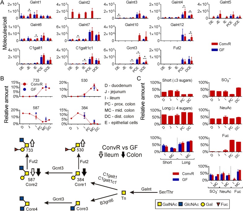
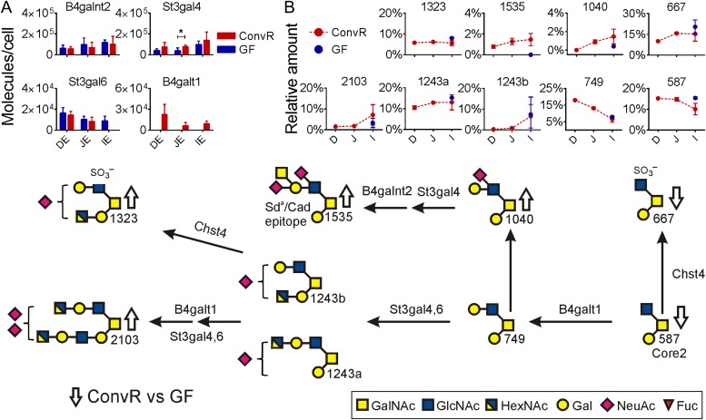
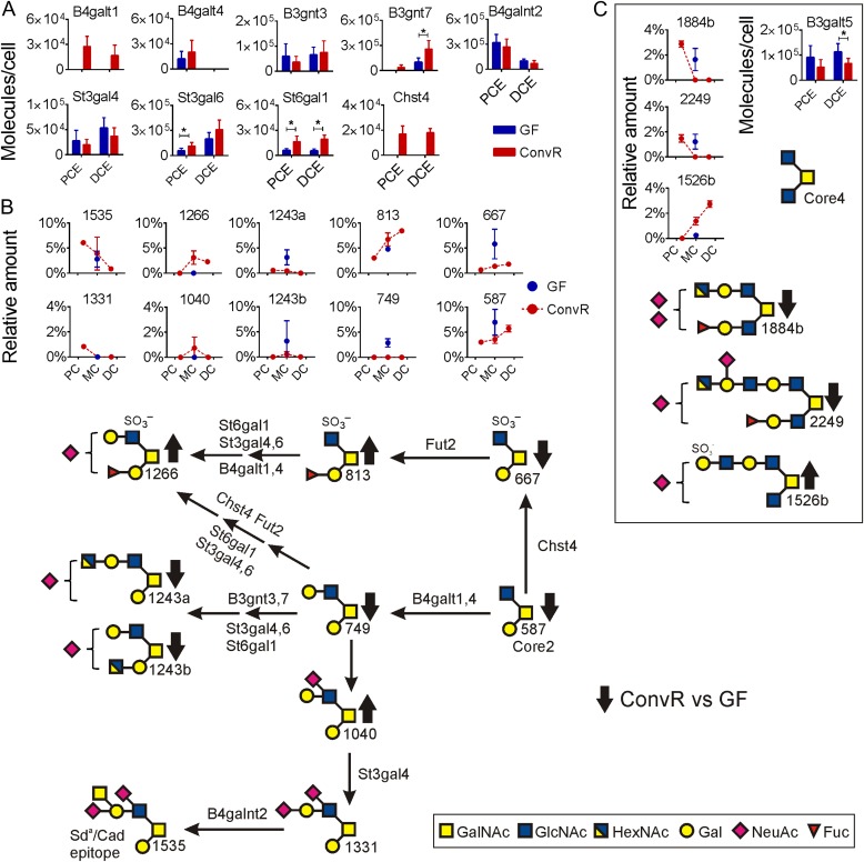
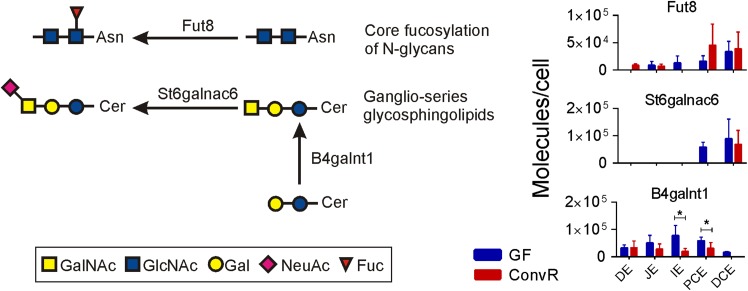
Intestinal cells are covered by mucus. In the small intestine, a single unattached mucus is present whereas the colon has both an inner attached mucus layer and an outer loose mucus. The attached mucus of the colon is impenetrable to bacteria while the loose mucus acts as a habitat for commensal bacteria. In germ-free (GF) mice, small intestinal mucus is attached to the epithelium and the inner colon mucus is penetrable. O-glycosylation plays an important role in the host-microbiota interactions as the commensal bacteria use glycans as nutrient sources and attachment sites. While mucus protein composition is relatively homogenous along the intestine, its main component the Muc2 mucin shows regiospecific O-glycan patterns. We have now analyzed the glycosyltransferase relative concentrations in the epithelial cells along the intestine in GF and conventionally raised mice and compared this with the O-glycans formed. As Muc2 is the main O-glycosylated product in mucus, we made the simplified assumption that most of the glycosyltransferases found in the epithelial cells are involved in Muc2 O-glycan biosynthesis. The O-glycosyltransferase abundances along the intestine correlated well with the Muc2 O-glycan patterns. Some of the glycosyltransferases involved in the O-glycan elongation were decreased in GF mice, something that is in concordance with the observed shorter Muc2 O-glycans.

Keywords: colon; mass spectrometry; mouse; mucus; small intestine.


Figures

Fig. 1.
Fig. 1.
The main mucin-type O-glycosylation pathways in mouse intestine. (A) Relative protein amounts for GalNAc transferases and for the enzymes responsible forming core structures along the intestine in ConvR and GF mice. Significance changes based on multiple t-test are shown with an asterisk. (B) Relative amounts of major core glycans are shown along the intestine in ConvR mice and compared with GF mice in ileum and middle colon, glycans are named by their molecular mass. (C) Relative amounts of short and long Muc2 glycans along ConvR mice intestine and comparison ConvR vs GF (left panel); relative amounts of sulfated, sialylated and fucosylated Muc2 glycans along ConvR mice intestine and comparison ConvR vs GF mice (right panel). Samples for Muc2 glycan analysis and proteomics from intestinal epithelial cells (E) are marked as follows: D, duodenum; J, jejunum; I, ileum; PC, proximal colon; MC, middle colon; DC, distal colon. Arrows (up/down) on biosynthesis pathway show the direction of the alteration in O-glycan amounts in the presence of microbiota. This figure is available in black and white in print and in color at Glycobiology online.
Fig. 2.
Fig. 2.
Main mucin-type O-glycosylation pathways in mouse small intestine. (A) Relative concentrations for glycosyltransferases detected in small intestine of ConvR and GF mice. Significant protein changes based on multiple t-test are shown with an asterisk. (B) Relative amounts of most abundant glycans are shown in ConvR mice small intestine and compared with GF mice in ileum, identified glycans are named by their molecular mass. Arrows (up/down) on biosynthesis pathway show the alteration in O-glycan amounts in the presence of microbiota. This figure is available in black and white in print and in color at Glycobiology online.
Fig. 3.
Fig. 3.
The main mucin-type O-glycosylation pathways in mouse colon. (A) Relative concentrations for glycosyltransferases detected in colon of ConvR and GF mice. Significant protein changes based on multiple t-test are shown with an asterisk. (B) Relative amounts of most abundant glycans are shown in ConvR mice colon and compared with GF mice. Identified glycans are named by their molecular mass. (C) Relative amounts of Core4 glycans and the B3galt5 transferase. Arrows (up/down) on biosynthesis pathway show the alteration in O-glycan amounts in the presence of microbiota. This figure is available in black and white in print and in color at Glycobiology online.
Fig. 4.
Fig. 4.
Non-O-glycan transferases altered in mouse intestine by microbiota. Examples of transferases involved in N-glycan and glycosphingolipid biosynthesis. The relative amounts of these glycans were not analyzed. Significant protein changes based on multiple t-test are shown with an asterisk. This figure is available in black and white in print and in color at Glycobiology online.

References

    1. Asker N, Axelsson MAB, Olofsson SO, Hansson GC. 1998. Dimerization of the human MUC2 mucin in the endoplasmic reticulum is followed by a N-glycosylation-dependent transfer of the mono- and dimers to the golgi apparatus. J Biol Chem. 273:18857–18863. - PubMed
    1. Atuma C, Strugala V, Allen A, Holm L. 2001. The adherent gastrointestinal mucus gel layer: thickness and physical state in vivo. Am J Physiol Gastrointest Liver Physiol. 280:G922–G929. - PubMed
    1. Bennett EP, Hassan H, Hollingsworth MA, Clausen H. 1999. A novel human UDP-N-acetyl-D-galactosamine:polypeptide N-acetylgalactosaminyltransferase, GalNAc-T7, with specificity for partial GalNAc-glycosylated acceptor substrates. FEBS Lett. 460:226–230. - PubMed
    1. Bennett EP, Mandel U, Clausen H, Gerken TA, Fritz TA, Tabak LA. 2012. Control of mucin-type O-glycosylation: A classification of the polypeptide GalNAc-transferase gene family. Glycobiology. 22:736–756. - PMC - PubMed
    1. Bergstrom K, Fu J, Johansson MEV, Liu X, Gao N, Wu Q, Song J, McDaniel JM, McGee S, Chen W et al. . 2016. Core 1- and 3-derived O-glycans collectively maintain the colonic mucus barrier and protect against spontaneous colitis in mice. Mucosal Immunol. 1–13. - PMC - PubMed