. 2017 Mar 10;292(10):4089-4098.
doi: 10.1074/jbc.M117.775916. Epub 2017 Jan 25.

Localization of Protein Kinase NDR2 to Peroxisomes and Its Role in Ciliogenesis

Affiliations

Localization of Protein Kinase NDR2 to Peroxisomes and Its Role in Ciliogenesis

Shoko Abe et al. J Biol Chem. .

Abstract

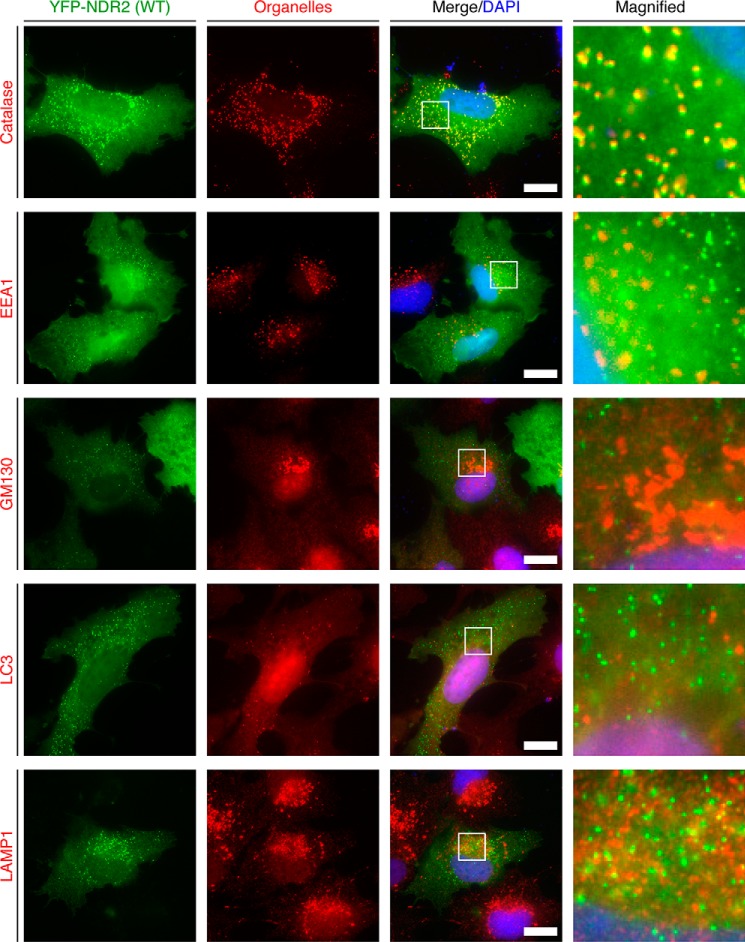
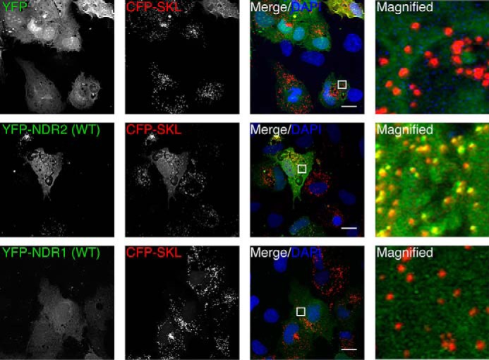
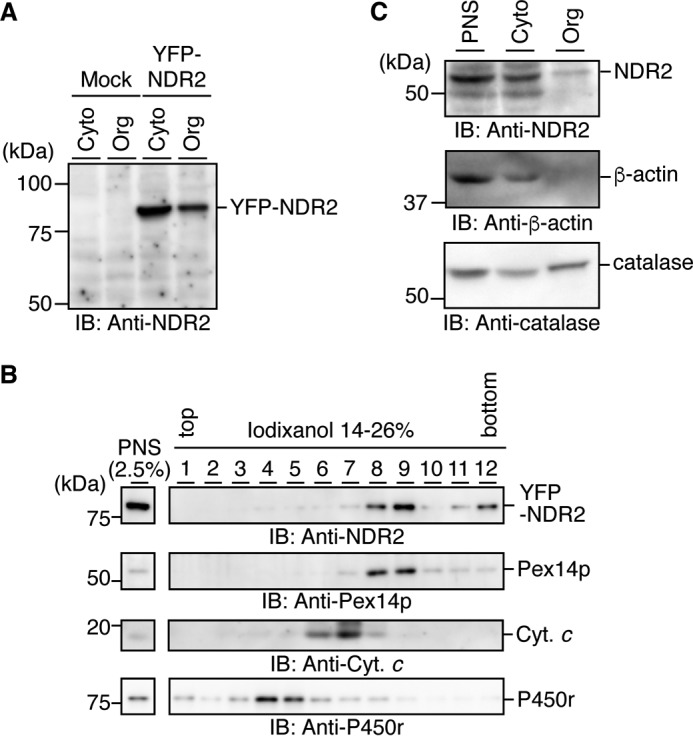
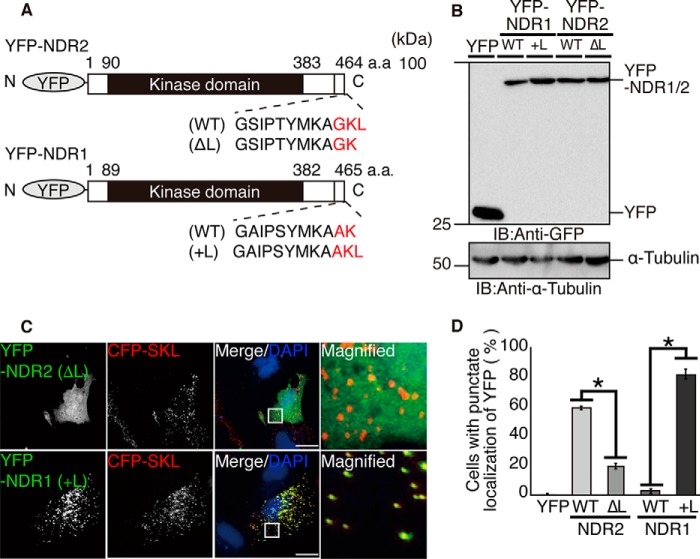
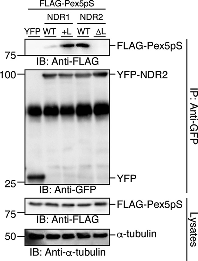
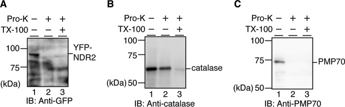
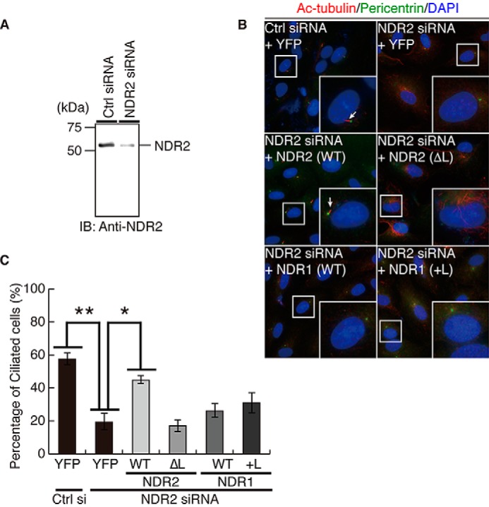
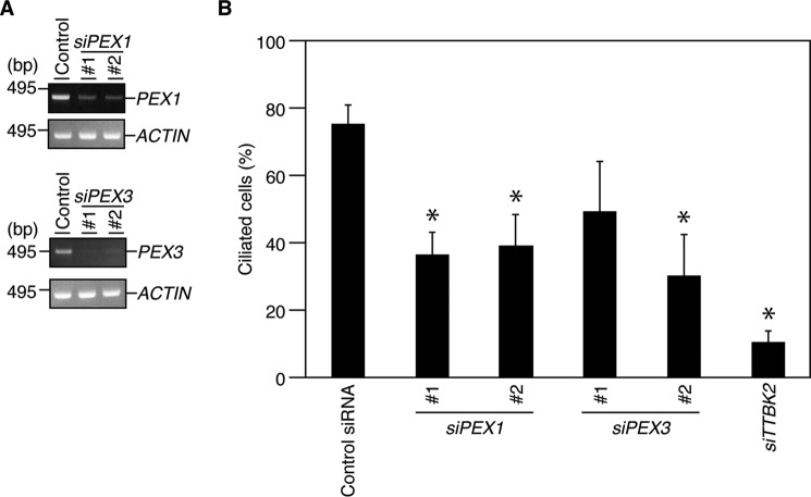
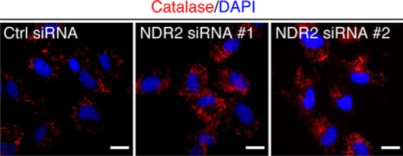
Nuclear Dbf2-related (NDR) kinases, comprising NDR1 and NDR2, are serine/threonine kinases that play crucial roles in the control of cell proliferation, apoptosis, and morphogenesis. We recently showed that NDR2, but not NDR1, is involved in primary cilium formation; however, the mechanism underlying their functional difference in ciliogenesis is unknown. To address this issue, we examined their subcellular localization. Despite their close sequence similarity, NDR2 exhibited punctate localization in the cytoplasm, whereas NDR1 was diffusely distributed within the cell. Notably, NDR2 puncta mostly co-localized with the peroxisome marker proteins, catalase and CFP-SKL (cyan fluorescent protein carrying the C-terminal typical peroxisome-targeting signal type-1 (PTS1) sequence, Ser-Lys-Leu). NDR2 contains the PTS1-like sequence, Gly-Lys-Leu, at the C-terminal end, whereas the C-terminal end of NDR1 is Ala-Lys. An NDR2 mutant lacking the C-terminal Leu, NDR2(ΔL), exhibited almost diffuse distribution in cells. Additionally, NDR2, but neither NDR1 nor NDR2(ΔL), bound to the PTS1 receptor Pex5p. Together, these findings indicate that NDR2 localizes to the peroxisome by using the C-terminal GKL sequence. Intriguingly, topology analysis of NDR2 suggests that NDR2 is exposed to the cytosolic surface of the peroxisome. The expression of wild-type NDR2, but not NDR2(ΔL), recovered the suppressive effect of NDR2 knockdown on ciliogenesis. Furthermore, knockdown of peroxisome biogenesis factor genes (PEX1 or PEX3) partially suppressed ciliogenesis. These results suggest that the peroxisomal localization of NDR2 is implicated in its function to promote primary cilium formation.

Keywords: NDR (nuclear dbf2-related) kinase; NDR2; cilia; ciliogenesis; peroisome targeting signal 1; peroxisome; protein kinase; protein targeting; protein translocation; serine/threonine protein kinase.


Conflict of interest statement

The authors declare that they have no conflicts of interest with the contents of this article

Figures

FIGURE 1.
FIGURE 1.
NDR2 co-localizes with catalase. RPE1 cells were transfected with the plasmid for YFP-NDR2 and cultured for 24 h. Cells were fixed and then imaged by YFP fluorescence (green) and stained with the antibodies against catalase, EEA1, GM130, LC3, and LAMP1, as indicated (red). DNA was stained with DAPI (blue). Merged fluorescence images are shown in the third column. Magnified images of the white boxes in the third column are shown in the fourth column. Scale bar, 20 μm.
FIGURE 2.
FIGURE 2.
NDR2, but not NDR1, co-localizes with CFP-SKL. RPE1 cells were co-transfected with the plasmid for CFP-SKL and the plasmids for YFP, YFP-NDR2, or YFP-NDR1 and cultured for 24 h. Cells were fixed and imaged by YFP (green) and CFP (red) fluorescence. DNA was stained with DAPI (blue). Merged fluorescence images are shown in the third column. Magnified images of the white boxes in the third column are shown in the fourth column. Scale bar, 20 μm.
FIGURE 3.
FIGURE 3.
Subcellular fractionation analysis of NDR2. A, subcellular fractionation of YFP-NDR2. PNS fractions of HeLa cells (Mock) or YFP-NDR2-expressing HeLa cells were separated into the cytosolic (Cyto) and organellar (Org) fractions. Equal aliquots of each fraction were analyzed by immunoblotting (IB) with an anti-NDR2 antibody. B, localization of YFP-NDR2 in the peroxisomal fraction. PNS fractions of YFP-NDR2-expressing HeLa cells were separated by ultracentrifugation on a 14–26% iodixanol density gradient. The gradient was collected into 12 fractions. Equal aliquots of each fraction and PNS fraction (2.5% of the loaded amount) were analyzed by immunoblotting with antibodies against NDR2, Pex14p, cytochrome c (Cyt. c), and P450 reductase (P450r). C, subcellular fractionation of endogenous NDR2. PNS fractions of RPE1 cells were separated into cytosolic and organellar fractions. Equal aliquots of each fraction were analyzed by immunoblotting with antibodies against NDR2, β-actin, and catalase.
FIGURE 4.
FIGURE 4.
The C-terminal GKL motif is required for the peroxisomal localization of NDR2. A, schematic structures of YFP-tagged mouse NDR2, NDR1, and their mutants used in this study. The C-terminal amino acid sequences of NDR2, NDR1, and their mutants are indicated. B, immunoblot analysis of the expression of YFP, YFP-NDR2, YFP-NDR1, and their mutants. RPE1 cells were transfected with the plasmids each encoding YFP, YFP-NDR2, YFP-NDR1, or their mutants and cultured for 24 h, and cell lysates were analyzed by immunoblotting (IB) with anti-GFP and anti-α-tubulin antibodies (as a loading control). C, subcellular localization of NDR2(ΔL) and NDR1(+L). RPE1 cells were co-transfected with the plasmids encoding CFP-SKL and YFP-NDR2(ΔL) or YFP-NDR1(+L) and analyzed by YFP (green) and CFP (red) fluorescence. DNA was stained with DAPI (blue). Merged fluorescence images are shown in the third column. Magnified images of the white boxes in the third column are shown in the fourth column. Scale bar, 20 μm. D, quantitative analysis of the percentage of cells in which YFP fluorescence exhibits punctate co-localization with CFP-SKL. Data are expressed as the means ± S.E. (error bars) of three independent experiments (>100 cells/experiment, one-way ANOVA followed by Dunnett's test). *, p < 0.01.
FIGURE 5.
FIGURE 5.
NDR2 and NDR1(+L), but neither NDR2(ΔL) nor NDR1, bind to Pex5p. HEK293T cells were co-transfected with the plasmids encoding FLAG-tagged Pex5pS and YFP-tagged NDR2, NDR1, or their mutants. Cell lysates were subjected to immunoprecipitation (IP) with an anti-GFP antibody and analyzed by immunoblotting (IB) with anti-FLAG and anti-GFP antibodies.
FIGURE 6.
FIGURE 6.
Topology analysis of NDR2. RPE1 cells were transfected with the plasmid for YFP-NDR2, and the PNS fraction was prepared. The PNS fraction was mock-treated (−) or treated with proteinase K (Pro-K) (+) in the absence (−) or presence (+) of Triton X-100 (TX-100). The reaction mixture was ultracentrifuged, and the resulting pellets were analyzed by SDS-PAGE and immunoblotting (IB) with antibodies against GFP (A), catalase (B), and PMP70 (C).
FIGURE 7.
FIGURE 7.
The C-terminal GKL motif is required for NDR2 to promote ciliogenesis. A, effect of NDR2-targeting siRNA on the expression of endogenous NDR2. RPE1 cells were transfected with human NDR2-targeting siRNA and cultured for 72 h, and lysates were analyzed by immunoblotting (IB) with an anti-NDR2 antibody. B, effects of expression of YFP, YFP-NDR2 (WT or ΔL), or YFP-NDR1 (WT or +L) on the ciliogenesis of NDR2 knockdown cells. RPE1 cells were transfected with the plasmids coding for YFP or YFP-tagged mouse NDR2, NDR1, or their mutants and cultured for 4 h. Then cells were transfected with human NDR2-targeting siRNA, cultured for 24 h, and serum-starved for 48 h. Cells were fixed and stained with anti-Ac-tubulin (red) and anti-pericentrin (green) antibodies. DNA was stained with DAPI (blue). Insets, magnified images (× 2.7) of the white boxes. Arrows, primary cilia. C, quantitative analysis of the percentage of ciliated cells. RPE1 cells were treated as in B. Data are expressed as the means ± S.E. (error bars) of three independent experiments (>100 cells/experiment, one-way ANOVA followed by a Dunnett's test). *, p < 0.05; **, p < 0.01.
FIGURE 8.
FIGURE 8.
Knockdown of PEX1 or PEX3 suppresses ciliogenesis. A, knockdown efficiency of PEX1- or PEX3-targeting siRNAs, as measured by RT-PCR. RPE1 cells were transfected with control siRNA or PEX1- or PEX3-targeting siRNAs and cultured for 72 h, and total RNAs were subjected to RT-PCR analysis. B, effects of PEX1 or PEX3 knockdown on ciliogenesis. RPE1 cells were transfected with control siRNA or siRNAs targeting PEX1, PEX3, or TTBK2, cultured for 24 h, and then serum-starved for 48 h. The percentages of ciliated cells were counted as in Fig. 7C. Data are expressed as the means ± S.E. (error bars) of three independent experiments (>100 cells/experiment, one-way ANOVA followed by Dunnett's test). *, p < 0.05, compared with control siRNA-transfected cells.
FIGURE 9.
FIGURE 9.
Knockdown of NDR2 has no apparent effect on peroxisome formation. RPE1 cells were treated with 10 nm control or NDR2-targeting siRNAs and cultured for 48 h. Cells were fixed and stained with an anti-catalase antibody (red). DNA was stained with DAPI (blue). Scale bar, 20 μm.

Similar articles

Cited by

References

    1. Hergovich A., Stegert M. R., Schmitz D., and Hemmings B. A. (2006) NDR kinases regulate essential cell processes from yeast to humans. Nat. Rev. Mol. Cell Biol. 7, 253–264 - PubMed
    1. Hergovich A. (2013) Regulation and functions of mammalian LATS/NDR kinases: looking beyond canonical Hippo signalling. Cell Biosci. 3, 32. - PMC - PubMed
    1. Emoto K. (2011) The growing role of the Hippo-NDR kinase signaling in neuronal development and disease. J. Biochem. 150, 133–141 - PubMed
    1. Nagai T., and Mizuno K. (2014) Multifaceted roles of Furry proteins in invertebrates and vertebrates. J. Biochem. 155, 137–146 - PubMed
    1. Cornils H., Kohler R. S., Hergovich A., and Hemmings B. A. (2011) Human NDR kinases control G1/S cell cycle transition by directly regulating p21 stability. Mol. Cell Biol. 31, 1382–1395 - PMC - PubMed

MeSH terms

Substances

LinkOut - more resources