. 2017 Feb;216(2):425-439.
doi: 10.1083/jcb.201606078. Epub 2017 Jan 25.

The Vps13p-Cdc31p complex is directly required for TGN late endosome transport and TGN homotypic fusion

Affiliations

The Vps13p-Cdc31p complex is directly required for TGN late endosome transport and TGN homotypic fusion

Mithu De et al. J Cell Biol. 2017 Feb.

Abstract

Yeast VPS13 is the founding member of a eukaryotic gene family of growing interest in cell biology and medicine. Mutations in three of four human VPS13 genes cause autosomal recessive neurodegenerative or neurodevelopmental disease, making yeast Vps13p an important structural and functional model. Using cell-free reconstitution with purified Vps13p, we show that Vps13p is directly required both for transport from the trans-Golgi network (TGN) to the late endosome/prevacuolar compartment (PVC) and for TGN homotypic fusion. Vps13p must be in complex with the small calcium-binding protein Cdc31p to be active. Single-particle electron microscopic analysis of negatively stained Vps13p indicates that this 358-kD protein is folded into a compact rod-shaped density (20 × 4 nm) with a loop structure at one end with a circular opening ∼6 nm in diameter. Vps13p exhibits ATP-stimulated binding to yeast membranes and specific interactions with phosphatidic acid and phosphorylated forms of phosphatidyl inositol at least in part through the binding affinities of conserved N- and C-terminal domains.


Figures

Figure 1.
Figure 1.
MSS from vps13Δ cells is inactive for TGN–PVC transport and TGN homotypic fusion. (A) Diagram of the cell-free TGN–PVC transport reaction. (B) TGN–PVC transport reactions using K-V donor MSS and PSHA acceptor MSS from VPS13+ and vps13Δ strains. Black triangles, VPS13+ donor and acceptor; blue circles, vps13Δ donor and acceptor; green squares, VPS13+ donor and vps13Δ acceptor; red diamonds, vps13Δ donor and VPS13+ acceptor. (C) Diagram of the cell-free TGN-homotypic fusion reaction. (D) TGN homotypic fusion reactions using K-V donor MSS and SHA acceptor MSS from VPS13+ and vps13Δ strains. Black triangles, VPS13+ donor and acceptor; blue circles, vps13Δ donor and acceptor; green squares, VPS13+ donor and vps13Δ acceptor; red diamonds, vps13Δ donor and VPS13+ acceptor.
Figure 2.
Figure 2.
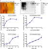
Addition of purified Vps13p rescues cell-free TGN–PVC transport and TGN homotypic fusion reactions performed with vps13Δ extracts. Silver-stained SDS-PAGE (A) and anti-TAP immunoblot (B) after SDS-PAGE of fractions from Vps13p purification. Lane 1, markers, lane 2, S13; lane 3, S55; lane 4, IgG Sepharose flow through; lane 5, eluate after TEV cleavage; lane 6, calmodulin Sepharose flow-through; lanes 7–10, calmodulin Sepharose eluate fractions 2–5. (C) Calmodulin Sepharose fraction 3 was analyzed by Coomassie staining after SDS-PAGE. (D) Purified Vps13p was titrated into TGN–PVC transport reactions using K-V donor MSS and PSHA acceptor MSS from vps13Δ strains. (E) Purified Vps13p was titrated into TGN–TGN homotypic fusion reactions using K-V donor MSS and SHA acceptor MSS from vps13Δ strains. (F) Time course of the TGN–PVC transport reaction using K-V donor MSS and PSHA acceptor MSS from vps13Δ strains. Each time point contained 1 µg purified Vps13p. (G) Time course of the TGN-homotypic fusion reaction using K-V donor MSS and SHA acceptor MSS from vps13Δ strains. Each time point contained 0.75 µg purified Vps13p.
Figure 3.
Figure 3.
3D architecture of Vps13p. (A) 2D class averages of Vps13 reveal an elongated rod with a loop structure on one end and a hook-like density on the opposite end. The rod can be observed in straight (left) or kinked configurations (right). The loop and hook structures (indicated by arrows) can be observed on the same or opposite sides, suggesting rotational flexibility between the top and bottom half of the protein. (B) 3D reconstructions of Vps13 with the loop and hook structures on the opposite (left) or the same side (right) of Vps13. The 3D maps confirm that the loop structure is hollow.
Figure 4.
Figure 4.
Membrane and lipid binding of Vps13p. (A) Membrane-binding reactions were conducted as described in Materials and methods. Lane 1, input (0.3 µg purified Vps13p); lanes 2–4, without ATP-regenerating system; lanes 5–7, with ATP-regenerating system, lanes 2 and 5, supernatant fractions (S); lanes 3 and 6, pellet fractions (P); lanes 4 and 7, sample of reactions before centrifugation (T). (B) Liposome-binding assays were conducted as described in Materials and methods. The figure shows data from one representative experiment. B, bottom fraction; M, middle fraction; P, pellet fraction; T, top fraction. Table 1 presents quantification of results from three independent experiments.
Figure 5.
Figure 5.
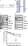
Lipid binding by purified Vps13p N and C domains. (A) Schematic representation of N domain and C domains. (B) Coomassie-stained SDS-PAGE of purified N domain (N) and C domain (C). (C and D) Liposome float-up assays were conducted as described in Materials and methods. (C) Binding assays for purified N domain. (D) Binding assays for purified C domain. The figure shows data from representative experiments. B, bottom fraction; M, middle fraction; P, pellet fraction; T, top fraction. Table 2 presents quantification of results from three independent experiments for each domain. (E) Inhibition of TGN–PVC transport by addition of purified N and C terminal domains. Purified domains were added to assembled reactions on ice before incubation at 30°C. Data are plotted as percent activity remaining and represent mean and SD of three independent experiments.
Figure 6.
Figure 6.
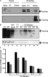
Coisolation of Vps13-3XHA with Z domain–tagged Cdc31p. (A) Experiments to measure coisolation of Vps13-3XHA with ZCdc31p were conducted as described in Materials and methods. The top blot was probed with anti–TAP antibody to detect Vps13p. The bottom blot was probed with anti–Cdc31p antibody. Eluate, IgG Sepharose eluate from cells lacking ZCdc31p (vector) or expressing ZCdc31p; FT, flow-through of IgG Sepharose; input, sample of homogenate. The input and flow-through samples represent 0.3% of the total, whereas the eluate represents 8% of the total. The ZCdc31p plasmid results in elevated expression of the 25 kD ZCdc31p relative to 18 kD endogenous Cdc31p, the latter of which is too low to be detected in samples from either the vector or ZCdc31p strains. (B) Copurification of Cdc31p with Vps13p. Fractions from Vps13p purification were immunoblotted with anti–TAP antibody to detect Vps13p (top blot) or anti–Cdc31p antibody (bottom blot). Lane 1, S13; lane 2, S55; lane 3, TEV eluate; lane 4, flow-through of calmodulin Sepharose column (CS FT); lane 5–9, eluted fractions 2–6 from calmodulin Sepharose (CS eluate). (C) Complementation by 5 µl of fractions 2–6 of calmodulin Sepharose elution shown in B of TGN–PVC transport reactions conducted with K-V donor and PSHA acceptor from vps13Δ strain (black columns) or cdc31-2ts strain (gray columns).
Figure 7.
Figure 7.
Cdc31p overexpression rescues toxicity of Vps13p overexpressed under GAL1 promoter control. pRS316 (vector), p416-GALpr-CDC31 expressing CDC31 under GAL1promoter control (GAL1pr-CDC31), and plasmid p426-GPDpr-CDC31 expressing CDC31 under TDH3 promoter control were transformed into VPS13+ strain CRY1 and EBY43, in which the GAL1 promoter was integrated in front of the VPS13 structural gene. Strains were grown in YPD liquid culture, and serial dilutions were pronged onto 2% (wt/vol) glucose plates (synthetic complete 2% [wt/vol] glucose medium with uracil) and 2% (wt/vol) galactose plates (synthetic complete 2% [wt/vol] galactose medium with uracil) and grown for 2 d at 30°C.
Figure 8.
Figure 8.
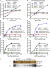
Analysis of Cdc31p function using cell-free TGN–PVC and TGN homotypic fusion reactions. (A) TGN–PVC transport reactions using K-V donor MSS and PSHA acceptor MSS from VPS13+ CDC31+ and VPS13+ cdc31-2ts strains. Black triangles, VPS13+ CDC31+ donor and acceptor; blue circles, VPS13+ cdc31-2ts donor and acceptor; green squares, VPS13+ CDC31+ donor and VPS13+ cdc31-2ts acceptor; red diamonds, VPS13+ cdc31-2ts donor and VPS13+ CDC31+ acceptor. (B) TGN-homotypic fusion reactions using K-V donor MSS and SHA acceptor MSS from VPS13+ CDC31+ and VPS13+ cdc31-2ts strains. Black triangles, VPS13+ CDC31+ donor and acceptor; blue circles, VPS13+ cdc31-2ts donor and acceptor; green squares, VPS13+ CDC31+ donor and VPS13+ cdc31-2ts acceptor; red diamonds, VPS13+ cdc31-2ts donor and VPS13+ CDC31+ acceptor. (C) Titration of Vps13p purified from CDC31+ strain (blue triangles) or from cdc31-2ts strain (red circles) into TGN–PVC transport reactions using K-V donor MSS and PSHA acceptor MSS from VPS13+ cdc31-2ts strains. (D) Titration of Vps13p purified from CDC31+ strain (blue triangles) or from cdc31-2ts strain (red circles) into TGN homotypic fusion reactions using K-V donor MSS and SHA acceptor MSS from VPS13+ cdc31-2ts strains. (E) Titration of Vps13p purified from CDC31+ strain (black triangles) or from cdc31-2ts strain (green circles) into TGN–PVC transport reactions using K-V donor MSS and PSHA acceptor MSS from vps13Δ CDC31+ strains. (F) Titration of Vps13p purified from CDC31+ strain (black triangles) or from cdc31-2ts strain (green circles) into TGN homotypic fusion reactions using K-V donor MSS and SHA acceptor MSS from vps13Δ CDC31+ strains. (G) Purification of Vps13-TAP from cdc31-2ts strain. Fractions from purification of Vps13-TAP from cdc31-2ts strain MDY9 (p416-ADH1pr-VPS13-TAP) were subjected to electrophoresis in SDS-PAGE and gels were silver-stained (top) or Western blotted (bottom) using anti–TAP antibody. Lane 1, S55; lane 2, IgG Sepharose flow-through (IgG FT); lane3, TEV eluate of IgG Sepharose; lane 4–9, eluted fractions 2–7 of calmodulin Sepharose (CS eluates).

Comment in

Similar articles

Cited by

References

    1. Abazeed M.E., and Fuller R.S.. 2008. Yeast Golgi-localized, gamma-Ear-containing, ADP-ribosylation factor-binding proteins are but adaptor protein-1 is not required for cell-free transport of membrane proteins from the trans-Golgi network to the prevacuolar compartment. Mol. Biol. Cell. 19:4826–4836. 10.1091/mbc.E07-05-0442 - DOI - PMC - PubMed
    1. Abazeed M.E., Blanchette J.M., and Fuller R.S.. 2005. Cell-free transport from the trans-Golgi network to late endosome requires factors involved in formation and consumption of clathrin-coated vesicles. J. Biol. Chem. 280:4442–4450. 10.1074/jbc.M412553200 - DOI - PubMed
    1. Balikova I., Lehesjoki A.E., de Ravel T.J., Thienpont B., Chandler K.E., Clayton-Smith J., Träskelin A.L., Fryns J.P., and Vermeesch J.R.. 2009. Deletions in the VPS13B (COH1) gene as a cause of Cohen syndrome. Hum. Mutat. 30:E845–E854. 10.1002/humu.21065 - DOI - PubMed
    1. Bankaitis V.A., Johnson L.M., and Emr S.D.. 1986. Isolation of yeast mutants defective in protein targeting to the vacuole. Proc. Natl. Acad. Sci. USA. 83:9075–9079. 10.1073/pnas.83.23.9075 - DOI - PMC - PubMed
    1. Bigay J., and Antonny B.. 2005. Real-time assays for the assembly-disassembly cycle of COP coats on liposomes of defined size. Methods Enzymol. 404:95–107. 10.1016/S0076-6879(05)04010-3 - DOI - PubMed

MeSH terms