. 2017 Mar 1;198(5):2028-2037.
doi: 10.4049/jimmunol.1601199. Epub 2017 Jan 25.

Mycobacterium tuberculosis Membrane Vesicles Inhibit T Cell Activation

Affiliations

Mycobacterium tuberculosis Membrane Vesicles Inhibit T Cell Activation

Jaffre J Athman et al. J Immunol. .

Abstract

Mycobacterium tuberculosis utilizes multiple mechanisms to evade host immune responses, and inhibition of effector CD4+ T cell responses by M. tuberculosis may contribute to immune evasion. TCR signaling is inhibited by M. tuberculosis cell envelope lipoglycans, such as lipoarabinomannan and lipomannan, but a mechanism for lipoglycans to traffic from M. tuberculosis within infected macrophages to reach T cells is unknown. In these studies, we found that membrane vesicles produced by M. tuberculosis and released from infected macrophages inhibited the activation of CD4+ T cells, as indicated by reduced production of IL-2 and reduced T cell proliferation. Flow cytometry and Western blot demonstrated that lipoglycans from M. tuberculosis-derived bacterial vesicles (BVs) are transferred to T cells, where they inhibit T cell responses. Stimulation of CD4+ T cells in the presence of BVs induced expression of GRAIL, a marker of T cell anergy; upon restimulation, these T cells showed reduced ability to proliferate, confirming a state of T cell anergy. Furthermore, lipoarabinomannan was associated with T cells after their incubation with infected macrophages in vitro and when T cells were isolated from lungs of M. tuberculosis-infected mice, confirming the occurrence of lipoarabinomannan trafficking to T cells in vivo. These studies demonstrate a novel mechanism for the direct regulation of CD4+ T cells by M. tuberculosis lipoglycans conveyed by BVs that are produced by M. tuberculosis and released from infected macrophages. These lipoglycans are transferred to T cells to inhibit T cell responses, providing a mechanism that may promote immune evasion.


Figures

Fig. 1
Fig. 1. Extracellular vesicles (EVs) from M. tuberculosis-infected macrophages inhibit TCR activation of CD4+ T cells
(A) EVs were purified from uninfected or M. tuberculosis-infected macrophages to produce Ctr EVs or Inf EVs, respectively. EVs (5 µg protein content) were assessed by western blot for host CD9 and CD63 (left) and M. tuberculosis lipoglycans (right, top band represents LAM and bottom band represents LM). (B) CD4+ T cells from spleens of C57BL/6 mice were treated for 1 h with Inf EVs or Ctr EVs (100 µg/ml EV protein concentration) and activated for 24 h with 1 µg/ml plate bound anti-CD3ε and 1 µg/ml soluble anti-CD28 in the continued presence of the EVs. IL-2 was measured by ELISA. Data are shown as mean ± SD of triplicate samples from one representative experiment (n>3 experiments). Student t test with Welch’s correction was used to assess statistical significance (*≤0.05).
Fig. 2
Fig. 2. BVs from axenic M. tuberculosis inhibit TCR-induced activation of CD4+ T cells
(A) BVs from 2 l axenic M. tuberculosis culture were purified by differential centrifugation and size exclusion chromatography. Western blot with polyclonal anti-M. tuberculosis Ab shows LAM and LM in size exclusion chromatography fractions. (B) BV-containing fractions (6–11) from (A) were pooled, concentrated and analyzed by western blot with CS-35 Ab (anti-LAM) in comparison to a purified LAM standard used for quantification. (C) CD4+ T cells derived from spleens of C57BL/6 mice were treated for 1 h with BVs or purified LAM and then stimulated for 24 h with 1 µg/ml plate bound anti-CD3ε and 1 µg/ml soluble anti-CD28 in the continued presence or absence of BVs or LAM. IL-2 was measured by ELISA. (D-E) Proliferation was measured by dye dilution in 72 h cultures by flow cytometry. Data are shown as mean ± SD of triplicate samples from one representative experiment (n=3 experiments).
Fig. 3
Fig. 3. Multiple M. tuberculosis lipoglycans can contribute to BV inhibition of CD4+ T cells
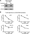
(A) CD4+ T cells from spleens of C57BL/6 mice were incubated for 24 h with BVs (10 µg/ml LAM equivalent concentration) or LAM (10 µg/ml), re-purified by magnetic sorting and assessed for LAM content by western blot with CS-35 Ab. (B) CD4+ T cells were treated with lipoglycans LAM, LM, PIM1/2 or PIM6 for 1 hour prior to activation with plate bound anti-CD3ε (1 µg/ml) and soluble anti-CD28 (1 µg/ml) for 24 h. IL-2 was measured by ELISA. Data are shown as mean ± SD of triplicate samples from one representative experiment (n=3 experiments).
Fig. 4
Fig. 4. BVs facilitate the trafficking of LAM and LM from infected macrophages to CD4+ T cells
(A) CD4+ T cells from spleens of C57L/6 mice were incubated with or without BVs (10 µg/ml LAM equivalent concentration) for 24 h, stained with anti-CD3ε and anti-M. tuberculosis Abs or isotype control Abs, fixed, and assessed by flow cytometry with polyclonal anti-M. tuberculosis Ab. Open outline: BV-treated T cells anti-M. tuberculosis Ab; grey outline: untreated T cells anti-M. tuberculosis Ab; black outline: BV-treated T cells with isotype control Ab. (B) P25 CD4+ T cells were cultured with M. tuberculosis-infected or uninfected macrophages for 24 h with the addition of P25 peptide (1 µg/ml). CD4+ T cells were then purified from macrophages by FACS with exclusion of CD11b+ cells and selection of CD3ε+ cells. Purified T cells were assessed for M. tuberculosis lipoglycan content by western blotting with polyclonal anti-M. tuberculosis Ab.
Fig. 5
Fig. 5. LAM trafficking to CD4+ T cells in vivo
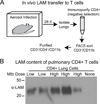
(A) Schematic of experimental design. Mice were aerosol infected with M. tuberculosis H37Rv at low or high dose. After 28 d of infection, lungs were harvested and CD4+ T cells were isolated by immunoaffinity purification. CD4+ T cells were further purified by FACS to select CD3+CD11b cells (≥99% purity), providing the preparation of CD3+CD4+CD11b cells for LAM analysis. (B) Western blot analysis of LAM content in lysates of purified CD3+CD4+CD11b lung cells after low dose or high dose aerosol infection of mice with M. tuberculosis H37Rv (“None” indicates lysate of immunoaffinity-purified CD4+ T cells from uninfected mouse lung). Each lane represents lysate from 105 cells derived from a single animal (the two low dose infections are from a single experiment, and the three high dose infections are from another experiment). LAM was detected in CD3+CD4+CD11b cells from lungs of mice after both low and high dose infections, although with some variability in level.
Fig. 6
Fig. 6. BVs inhibit the activation of both naïve and Th1 effector CD4+ T cells
(A-B) Naïve CD4+ T cells from C57BL/6 mice were incubated for 1 h with BVs at 10 µg/ml LAM equivalent concentration and stimulated with anti-CD3ε/anti-CD28 in the continued presence of BVs. After 24 h, IL-2 was measured in the supernatant by ELISA, and proliferation was measured after 48 h by [3H]-thymidine incorporation. (C) Naïve P25 CD4+ T cells were activated with and without BVs as in panel A, except T cells were stimulated with fixed macrophages presenting Ag85B peptide. (D-E) Th1 effector CD4+ T cells were prepared from splenocytes of C57BL/6 mice and activated as in panel A; after 24 h supernatants were harvested, and IFNγ (D) and IL-2 (E) were measured by ELISA. Data are shown as mean ± SD of triplicate samples from one representative experiment (n=3 experiments, except n=2 for panel C). Students t test with Welch’s correction was used to assess statistical significance (*, p≤0.05; **, p≤0.01).
Fig. 7
Fig. 7. BVs induce GRAIL expression and functional anergy in naïve CD4+ T cells
(A) CD4+ T cells were isolated from spleens of C57BL/6 mice and activated with anti-CD3ε/anti-CD28 in the presence or absence of BVs (10 µg/ml LAM equivalent concentration) or LAM (10 µg/ml). GRAIL expression was measured by qPCR after 24 h. (B) Western blot analysis of GRAIL protein expression in CD4+ T cells treated as in (A). (C-D) Naïve CD4+ T cells were activated as in Fig. 6A in the presence or absence of BVs, rested for 5 d with 10 ng/ml IL-7, and then re-stimulated with anti-CD3ε/anti-CD28. After 48 h of restimulation, proliferation was measured by addition of [3H]-thymidine for an additional 16 h (C). After 24 h of restimulation, supernatants were harvested and IL-2 was measured by ELISA (D). (E-F) Naïve P25 CD4+ T cells were activated for 48 h with fixed macrophages presenting Ag85B peptide in the presence or absence of BVs or ionomycin, rested for 5 d with 10 ng/ml IL-7, and restimulated with fixed macrophages and peptide. Proliferation and IL-2 were measured as in panels C and D. Data are shown as mean ± SD of triplicate samples from one representative experiment (n>3; except panels E and F, n = 2). Student t test with Welch’s correction was used to assess statistical significance (*, p≤0.05; **, p≤0.01; ***, p≤0.001).

References

    1. Giri PK, Kruh NA, Dobos KM, Schorey JS. Proteomic analysis identifies highly antigenic proteins in exosomes from M. tuberculosis-infected and culture filtrate protein-treated macrophages. Proteomics. 2010;10:3190–3202. - PMC - PubMed
    1. Ramachandra L, Qu Y, Wang Y, Lewis CJ, Cobb B, Takatsu K, Boom WH, Dubyak GR, Harding CV. Mycobacterium tuberculosis synergizes with ATP to induce release of microvesicles and exosomes containing major histocompatibility complex class II molecules capable of antigen presentation. Infect. Immun. 2010;78:5116–5125. - PMC - PubMed
    1. Athman JJ, Wang Y, McDonald DJ, Boom WH, Harding CV, Wearsch PA. Bacterial Membrane Vesicles Mediate the Release of Mycobacterium tuberculosis Lipoglycans and Lipoproteins from Infected Macrophages. J. Immunol. 2015;195:1044–1053. - PMC - PubMed
    1. Bhatnagar S, Shinagawa K, Castellino FJ, Schorey JS. Exosomes released from macrophages infected with intracellular pathogens stimulate a proinflammatory response in vitro and in vivo. Blood. 2007;110:3234–3244. - PMC - PubMed
    1. Singh PP, Smith VL, Karakousis PC, Schorey JS. Exosomes isolated from mycobacteria-infected mice or cultured macrophages can recruit and activate immune cells in vitro and in vivo. J. Immunol. 2012;189:777–785. - PMC - PubMed

Publication types

MeSH terms