. 2017 Feb;155(2):363-378.
doi: 10.1093/toxsci/kfw213. Epub 2016 Oct 23.

Dual Role of Epidermal Growth Factor Receptor in Liver Injury and Regeneration after Acetaminophen Overdose in Mice

Affiliations

Dual Role of Epidermal Growth Factor Receptor in Liver Injury and Regeneration after Acetaminophen Overdose in Mice

Bharat Bhushan et al. Toxicol Sci. 2017 Feb.

Abstract

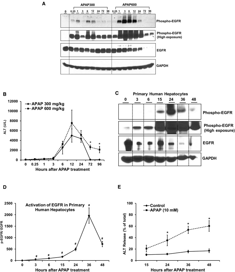
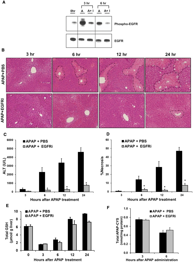
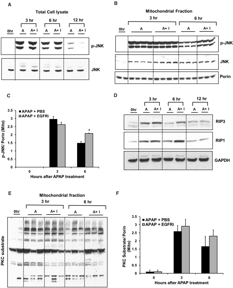
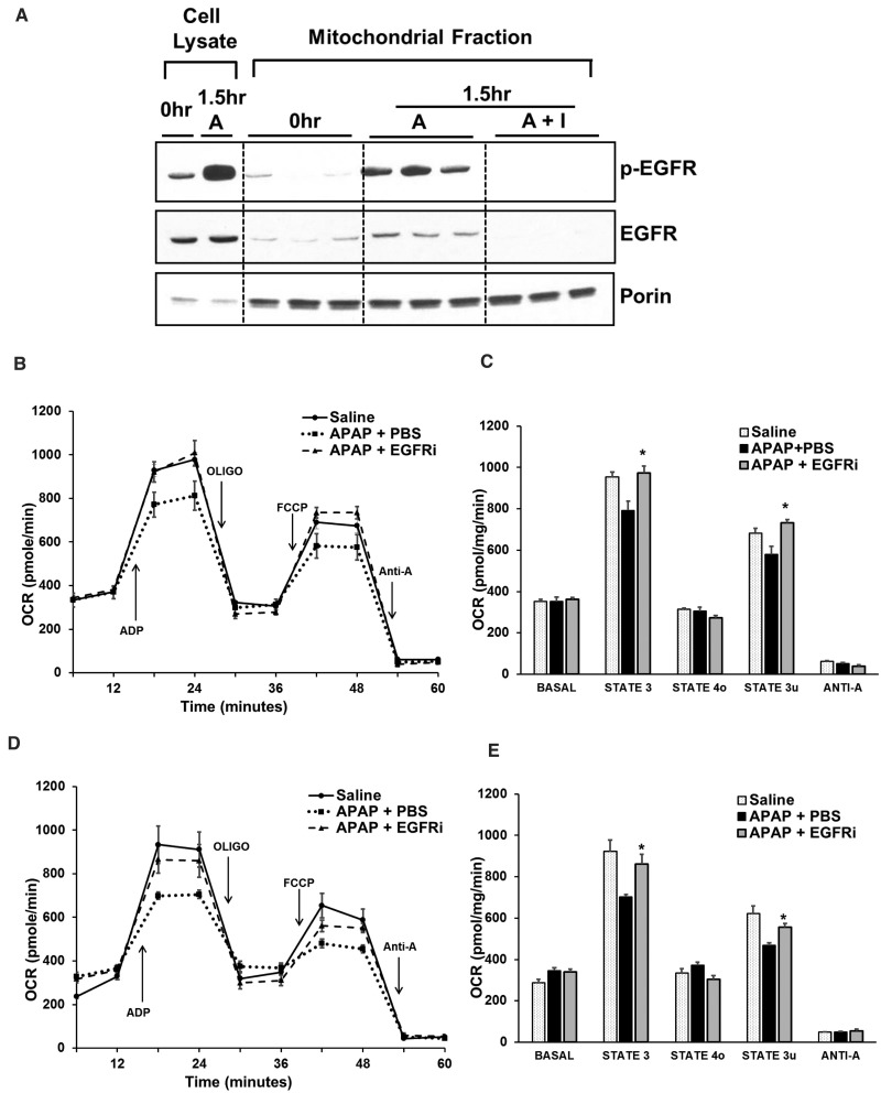
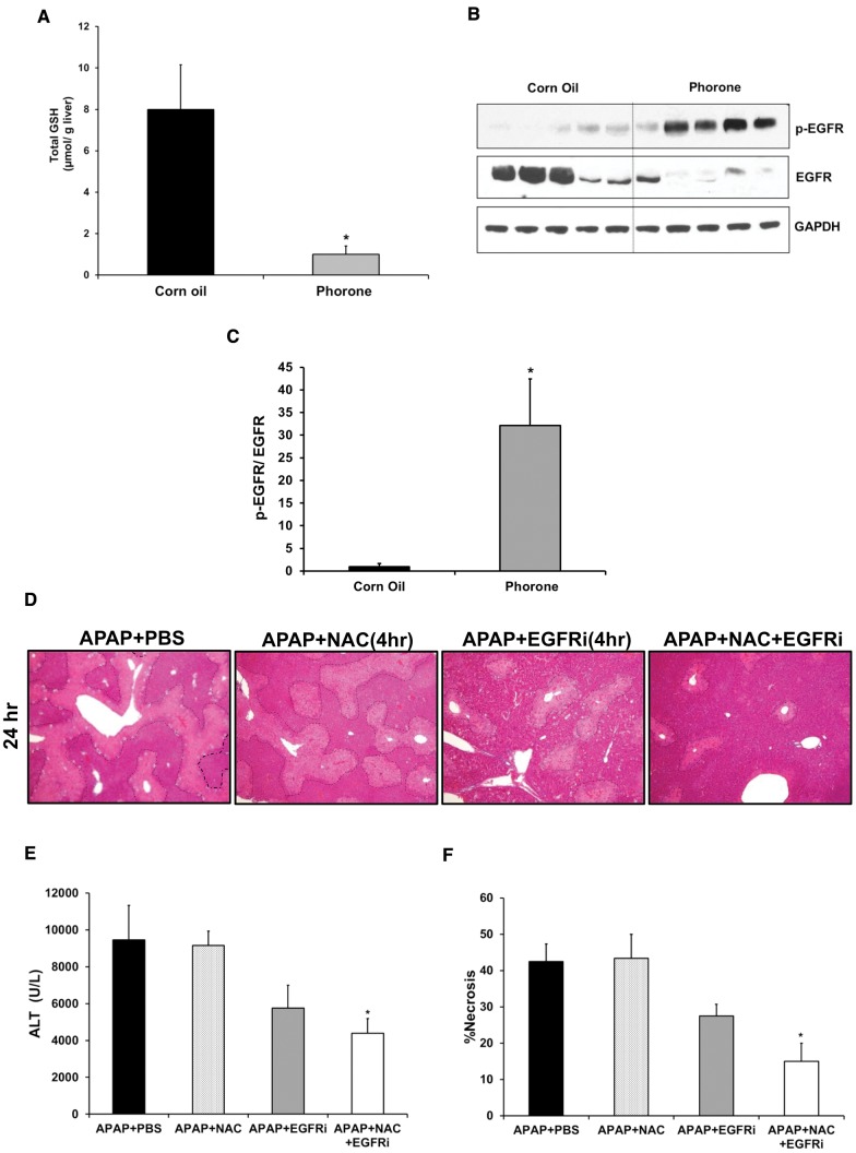
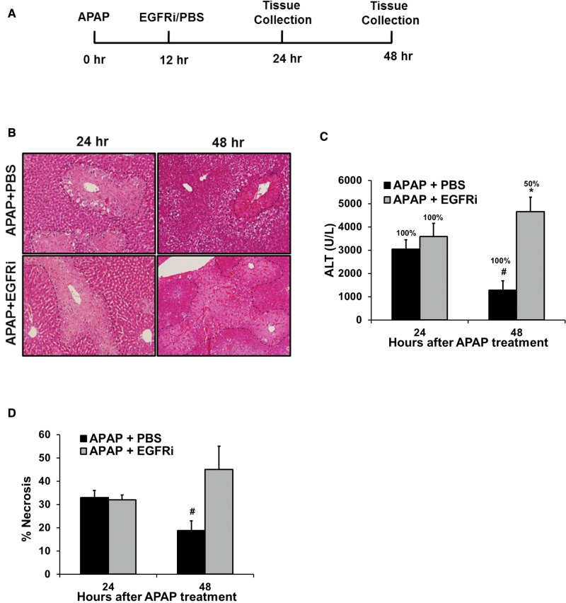
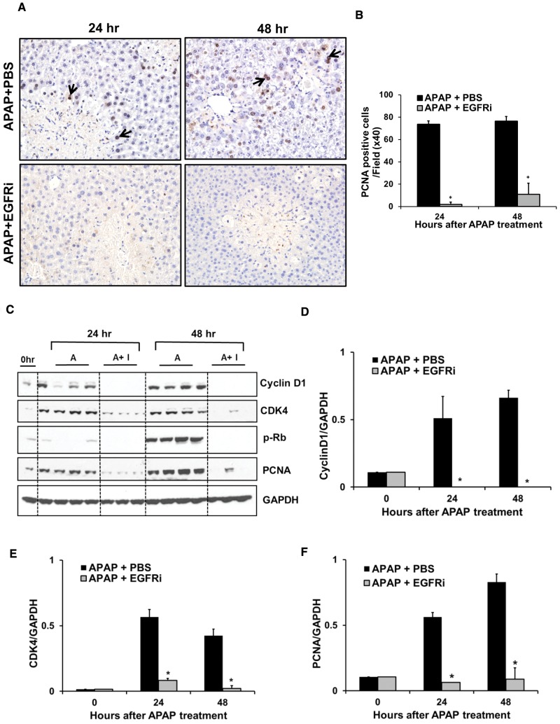
Epidermal growth factor receptor (EGFR) plays a crucial role in hepatocyte proliferation. Its role in acetaminophen (APAP)-mediated hepatotoxicity and subsequent liver regeneration is completely unknown. Role of EGFR after APAP-overdose in mice was studied using pharmacological inhibition strategy. Rapid, sustained and dose-dependent activation of EGFR was noted after APAP-treatment in mice, which was triggered by glutathione depletion. EGFR-activation was also observed in primary human hepatocytes after APAP-treatment, preceding elevation of toxicity markers. Treatment of mice with an EGFR-inhibitor (EGFRi), Canertinib, 1h post-APAP resulted in robust inhibition of EGFR-activation and a striking reduction in APAP-induced liver injury. Metabolic activation of APAP, formation of APAP-protein adducts, APAP-mediated JNK-activation and its mitochondrial translocation were not altered by EGFRi. Interestingly, EGFR rapidly translocated to mitochondria after APAP-treatment. EGFRi-treatment abolished mitochondrial EGFR activity, prevented APAP-mediated mitochondrial dysfunction/oxidative-stress and release of endonucleases from mitochondria, which are responsible for DNA-damage/necrosis. Treatment with N-acetylcysteine (NAC), 4h post-APAP in mice did not show any protection but treatment of EGFRi in combination with NAC showed decrease in liver injury. Finally, delayed treatment with EGFRi, 12-h post-APAP, did not alter peak injury but caused impairment of liver regeneration resulting in sustained injury and decreased survival after APAP overdose in mice. Impairment of regeneration was due to inhibition of cyclinD1 induction and cell cycle arrest. Our study has revealed a new dual role of EGFR both in initiation of APAP-injury and in stimulation of subsequent compensatory regeneration after APAP-overdose.

Keywords: Cyclin D1; Hepatotoxicity; N-acetylcysteine; hepatocyte proliferation.; mitochondrial dysfunction.


Figures

FIG. 1
FIG. 1
Rapid and sustained activation of EGFR in mice and primary human hepatocytes after APAP-treatment. (A) Western blot analysis of phospho-EGFR (Tyr1068) and EGFR in total cell lysate and (B) ALT levels in serum, at various time points after administration of 300 mg/kg and 600 mg/kg APAP in mice (n=3-8). (C) Western blot analysis of phospho-EGFR and EGFR in total cell lysate and (E) ALT release in medium (represented as percentage of total ALT levels), at various time points after treatment of primary human hepatocytes (PHH) with 10 mM APAP. (D) Densitometric analysis showing activation of EGFR in PHH with data representing mean ± SEM of independent western blot analysis from 4 liver donors. * indicate significant difference between groups at p<0.05. # indicates significant difference w.r.t. 0 hr time point wherever indicated.
FIG. 2
FIG. 2
Early treatment with EGFRi (1-h post-APAP) remarkably attenuated APAP-induced hepatotoxicity without altering APAP bioactivation and APAP-protein adducts formation. A, Western blot analysis of phospho-EGFR and EGFR in liver lysate, B, representative photomicrographs of H&E stained liver sections with necrotic area outlined, C, serum ALT levels, D, percentage necrosis area based on H&E stained liver sections, E, total glutathione levels in liver extract, and F, APAP-protein adducts levels in liver as measured by HPLC-ECD method. For all experiments mice (n = 3–12) were treated with 300 mg/kg APAP followed by canertinib (80 mg/kg) or PBS, 1-h post-APAP. All samples were collected at various time points up-to 24-h after APAP treatment. * indicate significant difference between groups at P < .05.
FIG. 3
FIG. 3
JNK activation, its mitochondrial translocation and signaling through other protein kinases not altered by EGFRi. Western blot analysis of phospho-JNK and JNK in (A) total cell lysate and (B) mitochondrial fraction with densitometric analysis of mitochondrial activated JNK (p-JNK) shown in (C). D, Western blot analysis of RIP3 and RIP1 in total cell lysate. E, Western blot analysis showing PKC activation (studied using antibody against phosphorylated PKC-substrates in mitochondria) with its densitometric analysis shown in (F). All analysis were done on liver samples collected at various time points after treatment with 300 mg/kg APAP + PBS or APAP + canertinib (80 mg/kg, 1-h post-APAP) (n = 3–4). *indicate significant difference between groups at P < .05.
FIG. 4
FIG. 4
APAP caused rapid mitochondrial translocation of EGFR and mitochondrial dysfunction, which was inhibited by EGFRi. A, Western blot analysis of phospho-EGFR and EGFR in mitochondrial fraction at 1.5 h after administration of APAP (300 mg/kg) in mice pretreated (2-h prior to APAP) with canertinib (80 mg/kg). B-E, Oxygen consumption rate analysis in freshly isolated mitochondria from mice (n = 3) treated with APAP (300 mg/kg) + PBS, APAP (300 mg/kg) + canertinib (80 mg/kg) or saline (control), using Seahorse extracellular flux analyzer. B and C, Canertinib was administered 2hr before APAP and analysis was done 1.5 h after APAP treatment. D and E, Canertinib was administered 1-h post-APAP and analysis was done 3 h after APAP treatment. Oxidative phosphorylation was manipulated with injection of ADP, oligomycin (oligo), FCCP, and antimycin A (anti-A). Respiration was sequentially measured in a coupled state with substrate present (basal respiration), followed by State 3 (phosphorylating respiration, in the presence of ADP and substrate), State 4o (leak state induced with the addition of oligomycin - inhibitor of ATP synthase), and then maximal uncoupler (FCCP)-stimulated respiration (State 3u). At the end, antimycin A (complex III inhibitor) was added to inhibit mitochondrial respiration completely * indicate significant difference w.r.t. to APAP + PBS group at P < .05.
FIG. 5
FIG. 5
Decreased oxidative stress, mitochondrial protein nitration and release of endonucleases from mitochondria by EGFRi. A, Oxidized glutathione levels in total liver extract. B, Representative photomicrographs of liver sections stained for nitrotyrosine-protein adducts. C, Western blot analysis of nitrotyrosine adducts in mitochondrial fraction with its densitometric analysis shown in (D). E, Western blot analysis of AIF, endonuclease G and SMAC in cytosolic fraction with densitometric analysis of AIF as shown in (F). All analysis were done on liver samples collected at various time points after treatment with APAP (300 mg/kg) + PBS or APAP (300 mg/kg) + canertinib (80 mg/kg, 1-h post-APAP) (n = 3–5). *indicate significant difference between groups at P < .05.
FIG. 6
FIG. 6
(A–C) Role of glutathione depletion in rapid activation of EGFR by APAP. (D–F) Enhanced protection against APAP hepatotoxicity by combination of NAC and EGFRi (administered 4-h post-APAP). (A) Total glutathione levels in liver extract and (B) western blot analysis of phospho-EGFR and EGFR in total cell lysate from liver samples obtained 2 h after treatment with Phorone (200 mg/kg in corn oil) or corn oil (control) in mice (n = 5). (C) Densitomertic analysis showing EGFR activation based on western blot image shown in (B). (D) Representative photomicrographs of H&E stained liver sections with necrotic area outlined, (E) serum ALT levels and (F) percentage necrosis area based on H&E stained liver sections of mice treated with 300 mg/kg APAP followed by canertinib (80 mg/kg), NAC (500 mg/kg), combination of canertinib (80 mg/kg) and NAC (500 mg/kg) or PBS (control), 4-h post-APAP (n = 5). Samples were collected 24 h after APAP treatment. *indicate significant difference w.r.t. control group at P < .05.
FIG. 7
FIG. 7
Increased progression of injury, impaired recovery and decreased survival after delayed (12 hr post-APAP) treatment with EGFRi. (A) Schematic showing experimental design. (B) Representative photomicrographs of H&E stained liver sections with necrotic area outlined, (C) serum ALT levels with percentage survival specified over bars and (D) percentage necrosis area based on H&E stained liver sections of mice treated with 300 mg/kg APAP followed by treatment with canertinib (80 mg/kg) or PBS, 12 hr post-APAP. All samples were collected and survival was recorded at 24 and 48 hr after APAP treatment (n=5-6). * indicate significant difference between groups at p<0.05. # indicates significant difference w.r.t. 24 hr time point within same treatment group.
FIG. 8
FIG. 8
Impaired liver regeneration and cell cycle arrest by delayed (12-h post-APAP) treatment with EGFRi. A, Representative photomicrographs of PCNA stained liver sections with arrows indicating cells in S-phase with nuclear PCNA staining (brown). B, Total number of PCNA-positive cell per high power field (×40). C, Western blot analysis of cyclinD1, CDK4, phospho-Rb, and PCNA using total liver extract. Densitometric analysis of (D) cyclinD1, (E) CDK4, and (F) PCNA. Mice were treated with 300 mg/kg APAP. Canertinib (80 mg/kg) or PBS was administered 12-h post-APAP (n = 3–5). All samples were collected at 24 and 48 h after APAP treatment. * indicate significant difference between groups at P < .05.

References

    1. Arany I. (2008). Dual role of the activated epidermal growth factor receptor in renal tubular cells during stress. Kidney Int. 73, 5–7. - PubMed
    1. Arany I., Megyesi J. K., Kaneto H., Price P. M., Safirstein R. L. (2004). Cisplatin-induced cell death is EGFR/src/ERK signaling dependent in mouse proximal tubule cells. Am. J. Physiol. Renal Physiol. 287, F543–F549. - PubMed
    1. Bernal W., Lee W. M., Wendon J., Larsen F. S., Williams R. (2015). Acute liver failure: A curable disease by 2024?. J. Hepatol. 62(1 Suppl), S112–S120., - PubMed
    1. Bhushan B., Borude P., Edwards G., Walesky C., Cleveland J., Li F., Ma X., Apte U. (2013). Role of bile acids in liver injury and regeneration following acetaminophen overdose. Am. J. Pathol. 183, 1518–1526. - PMC - PubMed
    1. Bhushan B., Edwards G., Desai A., Michalopoulos G., Apte U. (2016). Liver-specific deletion of integrin-linked kinase in mice attenuates hepatotoxicity and improves liver regeneration after acetaminophen overdose. Gene Exp. - PMC - PubMed

MeSH terms