. 2017 Jan 25;37(4):757-767.
doi: 10.1523/JNEUROSCI.2659-16.2016.

Loss of Plasticity in the D2-Accumbens Pallidal Pathway Promotes Cocaine Seeking

Affiliations

Loss of Plasticity in the D2-Accumbens Pallidal Pathway Promotes Cocaine Seeking

Jasper A Heinsbroek et al. J Neurosci. .

Abstract

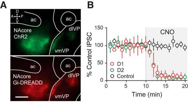
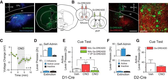
Distinct populations of D1- and D2-dopamine receptor-expressing medium spiny neurons (D1-/D2-MSNs) comprise the nucleus accumbens, and activity in D1-MSNs promotes, whereas activity in D2-MSNs inhibits, motivated behaviors. We used chemogenetics to extend D1-/D2-MSN cell specific regulation to cue-reinstated cocaine seeking in a mouse model of self-administration and relapse, and found that either increasing activity in D1-MSNs or decreasing activity in D2-MSNs augmented cue-induced reinstatement. Both D1- and D2-MSNs provide substantial GABAergic innervation to the ventral pallidum, and chemogenetic inhibition of ventral pallidal neurons blocked the augmented reinstatement elicited by chemogenetic regulation of either D1- or D2-MSNs. Because D1- and D2-MSNs innervate overlapping populations of ventral pallidal neurons, we next used optogenetics to examine whether changes in synaptic plasticity in D1- versus D2-MSN GABAergic synapses in the ventral pallidum could explain the differential regulation of VP activity. In mice trained to self-administer cocaine, GABAergic LTD was abolished in D2-, but not in D1-MSN synapses. A μ opioid receptor antagonist restored GABA currents in D2-, but not D1-MSN synapses of cocaine-trained mice, indicating that increased enkephalin tone on presynaptic μ opioid receptors was responsible for occluding the LTD. These results identify a behavioral function for D1-MSN innervation of the ventral pallidum, and suggest that losing LTDGABA in D2-MSN, but not D1-MSN input to ventral pallidum may promote cue-induced reinstatement of cocaine-seeking.

Significance statement: More than 90% of ventral striatum is composed of two cell types, those expressing dopamine D1 or D2 receptors, which exert opposing roles on motivated behavior. Both cell types send GABAergic projections to the ventral pallidum and were found to differentially promote cue-induced reinstatement of cocaine seeking via the ventral pallidum. Furthermore, after cocaine self-administration, synaptic plasticity was selectively lost in D2, but not D1 inputs to the ventral pallidum. The selective impairment in D2 afferents may promote the influence of D1 inputs to drive relapse to cocaine seeking.

Keywords: GABA; LTD; accumbens; cocaine; pallidum; relapse.


Figures

Figure 1.
Figure 1.
Cocaine self-administration and viral infection with optogenetic and chemogenetic transgenes in D1-Cre and D2-Cre mouse lines. A, D1-Cre (n = 27) and D2-Cre (n = 29) mice were trained to self-administer cocaine followed by extinction training. Note the burst of active lever responding on day 1 of extinction. B, Coronal slice showing representative Gi-DREADD hM4D expression in the accumbens core, and a higher-magnification inset showing that expression colabeled with the MSN marker DARPP-32. Left scale bar, 300 μm. Right scale bar, 50 μm. ac, Anterior commissure. Arrows indicate dorsoventral (DV) and mediolateral (ML) orientation.
Figure 2.
Figure 2.
Chemogenetic inhibition of D1 and D2 terminals abolished optical eIPSC in VP neurons. A, Sagittal sections showing Gi-DREADD and ChR2 expression in the projection from the accumbens to the VP, in particular to the dorsolateral VP subcompartment (dlVP) and to a lesser extent the ventromedial subcompartment (vmVP). Scale bar, 300 μm. ac, Anterior commissure. Arrow and letters indicate anteroposterior (AP) and dorsoventral (DV) orientation. B, Electrophysiological demonstration that optically evoked eIPSCs from D1 and D2 projections were inhibited by bath applying CNO (1 μm). D1 projection (n = 6), D2 projection (n = 5), and Control (n = 4) cells from non–DREADD-infected D1- and D2-MSN.
Figure 3.
Figure 3.
Augmented cocaine seeking produced by activating D1 MSNs depends on activity in the VP. D1-Cre mice were injected either with Gs-DREADD rM3D in the accumbens core or with a combination of Gs-DREADD in the accumbens core and Gi-DREADD hM4D in the VP. A, Middle, Low-magnification section of Gi DREADD expression in the VP. Left, Higher magnification with colabeled immunostaining for met-enkephalin, a histological marker for the VP. Scale bar, 300 μm. ac, Anterior commissure; cc, corpus callosum; ic, internal capsule; lv, lateral ventricle; BST, bed nuclei of the stria terminalis; DBH, horizontal nucleus of the diagonal band; LH, lateral hypothalamus; SI, substantia innominata. Arrows indicate dorsoventral (DV) and mediolateral (ML) orientation. B, Example of accumbens terminals in the dorsolateral VP expressing mCherry (red)-labeled Gs-DREADD surrounded by mCitrine-labeled Gi DREADD (green) infected VP neurons (for coronal example of accumbens core injection site, see Fig. 1B). Scale bars: left, 300 μm; right, 60 μm. CPu, Caudate-putamen; dlVP, dorsolateral part of the VP. C, Bath application of CNO (1 μm) hyperpolarized VP neurons infected with Gi-DREADD. D, Average active lever, inactive lever presses, and cocaine infusions earned over last 3 d of self-administration (top) and active and inactive lever pressing over last 2 d of extinction training (bottom) for D1-Cre mice. E, Stimulation of Gs-DREADD in D1-MSNs augmented cue-induced reinstatement, which was reversed by the simultaneous inhibition of the VP. Numbers in all bars indicate animals. *p < 0.05, Tukey post hoc test. F, Summary of average active lever, inactive lever presses during self-administration, and extinction data for D2-Cre mice infected with Gs-DREADD in accumbens D2-MSNs. G, Stimulating Gs-DREADD in D2-MSNs had no effect on cue-induced reinstatement of cocaine seeking.
Figure 4.
Figure 4.
Augmented cocaine seeking produced by inhibiting D2 MSNs depends on activity in the VP. A, D1- and D2-Cre mice were infected with Gi-DREADD hM4D in the accumbens alone or a combination of Gi-DREADD in the accumbens and Gi-DREADD in the VP. B, Sagittal image showing coexpression of mCherry-labeled Gi-DREADD-expressing neurons in the accumbens core (NAcore, red) and mCitrine-labeled Gi-DREADD-expressing neurons in the VP (green). Scale bar, 200 μm. IPAC, Interstitial nuclei of the posterior limb of the anterior commissure; CPu, caudate-putamen; dlVP, dorsolateral part of the VP. NAshell, nucleus accumbens shell. Arrows indicate anteroposterior (AP) and dorsoventral (DV) orientation. C, Average responding on the active lever and inactive lever, as well as infusions earned over the last 3 d of self-administration and 2 d of extinction in D2-Cre mice infected with Gi-DREADD in the accumbens and dorsolateral VP. D, Accumbens D2-MSN inhibition augmented cue-induced reinstatement, which was reversed by the simultaneous inhibition of the VP. Numbers in all bars indicate animals. *p < 0.001, Tukey post hoc. E, Average active lever and inactive lever presses and infusions during the last 3 d of self-administration and 2 d of extinction for D1-Cre mice transduced with Gi-DREADD in the accumbens. F, Inhibition of accumbens D1-MSN had no effect on cue-induced reinstatement of cocaine seeking. Numbers in all bars indicate animals.
Figure 5.
Figure 5.
Stimulation of D1-MSNs did not affect cue-induced sucrose seeking but increased responding in the presence of both cues and noncontingent sucrose pellets. A, Self-administration and extinction of sucrose in D1-Cre mice (n = 6). B, Stimulation of Gs-DREADD rM3D in accumbens D1-MSNs with CNO did not affect cue-induced reinstatement of sucrose seeking but significantly increased reinstatement to both cues and noncontingently delivered sucrose pellets. *p < 0.05, vehicle compared with CNO (t test). Bars represent number of mice. C, Sucrose self-administration and extinction in D2-Cre mice (n = 6). D, Stimulation of Gs-DREADD in D2-MSNs did not affect cue-induced reinstatement of sucrose seeking in the presence or absence of noncontingent sucrose pellets. Bars represent number of mice tested.
Figure 6.
Figure 6.
Induction of LTDGABA was selectively abolished in accumbens-pallidal synapses of the dorsolateral VP after cocaine self-administration. A, Experimental setup for slice electrophysiology experiments illustrated on a sagittal section. Channelrhodopsin injected in the accumbens of D1-Cre or D2-Cre mice was used for eliciting optical eIPSCs in VP neurons. Following baseline recordings, HFS was applied by electrically stimulating the accumbens-pallidal pathway in current clamp, after which optically evoked IPSCs were recorded from dorsolateral VP neurons. IPAC, Interstitial nuclei of the posterior limb of the anterior commissure; ac, anterior commissure; dlVP, dorsolateral part of the VP; vmVP, ventromedial part of the VP. B, Location of recordings in subcommissural dorsolateral VP and example of a patched VP neuron (insert right). ic, Internal capsule. C, Time course of optically evoked eIPSCs recorded from VP neurons induced by the selective optical stimulation of D1 and D2 terminals in yoked saline and naive mice before and after electrical HFS. Inserts (above graph), Example traces of optically evoked current traces before (gray) and after (colored) electrical HFS. *p < 0.001, main effect of time. D, Boxplots for averaged normalized eIPSC responses. *p < 0.05, t test comparing baseline (100%) with the average of 6–15 min after HFS in control mice. E, HFS increased PPR compared with baseline. Traces represent optical eIPSCs recorded from VP neurons before and after HFS. *p < 0.05, t test comparing prestimulation with poststimulation. F, Time course of optically evoked eIPSCs before and after HFS from cocaine self-administering mice. *p < 0.001, interaction D1 and D2 projection and time. G, Cocaine self-administration and extinction abolished HFS-induced LTDGABA in D2 pathway synapses. *p < 0.05, t test comparing baseline to the average 6–15 min after HFS. H, Cocaine self-administration had no effect on the increase in PPR in the D1 pathway, but abolished this increase in the D2 pathway to the VP. *p < 0.05, t test comparing prestimulus with poststimulus. Numbers in all bars indicate cells/animals.
Figure 7.
Figure 7.
Constitutive tone onto MORs after cocaine exposure in D2 pathway synapses masks LTDGABA. A, Bath application of the selective MOR antagonist CTOP blocked HFS-induced LTDGABA in both D1 and D2 pathway synapses. Gray background fill represents application of CTOP. Arrowhead indicates application of the electrical HFS protocol. B, Summary of optically evoked IPSCs after HFS from control mice. CTOP blocked LTDGABA in both the D1 and D2 pathway to the VP. Dashed line indicates baseline recordings averaging 100%. Traces represent typical eIPSC before (gray) and after (colored) LTD protocol application. C, Bath application of CTOP had no effect on brain slices from control mice in the D1 pathway or the D2 pathway to the VP, indicating no endogenous MOR tone. D, CTOP did not affect eIPSCs in D1 or D2 pathway synapses of control mice. Gray line and fill indicates the start of CTOP application. Traces represent typical eIPSCs before (gray) and after (colored) CTOP application. E, CTOP application restored eIPSC amplitude in the D2 pathway by unmasking MOR tone in the VP, without affecting eIPSC amplitude in the D1 pathway to the VP. Gray line and fill indicate the start of CTOP application. *p < 0.001, interaction between D1 and D2 pathway and time. F, Bar graphs summarizing change in eIPSC after CTOP application. *p < 0.05, t test comparing baseline to the average of min 6–15 during CTOP application. Traces represent typical eIPSCs before (gray) and after (colored) CTOP application. Numbers in all bars represent cells/animals.

Similar articles

Cited by

References

    1. Armbruster BN, Li X, Pausch MH, Herlitze S, Roth BL. Evolving the lock to fit the key to create a family of G protein-coupled receptors potently activated by an inert ligand. Proc Natl Acad Sci U S A. 2007;104:5163–5168. doi: 10.1073/pnas.0700293104. - DOI - PMC - PubMed
    1. Atwood BK, Kupferschmidt DA, Lovinger DM. Opioids induce dissociable forms of long-term depression of excitatory inputs to the dorsal striatum. Nat Neurosci. 2014;17:540–548. doi: 10.1038/nn.3652. - DOI - PMC - PubMed
    1. Bell K, Churchill L, Kalivas PW. GABAergic projection from the ventral pallidum and globus pallidus to the subthalamic nucleus. Synapse. 1995;20:10–18. doi: 10.1002/syn.890200103. - DOI - PubMed
    1. Bertran-Gonzalez J, Bosch C, Maroteaux M, Matamales M, Hervé D, Valjent E, Girault JA. Opposing patterns of signaling activation in dopamine D1 and D2 receptor-expressing striatal neurons in response to cocaine and haloperidol. J Neurosci. 2008;28:5671–5685. doi: 10.1523/JNEUROSCI.1039-08.2008. - DOI - PMC - PubMed
    1. Bock R, Shin JH, Kaplan AR, Dobi A, Markey E, Kramer PF, Gremel CM, Christensen CH, Adrover MF, Alvarez VA. Strengthening the accumbal indirect pathway promotes resilience to compulsive cocaine use. Nat Neurosci. 2013;16:632–638. doi: 10.1038/nn.3369. - DOI - PMC - PubMed

Publication types

MeSH terms

Substances

LinkOut - more resources