Review
. 2017 Jan 25;81(1):e00048-16.
doi: 10.1128/MMBR.00048-16. Print 2017 Mar.

Prokaryotic Heme Biosynthesis: Multiple Pathways to a Common Essential Product

Affiliations
Review

Prokaryotic Heme Biosynthesis: Multiple Pathways to a Common Essential Product

Harry A Dailey et al. Microbiol Mol Biol Rev. .

Abstract

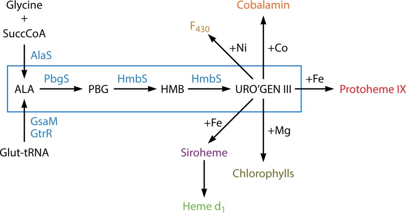
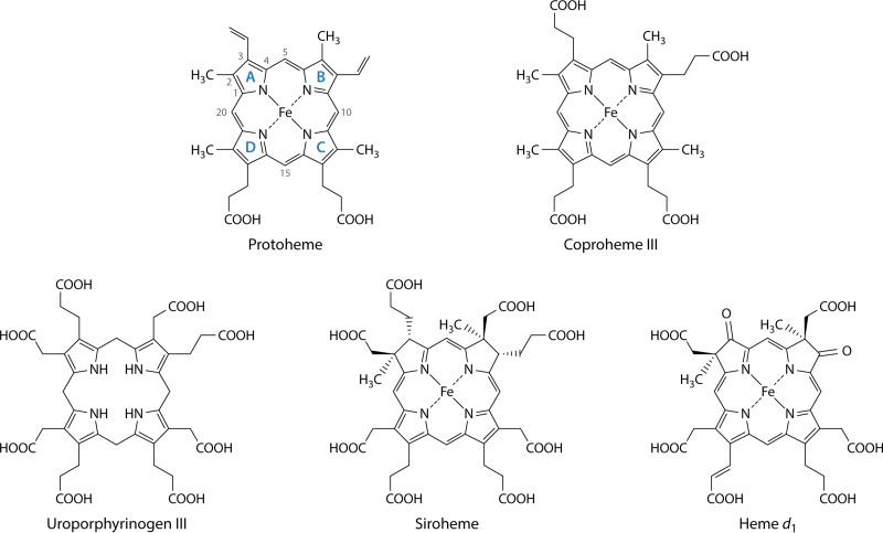
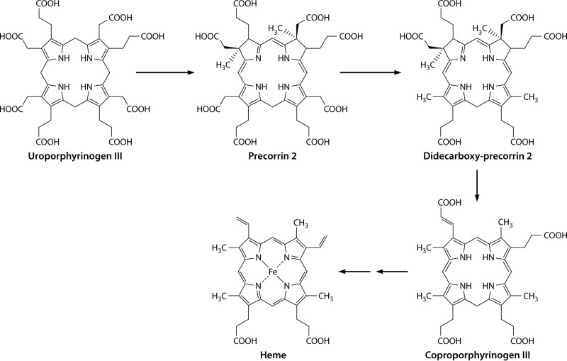
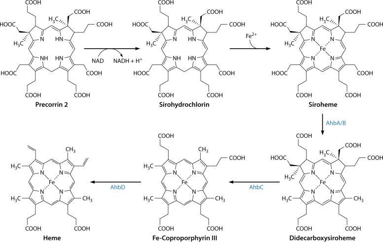
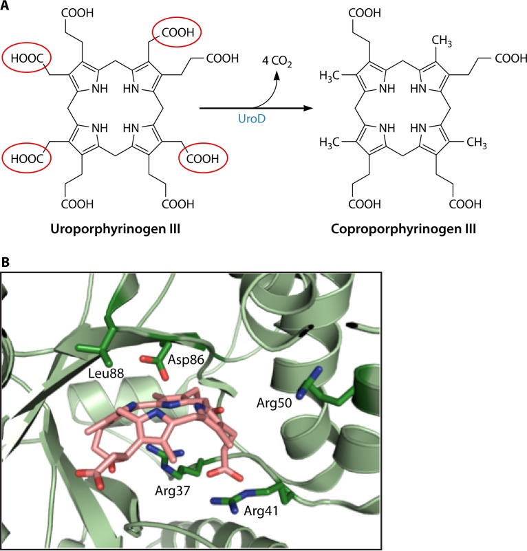
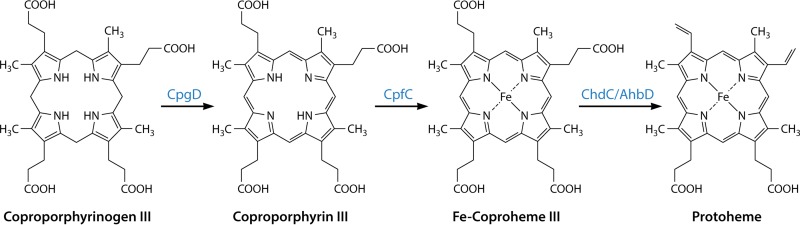
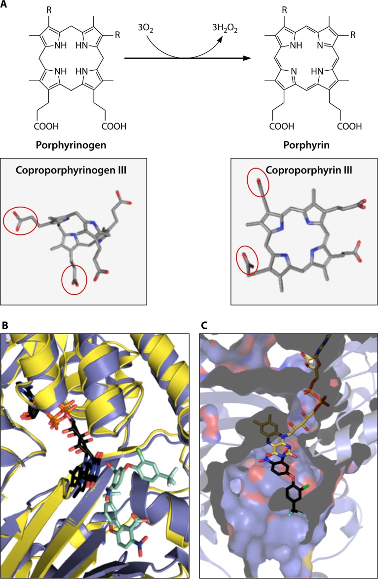
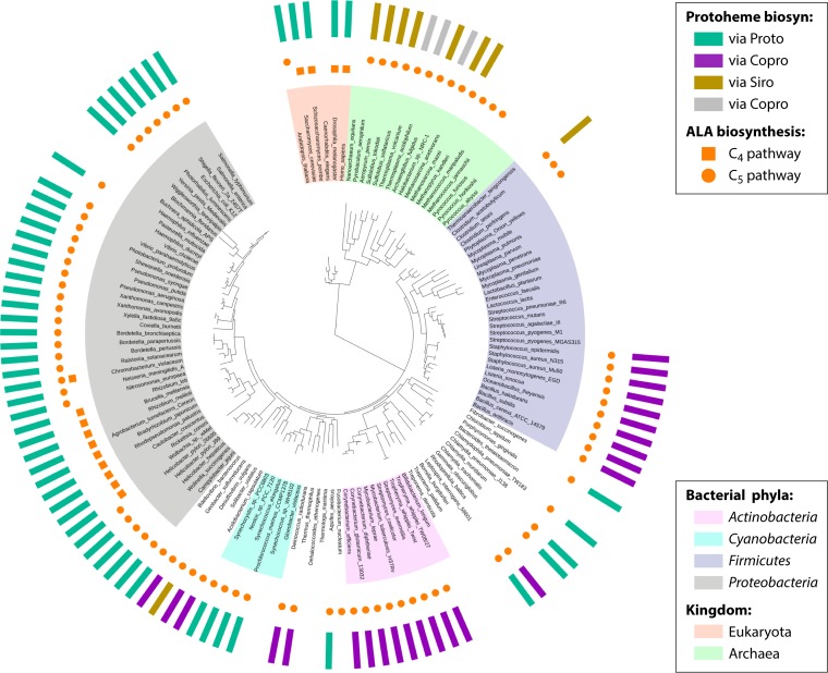
The advent of heme during evolution allowed organisms possessing this compound to safely and efficiently carry out a variety of chemical reactions that otherwise were difficult or impossible. While it was long assumed that a single heme biosynthetic pathway existed in nature, over the past decade, it has become clear that there are three distinct pathways among prokaryotes, although all three pathways utilize a common initial core of three enzymes to produce the intermediate uroporphyrinogen III. The most ancient pathway and the only one found in the Archaea converts siroheme to protoheme via an oxygen-independent four-enzyme-step process. Bacteria utilize the initial core pathway but then add one additional common step to produce coproporphyrinogen III. Following this step, Gram-positive organisms oxidize coproporphyrinogen III to coproporphyrin III, insert iron to make coproheme, and finally decarboxylate coproheme to protoheme, whereas Gram-negative bacteria first decarboxylate coproporphyrinogen III to protoporphyrinogen IX and then oxidize this to protoporphyrin IX prior to metal insertion to make protoheme. In order to adapt to oxygen-deficient conditions, two steps in the bacterial pathways have multiple forms to accommodate oxidative reactions in an anaerobic environment. The regulation of these pathways reflects the diversity of bacterial metabolism. This diversity, along with the late recognition that three pathways exist, has significantly slowed advances in this field such that no single organism's heme synthesis pathway regulation is currently completely characterized.

Keywords: biosynthetic pathways; heme; metabolic regulation; pathway evolution; tetrapyrroles.


Figures

FIG 1
FIG 1
Tetrapyrrole biosynthetic pathways in prokaryotes. An outline of prokaryotic tetrapyrrole biosynthetic pathways is shown with the conserved three-enzymatic-step core path from ALA (5-aminolevulinate) to uroporphyrinogen III boxed in blue. Among prokaryotes, one finds one of two generally nonoverlapping paths to ALA. The synthesis of ALA from glutamyl-tRNA (5-carbon pathway) is most common, with a limited number of bacteria possessing the enzyme AlaS to form ALA from glycine and succinyl-CoA (4-carbon pathway). The 4-carbon pathway to ALA is found only in bacteria that possess the protoporphyrin-dependent pathway branch. Uroporphyrinogen III is the precursor to Ni-containing F430 in methanogens, Co-containing cobalamin, and Fe-containing siroheme and heme d1 as well as coproporphyrinogen, which is the common precursor of Fe-containing protoheme and Mg-containing chlorophylls. Abbreviations: PBG, porphobilinogen; HMB, hydroxymethylbilane; uro'gen III, uroporphyrinogen III.
FIG 2
FIG 2
Structures of protoheme IX, coproheme III, uroporphyrinogen III, siroheme, and heme d1. The structure of protoheme IX is shown with the four pyrrole rings labeled by convention as rings A, B, C, and D. The numbering of side chains is also shown. Of note is that the D ring is inverted to form the IX isomer of protoporphyrin, which corresponds to the III isomer of uroporphyrinogen and coproporphyrinogen. Siroheme is not a true porphyrin but rather is an isobacteriochlorin, since its B and C rings have methyl groups substituted on the A and B rings, which prevents full macrocycle desaturation. Likewise, heme d1 is also not a porphyrin but is a dioxoisobacteriochlorin.
FIG 3
FIG 3
5-Aminolevulinic acid synthase (AlaS). (A) Crystal structure of AlaS from R. capsulatus. (Left) AlaS is a homodimeric protein in which each monomer (shown in color or gray) consists of three domains (red, green, and blue). (Right) In the active site of the enzyme, the PLP cofactor is covalently attached as a Schiff base to a conserved lysine residue. (Adapted from reference .) (B) Proposed reaction mechanism by which AlaS catalyzes the PLP-dependent condensation of glycine and succinyl-CoA to form ALA (see the text for a detailed explanation). (Adapted from reference .)
FIG 4
FIG 4
Glutamyl-tRNA reductase (GtrR). (A, left) Crystal structure of M. kandleri GtrR showing the unusual V-shaped form of the dimeric enzyme. (Right) In the active site of GluTR, C-48 is ideally positioned to attack the α-carboxyl group of the substrate. (Adapted from reference .) (B) Proposed reaction mechanism by which GtrR catalyzes the NADPH-dependent reduction of glutamyl-tRNA to glutamate-1-semialdehyde (see the text for a detailed explanation).
FIG 5
FIG 5
Glutamate-1-semialdehyde-2,1-aminomutase (GsaM). (A, left) Crystal structure of T. elongatus GsaM. (Right) In the active site of Synechococcus GsaM, the inhibitor gabaculine was observed to bind covalently to the PLP cofactor (m-carboxyphenylpyridoxalamine [mCPP]). (Adapted from reference .) (B) Depending on the initial form of the pyridoxal phosphate cofactor, GsaM is able to convert GSA into ALA via either DAVA or DOVA (4,5-dioxovalerate) (see the text for a detailed explanation). Kinetic and other biochemical data suggested that formation via DAVA is used by the enzyme. (Adapted from reference .)
FIG 6
FIG 6
Porphobilinogen synthase (PbgS). (A, left) Crystal structure of P. aeruginosa PbgS showing the octameric assembly of the protein representing a tetramer of homodimers. (Top right) In the active site of P. aeruginosa PbgS, two molecules of the substrate analog 5-fluorolevulinic acid (5F-LA) were observed to bind covalently to the enzyme through Schiff bases, with the catalytically essential lysine residues supporting a double-Schiff-base mechanism. (Bottom right) The antibiotic alaremycin was also observed to bind covalently to the enzyme through a Schiff base with the P-site lysine. (Adapted from reference .) (B) Proposed reaction mechanism by which PbgS catalyzes the asymmetric condensation of two ALA molecules to the pyrrole porphobilinogen (see the text for a detailed explanation). (Adapted from reference .)
FIG 7
FIG 7
Transformation of porphobilinogen into uroporphyrinogen III. Four molecules of porphobilinogen are deaminated and polymerized in an ordered sequential fashion (rings A to D) into a linear tetrapyrrole called hydroxymethylbilane by the action of hydroxymethylbilane synthase. The unstable bilane is acted upon by the enzyme uroporphyrinogen synthase, which inverts ring D and cyclizes the macrocycle to give the type III isomer of uroporphyrinogen. In the absence of the enzyme, hydroxymethylbilane spontaneously cyclizes to give the type I isomer.
FIG 8
FIG 8
Mechanism and structure of hydroxymethylbilane synthase (HmbS). (A) Mechanism of action of HmbS. HmbS contains a dipyrromethane cofactor, which is attached to an active-site cysteine residue and constitutes the holoenzyme form. The two rings of the cofactor are termed C1 and C2. During the polymerization process, the first substrate PBG molecule, ring A of the final product, undergoes deamination to generate an azafulvene species. This is then attached to the C2 ring of the cofactor to generate the ES1 substrate complex. This process is then repeated three more times to generate the ES2, ES3, and ES4 substrate complexes. The ES4 complex then undergoes hydrolysis between the C2 cofactor ring and ring A of the final product to generate the holoenzyme and the hydroxymethylbilane product. The acetate and propionate side chains are designated A and P, respectively. (B) Structure of E. coli HmbS shown in cartoon format, colored according to secondary structure. The 312 amino acid residues of the enzyme are folded into three α/β-domains with a large active-site cavity formed in the space between the three domains. The dipyrromethane cofactor is seen attached to C242 with a key catalytic residue, D84, located just below the two N atoms of the dipyrromethane.
FIG 9
FIG 9
Mechanism and structure of uroporphyrinogen III synthase (UroS). (A) Mechanism of action of UroS. The transformation of hydroxymethylbilane into uroporphyrinogen III is thought to proceed via a spiro intermediate, which is itself formed from an azafulvene intermediate generated from the loss of the hydroxyl group. Rearrangement of the spiro intermediate is then able to produce the type III isomer of uroporphyrinogen. The acetate and propionate side chains are designated A and P, respectively. (B) Structure of UroS. The structure of uroporphyrinogen III synthase from T. thermophilus, together with its product uroporphyrinogen III, is shown in cartoon format. The 252 amino acid residues of the enzyme are folded into two α/β-domains with a large active site formed at their juncture. There are a few highly conserved amino acid residues found in the active site, suggesting that the binding of the substrate in the correct orientation promotes the chemistry outlined in panel A.
FIG 10
FIG 10
Transformation of uroporphyrinogen III (Uro'gen III) into precorrin-2, a key precursor in the biogenesis of heme d1, vitamin B12, coenzyme F430, siroheme, and heme. Precorrin-2 is synthesized from uroporphyrinogen III by the action of the enzyme uroporphyrinogen III methyltransferase, which adds two S-adenosylmethionine-derived methyl groups to C-2 and C-7 of the macrocycle.
FIG 11
FIG 11
Proposed alternative heme biosynthetic pathway. Initially, it was thought that the alternative heme pathway involved the decarboxylation of precorrin-2 to give a didecarboxy compound, which then underwent a loss of the acetic acid side chains on rings A and B to give coproporphyrinogen III. The latter could then be converted into heme via the classic pathway.
FIG 12
FIG 12
The alternative heme biosynthetic pathway via siroheme as an intermediate. Precorrin-2 is converted into siroheme via sirohydrochlorin by utilizing the siroheme biosynthetic enzyme system. Siroheme then undergoes decarboxylation of the acetic acid side chains attached to C-12 and C-18 to generate didecarboxysiroheme. The loss of the acetic acid side chains attached to C-2 and C-7 is mediated by a radical SAM enzyme, AhbC, to give Fe-coproporphyrin (coproheme). The final step in biosynthesis is mediated by another radical SAM enzyme, AhbD, which promotes the loss of the carboxylic acid groups on the propionate side chains attached to C-3 and C-8 to generate heme.
FIG 13
FIG 13
Uroporphyrinogen decarboxylase (UroD). (A) Overall reaction of four sequential decarboxylations. The acetic acid side chains that are removed are highlighted in red circles. Details of the reaction are outlined in the text. (B) Active site of human UROD with a bound product (coproporphyrinogen III). While structures of bacterial UroDs are available, there are none with a bound substrate or product. However, given the homology between the bacterial and human enzymes, one would assume that the reaction mechanism is the same. The positions of the essential Asp residue and four Arg residues are shown. Numbering is according to that of the human enzyme.
FIG 14
FIG 14
The coproporphyrin-dependent pathway to protoheme. The pathway from coproporphyrinogen III to protoheme is shown along with the structures of the intermediates and enzymes responsible for each reaction. Details for each individual step are outlined in the text and in figures below.
FIG 15
FIG 15
(A) Conversion of porphyrinogen to porphyrin. The conversion of coproporphyrinogen to coproporphyrin is catalyzed by CgoX, while the conversion of protoporphyrinogen to protoporphyrin is catalyzed by the homologous protein PgoX. The only difference between the two reactions is that CgoX acts upon coproporphyrinogen, while PgoX acts upon protoporphyrinogen. For coproporphyrinogen, “R” groups on the diagram are propionates, while for protoporphyrinogen, R groups are vinyls. The reaction in vitro utilizes three molecules of molecular oxygen and produces three molecules of hydrogen peroxide. Below the structure drawings are three-dimensional representations of coproporphyrinogen and coproporphyrin. The A- and B-ring propionates of coproporphyrinogen III, which are vinyl groups in protoporphyrinogen IX, are outlined in red. The diagram illustrates the flexibility of the porphyrinogen allowed by the presence of the four saturated methyl mesobridges in the tetrapyrrole compared to the planarity of the aromatic porphyrin macrocycle. (B) Cartoon structure of the active site of B. subtilis CgoX (blue) on M. xanthus PgoX (yellow) (PDB accession numbers 3I6D and 2IVD, respectively) showing the close similarity of the structures of the two proteins. The overlaid FAD molecules are shown in a black stick representation, and the positions of the two inhibitor acifluorfen molecules are shown in turquoise. Previously, others presented the B. subtilis CgoX structure with a molecule of protoporphyrinogen modeled into the active site based upon the positions of the acifluorfen molecules (211). However, in the absence of clearly identified catalytic residues along with a lack of knowledge about potential molecular rearrangements that may occur upon substrate binding, we did not reproduce that model. (C) Cutaway view through the active site of B. subtilis CgoX. The position of the acifluorfen is shown as black sticks, and FAD is in yellow. This view illustrates the spacious active-site pocket that can easily accommodate the tetrapyrrole molecule. Both panels B and C show the close proximity of the flavin and inhibitor ring structures.
FIG 16
FIG 16
Iron insertion by ferrochelatase. Two ferrochelatases are described in the text. One, CpfC, catalyzes the insertion of ferrous iron into coproporphyrin III, and the other, PpfC, inserts iron into protoporphyrin IX. Both ferrochelatases are structurally homologous and probably possess the same catalytic features. (A) The PpfC reaction. This reaction differs from the CpfC reaction in the presence of the vinyl groups on the A and B rings of protoporphyrin, which are propionates for coproporphyrin for the CpfC reaction. (B) Cartoon structures of B. subtilis CpfC, in wheat color on the left (PDB accession number 1C1H), and human ferrochelatase (the best-characterized PpfC enzyme), in yellow on the right (PDB accession number 2QD1). The red arrows point to the position of the bound porphyrin in the active sites. It should be noted that no structures of a CpfC enzyme with the bound substrate porphyrin or a product are available in the PDB, so the structure with the tight-binding, competitive inhibitor N-methylmesoporphyrin is shown. PpfC enzymes possess a larger lip on the active site that closes over the “mouth” when porphyrin is bound. This enclosure of the active site precludes the binding of coproporphyrin with its propionate groups on the A and B rings. (C) Cartoon representation of the active site of human ferrochelatase illustrating the positions of conserved active-site residues in relation to the bound porphyrin substrate. Current models for enzyme function propose that the essential His residue is the acceptor for the two pyrrole nitrogen protons, Lys and Tyr help align the porphyrin macrocycle in the active site, and Met (Tyr in some CpfC enzymes) is the site of iron donation. Details and alternative models are presented in the text. (D) Ribbon cartoon representation demonstrating the three structural conformations of human ferrochelatase. The substrate-free resting state is wheat colored. Upon binding of the porphyrin substrate, the upper lip (shown in green) closes the active-site pocket. (E) Following metalation, the lower lip/π-helix extends (shown in red) to facilitate product release. Upon metalation, the 14-propionate (C ring) of heme flips conformation. This is accompanied by a reorientation of the essential His residue, and this movement is proposed to cause pocket opening and π-helix extension.
FIG 17
FIG 17
Decarboxylation of coproheme III to form protoheme IX. (A) Reaction catalyzed by the terminal enzyme in the CPD pathway, ChdC. The two propionates that are converted to vinyl groups are circled in red. The reaction goes through a monovinyl monopropionate deuteroheme intermediate. (B) Structure of ChdC from Geobacillus stearothermophilus (PDB accession number 1T0T) with the putative active-site pocket shown in red. On the left is a space-filling model, and on the right is the same protein rotated 90° and shown in a ribbon format to illustrate the hole through the middle of the pentamer doughnut. (C) Structure of L. monocytogenes CpdC with bound coproheme (PDB accession number 5LOQ). The residues shown are the conserved His174 residue (which is ligated to the iron of the coproheme) as well as Arg133 and Lys151. The latter two residues are hydrogen bonded to the propionates of rings C and D (230). This pocket tightly binds heme, so there must be some structural difference for ChdC to allow the substrate to bind and the product to be released.
FIG 18
FIG 18
Protoporphyrin-dependent pathway to protoheme IX synthesis. An overview of the three-step reaction and intermediates from coproporphyrinogen to protoheme, along with the enzymes responsible for the reaction, is shown. Coproporphyrinogen III is oxidatively decarboxylated at two propionate groups to yield protoporphyrinogen IX. This intermediate is oxidized to protoporphyrin, which is converted into heme upon iron insertion.
FIG 19
FIG 19
Coproporphyrinogen III oxidase (CgdC). (A, left) Crystal structure of yeast CgdC (245). (Right) In the structure of human CgdC, a citrate molecule was observed to bind, indicating the localization of the active site. (Adapted from reference .) (B) Proposed reaction mechanism by which CgdC catalyzes the two oxidative decarboxylation reactions of coproporphyrinogen III to protoporphyrinogen IX with oxygen as the terminal electron acceptor (see the text for a detailed explanation). (Adapted from reference .)
FIG 20
FIG 20
Coproporphyrinogen III dehydrogenase (CgdH). (A, left) Crystal structure of E. coli CgdC. (Right) In the active site of the enzyme, the [4Fe-4S] cluster is coordinated by the three cysteine residues of the conserved CX3CX2C sequence motif and by one of the two bound SAM molecules. (Adapted from reference .) (B) Proposed reaction mechanism by which the radical SAM enzyme CgdH catalyzes two oxidative decarboxylation reactions of the coproporphyrinogen III propionate side chains on rings A and B to the corresponding vinyl groups of protoporphyrinogen without oxygen as the terminal electron acceptor (see the text for a detailed explanation).
FIG 21
FIG 21
Phylogenic distribution of the three currently characterized pathways for protoheme synthesis mapped onto the Tree of Life. The outside ring shows (as vertical rectangles) the presence or absence of the siroheme-dependent branch (via Siro), the coproporphyrin-dependent branch (via Copro), or the classic protoporphyrin-dependent branch (via Proto). Gray rectangles mark the organisms that contain unusual combinations of genes normally involved in different pathways for protoheme synthesis (hybrid paths). The distribution of the two routes used to synthesize 5-aminolevulinic acid (ALA) are also presented: the Shemin or C4 pathway and the C5 pathway. The absence of a circle or square shows the inability of an organism to produce tetrapyrroles of any kind. Likewise, the absence of a rectangle in the outside ring indicates the absence of any known route for protoheme synthesis in an organism. This illustration covers only 133 representative organisms (17 archaea, 110 eubacteria, and 6 eukaryotes) included in the Tree of Life (391, 392). A full analysis of the 978 representative microorganisms performed in this work is available in the SEED subsystem “Heme Biosynthesis: Protoporphyrin-, Coproporphyrin-, and Siroheme-Dependent Pathways” (see http://pubseed.theseed.org//SubsysEditor.cgi?page=ShowFunctionalRoles&subsystem=Heme_Biosynthesis%3A_protoporphyrin-%2C_coproporphyrin-_and_siroheme-dependent_pathways). Mapped onto this tree, the siroheme-dependent pathway occurs largely in the Archaea, in the Thermodesulfobacteria, and, rarely, in several other taxa (see Table S3 in the supplemental material). The CPD route (teal) is found primarily in slow-evolving monoderm Firmicutes and Actinobacteria and in evolutionarily early-branching (Acidobacteria, Planctomyces, and Aquificae) and transitional (Deinococcus-Thermus group) diderm phyla, while the PPD pathway has a wider distribution, occurring in Proteobacteria, cyanobacteria, the Bacteroidetes-Chlorobi and Chlamydiae-Verrucomicrobia groups, Aquificae, Gemmatimonadetes, Caldithrix, and several other taxa. This is the main route for protoheme production in the evolutionarily younger Proteobacteria, with an illuminating exception of the Deltaproteobacteria. Deltaproteobacteria and epsilonproteobacteria are believed to be the oldest phyla within the Proteobacteria, with the other main proteobacterial groups being derived from them linearly in a directional rather than in a tree-like manner in the order delta/epsilonproteobacteria → alphaproteobacteria → betaproteobacteria → gammaproteobacteria (393). Notably, the phylum Deltaproteobacteria is the only proteobacterial phylum in which all 3 routes leading to protoheme are represented in different species (Table S3). A complete reconstruction of the heme biosynthetic pathways in the 38 representative deltaproteobacterial genomes is available online in the SEED subsystem (limit view to “deltaproteobacteria”). As a few examples, genomes of Stigmatella aurantiaca and Myxococcus xanthus encode the PPD pathway, and genomes of Geobacter metallireducens and Desulfuromonas acetoxidans harbor the CPD pathway, while genomes of Desulfovibrio vulgaris, Desulfatibacillum alkenivorans, and Desulfobacula toluolica harbor the siroheme-dependent route.
FIG 22
FIG 22
Irr senses iron indirectly through the status of heme biosynthesis. Under high-iron conditions, Irr forms a complex with protoporphyrin ferrochelatase (FC), which inactivates Irr, followed by heme-dependent degradation. Heme is represented in red, and protoporphyrin is shown in blue. Under low-iron conditions, ferrochelatase is bound to protoporphyrin but not iron, and ferrochelatase cannot form a complex with Irr. In Rhizobium leguminosarum, Irr is not degraded in response to iron, but heme inhibits DNA binding.
FIG 23
FIG 23
Differential control of Irr-repressed genes by the variable affinity of Irr for target promoters. Low-affinity promoters such as those found in the housekeeping gene leuC or pbgS allow some expression (derepression) even under low-iron conditions, where Irr levels are highest. High-affinity promoters such as those found for mbfA and bfr repress expression strongly at low and intermediate levels of iron and are derepressed only at high iron levels.

Similar articles

Cited by

References

    1. Johnson MP. 2016. Photosynthesis. Essays Biochem 60:255–273. doi:10.1042/EBC20160016. - DOI - PMC - PubMed
    1. Ragsdale SW. 2014. Biochemistry of methyl-coenzyme M reductase: the nickel metalloenzyme that catalyzes the final step in synthesis and the first step in anaerobic oxidation of the greenhouse gas methane. Met Ions Life Sci 14:125–145. doi:10.1007/978-94-017-9269-1_6. - DOI - PubMed
    1. Rinaldo S, Sam KA, Castiglione N, Stelitano V, Arcovito A, Brunori M, Allen JW, Ferguson SJ, Cutruzzola F. 2011. Observation of fast release of NO from ferrous d(1) haem allows formulation of a unified reaction mechanism for cytochrome cd(1) nitrite reductases. Biochem J 435:217–225. doi:10.1042/BJ20101615. - DOI - PubMed
    1. Banerjee R, Ragsdale SW. 2003. The many faces of vitamin B12: catalysis by cobalamin-dependent enzymes. Annu Rev Biochem 72:209–247. doi:10.1146/annurev.biochem.72.121801.161828. - DOI - PubMed
    1. Jost M, Fernandez-Zapata J, Polanco MC, Ortiz-Guerrero JM, Chen PY, Kang G, Padmanabhan S, Elias-Arnanz M, Drennan CL. 2015. Structural basis for gene regulation by a B12-dependent photoreceptor. Nature 526:536–541. doi:10.1038/nature14950. - DOI - PMC - PubMed

Publication types

MeSH terms