. 2017 Jan 25;9(374):eaai9338.
doi: 10.1126/scitranslmed.aai9338.

RON kinase: A target for treatment of cancer-induced bone destruction and osteoporosis

Affiliations

RON kinase: A target for treatment of cancer-induced bone destruction and osteoporosis

Kelsi Andrade et al. Sci Transl Med. .

Abstract

Bone destruction occurs in aging and numerous diseases, including osteoporosis and cancer. Many cancer patients have bone osteolysis that is refractory to state-of-the-art treatments, which block osteoclast activity with bisphosphonates or by inhibiting the receptor activator of nuclear factor κB ligand (RANKL) pathway. We previously showed that macrophage-stimulating protein (MSP) signaling, which is elevated in about 40% of breast cancers, promotes osteolytic bone metastasis by activation of the MSP signaling pathway in tumor cells or in the bone microenvironment. We show that MSP signals through its receptor, RON tyrosine kinase, expressed on host cells, to activate osteoclasts directly by a previously undescribed pathway that is complementary to RANKL signaling and converges on proto-oncogene, non-receptor tyrosine kinase SRC (SRC). Genetic or pharmacologic inhibition of RON kinase blocked cancer-mediated bone destruction and osteoporosis in several mouse models. Furthermore, the RON kinase inhibitor BMS-777607/ASLAN002 altered markers of bone turnover in a first-in-human clinical cancer study, indicating the inhibitor's potential for normalizing bone loss in patients. These findings uncover a new therapeutic target for pathogenic bone loss and provide a rationale for treatment of bone destruction in various diseases with RON inhibitors.


Conflict of interest statement

Competing interests: M.M. is an employee of Aslan Pharmaceuticals. Other authors declare no competing interests.

Figures

Fig. 1
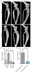
Fig. 1. MSP induction of bone osteolysis requires host RON kinase activity
(A) Representative microcomputed tomography (μCT) images of tibial bone lesions, 21 days after PyMT tumor cell injection. Scale bar, 2 mm. (B) Quantification of osteolytic area in tibias 42 days after PyMT-MSP tumor cell injection using high resolution ex vivo X-ray analysis (n=10 per group); **p=0.0013, ***p=0.0001, ns=not significant, two-tailed, unpaired Welch’s t-tests. (C) Quantification of osteolytic area in tibias 35 days after DU4475 tumor cell injection using high resolution ex vivo X-ray analysis (n=5 per group); *p=0.021, two-tailed, unpaired Welch’s t-test.
Fig. 2
Fig. 2. RON inhibition reduces osteolysis in prophylactic and therapeutic settings
(A) Representative X-rays of PyMT-MSP bone lesions from mice treated with OSI-296. Treatment began 3 days after tumor cell injection (pre-osteolysis) or 3 weeks after injection (post-osteolysis). Mice were sacrificed 42 days after injection for analysis. (B) Representative X-rays of PyMT-MSP bone lesions from mice treated with BMS-777607/ASLAN002. Treatment began 3 days after tumor cell injection. Mice were sacrificed 21 days after injection for analysis. (C) Quantification of osteolytic area in PyMT-MSP bone lesions from mice treated with OSI-296 pre- or post-osteolysis (n=5–9 per group); *p=0.024, **p=0.0091, two-tailed, unpaired Welch’s t-tests. (D) Quantification of osteolytic area in PyMT-MSP bone lesions from mice treated with BMS-777607/ASLAN002 (n=4–5 per group); *p = 0.011, two-tailed, unpaired Welch’s t-test (E) Representative X-rays of DU4475-induced bone lesions from mice treated with BMS-777607/ASLAN002. Treatment began 3 days after tumor cell injection. Mice were sacrificed 28 days after injection for analysis. (F) Quantification of osteolytic area in DU4475 bone lesions from mice treated with BMS-777607/ASLAN002 (n=5 per group); **p=0.0094, two-tailed, unpaired Welch’s t-test.
Fig. 3
Fig. 3. MSP tumor-induced osteolysis does not depend on RANKL or TGFβ signaling
(A) Serum RANKL concentration in mice (n=3 per group); ns=not significant, two-tailed, unpaired Welch’s t-test. (B) Quantification of osteolytic area in bone lesions from mice (n=11–16 per group for WT mice and n=3 (− muRANK-Fc) or 5 (+ mu-RANK-Fc) per group for RON TK−/− mice); ****p<0.0001, two-tailed, unpaired Welch’s t-test. (C) Quantification of osteolytic area in mice with MDA-MB-231 tumors (n=4–7 per group). Mice were treated with either mu-RANK-Fc or OSI-296 beginning 3 days after tumor cell injection and throughout the experiment; **p=0.0094, ***p=0.0004, ****p<0.0001, two-tailed, unpaired Welch’s t-tests. (D) Quantification of osteolytic area in mice injected with WT PyMT tumors +/− MSP expression or TGFβRII−/− PyMT tumors +/− MSP expression (n=5–6 per group); #p=0.027, *p=0.032, **p=0.0050, two-tailed, unpaired Welch’s t-tests.
Fig. 4
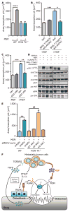
Fig. 4. MSP promotes osteoclast activity and survival through activation of SRC
(A) In vitro resorption area of WT or RON TK−/− osteoclasts +/− MSP (n=4 per group); ****p<0.0001, two-tailed, unpaired Welch’s t-test. (B–C) Quantification of resorption area +/− MSP and/or the RON inhibitors BMS-777607/ASLAN002 (B) or OSI-296 (C); n=3 per group; *p=0.016, **p=0.0020, ***p=0.0001, two-tailed, unpaired Welch’s t-tests. (D) Representative Western blots of osteoclast lysates from WT cells treated with MSP, muRANK-Fc, and/or BMS-777607/ASLAN002. (E) Quantification of resorption area from RON TK−/− bone marrow precursor cells transduced with empty vector, WT, constitutively active (CA), or kinase dead (KD) SRC, +/− MSP. WT osteoclasts served as a control for normal osteoclast activity (n = 3 per group). **p=0.0013, ##p=0.0060, two-tailed, unpaired Welch’s t-tests. (F) Model of signaling pathways in the bone microenvironment that control MSP-mediated osteolysis. We propose that MSP/RON signaling functions in parallel to the vicious cycle mediated by RANKL and TGFβ.
Fig. 5
Fig. 5. Loss of RON activity protects from osteoporotic bone loss
(A) Representative μCT images of the proximal tibia (axial view of the metaphyseal region) from WT and RON TK−/− mice 28 days after ovariectomy (OVX) or sham operation. Treatment with BMS-777607/ASLAN002 began 1 day after ovariectomy. Mice were euthanized 28 days after ovariectomy for analysis. (B) Quantification of bone mineral density (BMD) in the metaphyseal region of the tibia determined by dual-energy X-ray absorptiometry (DXA) (n=5 per group); *p=0.027, **p=0.0032, ***p=0.0007, two-tailed, unpaired Welch’s t-tests. (C) Quantification of trabecular bone volume (BV) in the tibia expressed as percent total volume (TV), determined by bone histomorphometry analysis (n=5 per group); *p=0.014, two-tailed, unpaired Welch’s t-test. (D) Quantification of trabecular separation (Tb. Sp.) in the tibia (n=5 per group), two-tailed, unpaired Welch’s t-tests. (E) Representative sections of the tibia from each group stained with hematoxylin and eosin; scale bar, 50 μm.
Fig. 6
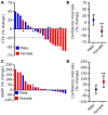
Fig. 6. The RON inhibitor BMS-777607/ASLAN002 reduces bone turnover in humans
(A) Percent change in plasma CTX levels (compared to baseline prior to drug treatment) in blood plasma of 21 patients that received BMS-777607/ASLAN002 for at least 15 days in a Phase 1 clinical trial. Most patients received BMS-777607/ASLAN002 for 28 days; values reported correspond to the difference between baseline (day 0) and the 28-day time point. Exceptions in duration of treatment and CTX testing are noted (*15 days). Blue bars=males, red bars=females. Arrowhead denotes a patient less than 50 years of age (42 years). (B) Ninety-five percent confidence intervals for percent change in CTX for males and females; **p=0.0035, one-sample t-test versus a hypothetical mean of 0 (no change following treatment compared to baseline). (C) Percent change in bone-specific alkaline phosphatase (BSAP) levels (compared to baseline) in blood plasma of 21 patients that received BMS-777607/ASLAN002 for 28 days. 18 of the patients are also shown in (A). (D) Ninety-five percent confidence intervals for percent change in BSAP for males and females; **p=0.0090, one-sample t-test versus a hypothetical mean of 0 (no change following treatment compared to baseline). Blue bars=males, red bars=females.

References

    1. National Osteoporosis Foundation report. 2002
    1. Coleman RE, Lipton A, Roodman GD, Guise TA, Boyce BF, Brufsky AM, Clezardin P, Croucher PI, Gralow JR, Hadji P, Holen I, Mundy GR, Smith MR, Suva LJ. Metastasis and bone loss: advancing treatment and prevention. Cancer Treat Rev. 2010;36:615–620. - PMC - PubMed
    1. Coleman RE. Future directions in the treatment and prevention of bone metastases. Am J Clin Oncol. 2002;25:S32–38. - PubMed
    1. Diel IJ, Solomayer EF, Bastert G. Treatment of metastatic bone disease in breast cancer: bisphosphonates. Clin Breast Cancer. 2000;1:43–51. - PubMed
    1. Melton LJ, 3rd, Kyle RA, Achenbach SJ, Oberg AL, Rajkumar SV. Fracture risk with multiple myeloma: a population-based study. J Bone Miner Res. 2005;20:487–493. - PubMed

MeSH terms

Substances