. 2017 Jan;6(1):71-74.
doi: 10.3892/mco.2016.1075. Epub 2016 Nov 8.

A giant and insidious subphrenic biloma formation due to gallbladder perforation mimicking biliary cystic tumor: A case report

Affiliations

A giant and insidious subphrenic biloma formation due to gallbladder perforation mimicking biliary cystic tumor: A case report

Guwei Ji et al. Mol Clin Oncol. 2017 Jan.

Abstract

Gallbladder perforation (GBP) represents a rare, but potentially life-threatening, complication of acute cholecystitis. GBP is subdivided into three categories whereas the development of biloma is extremely rare. The present case study reports on a 40-year-old man with a 10-year history of calculus cholecystitis, who was referred to The First Affiliated Hospital of Nanjing Medical University (Nanjing, China) for the surgical treatment of an emerging massive hepatic entity with insidious symptoms and normal laboratory tests. A preoperative imaging study demonstrated the collection with internal septations and mural nodules, but no visible communication with the biliary system. Given the suspected biliary cystic tumor, a laparotomy was performed and the lumen was scattered with papillae. An intraoperative frozen section examination illustrated a simple hepatic cyst. Biochemical analysis of the collection and histopathology of the gallbladder and capsule substantiated the diagnosis of biloma formation due to GBP. The purpose of the present case report was to demonstrate how a pinhole-sized perforation with extravasation of unconcentrated bile from the gallbladder may result in insidious clinical presentation and an undetected leak site. According to the clinicopathological characteristics and composition, formation of biloma should be classified as type IV GBP. To differentiate bilomas with intracystic septations and mural nodules from BCTs is difficult via a preoperative examination, and the definitive diagnosis should be based on a histological examination. Laparotomy with frozen section examination may be the optimal approach in such a case.

Keywords: biliary cystic tumor; biloma; calculus cholecystitis; gallbladder perforation; insidious symptoms.


Figures

Figure 1.
Figure 1.
Axial abdominal computed tomography scan, revealing a massive cystic lesion without identifiable capsule, distorting the liver configuration.
Figure 2.
Figure 2.
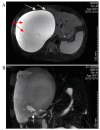
Magnetic resonance imaging and magnetic resonance choangiopancreatography scans of the cystic lesion. (A) The collection was revealed to be hyper-intensive on an axial T2-weighted image, with some iso-intensity septations (illustrated by the red arrows) and small mural nodules (illustrated by the white arrows). (B) A magnetic resonance cholangiopancreatography image illustrating a giant subphrenic collection and gallbladder stones (shown by the white arrow), although there was no visible communication between the lesion and gallbladder.
Figure 3.
Figure 3.
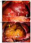
Intraoperative images (A) demonstrating the greater omentum adhered to the undersurface of the collection densely, and (B) scattered papilla inside the capsule (shown by the white arrow) and subphrenic portion of the lesion (shown by the yellow arrow).
Figure 4.
Figure 4.
Routine pathology demonstrated that the capsule comprised fibrous tissue without epithelial lining (hematoxylin and eosin stain; magnification, ×100).

Similar articles

Cited by

References

    1. Kannan U, Parshad R, Regmi SK. An unusual presentation of biloma five years following cholecystectomy: A case report. Cases J. 2009;2:8048. - PMC - PubMed
    1. Göbel T, Kubitz R, Blondin D, Häussinger D. Intrahepatic type II gall bladder perforation by a gall stone in a CAPD patient. Eur J Med Res. 2011;16:213–216. doi: 10.1186/2047-783X-16-5-213. - DOI - PMC - PubMed
    1. Derici H, Kara C, Bozdag AD, Nazli O, Tansug T, Akca E. Diagnosis and treatment of gallbladder perforation. World J Gastroenterol. 2006;12:7832–7836. doi: 10.3748/wjg.v12.i48.7832. - DOI - PMC - PubMed
    1. Ausania F, Suarez S Guzman, Garcia H Alvarez, Senra del, Rio P, Nuñez E Casal. Gallbladder perforation: Morbidity, mortality and preoperative risk prediction. Surg Endosc. 2015;29:955–960. doi: 10.1007/s00464-014-3765-6. - DOI - PubMed
    1. Seyal AR, Parekh K, Gonzalez-Guindalini FD, Nikolaidis P, Miller FH, Yaghmai V. Cross-sectional imaging of perforated gallbladder. Abdom Imaging. 2014;39:853–874. doi: 10.1007/s00261-014-0121-1. - DOI - PubMed