

Understanding early dementia: EEG, MRI, SPECT and memory evaluation
- PMID: 28123789
- PMCID: PMC4936613
- DOI: 10.1515/tnsci-2015-0005
Understanding early dementia: EEG, MRI, SPECT and memory evaluation
Abstract
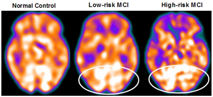
Background: An increase in the EEG upper/low α power ratio has been associated with mild cognitive impairment (MCI) due to Alzheimer's disease (AD) and to the atrophy of temporoparietal brain areas. Subjects with a higher α3/α2 frequency power ratio showed lower brain perfusion than in the low α3/α2 group. The two groups show significantly different hippocampal volumes and correlation with θ frequency activity.
Methods: Seventy-four adult subjects with MCI underwent clinical and neuropsychological evaluation, electroencephalogram (EEG) recording, and high resolution 3D magnetic resonance imaging (MRI). Twenty-seven of them underwent EEG recording and perfusion single-photon emission computed tomography (SPECT) evaluation. The α3/α2 power ratio and cortical thickness were computed for each subject. The difference in cortical thickness between the groups was estimated.
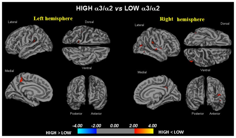
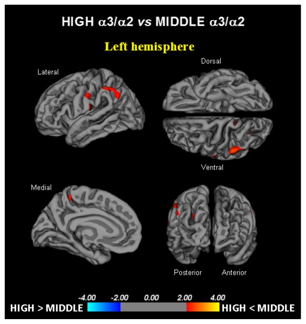
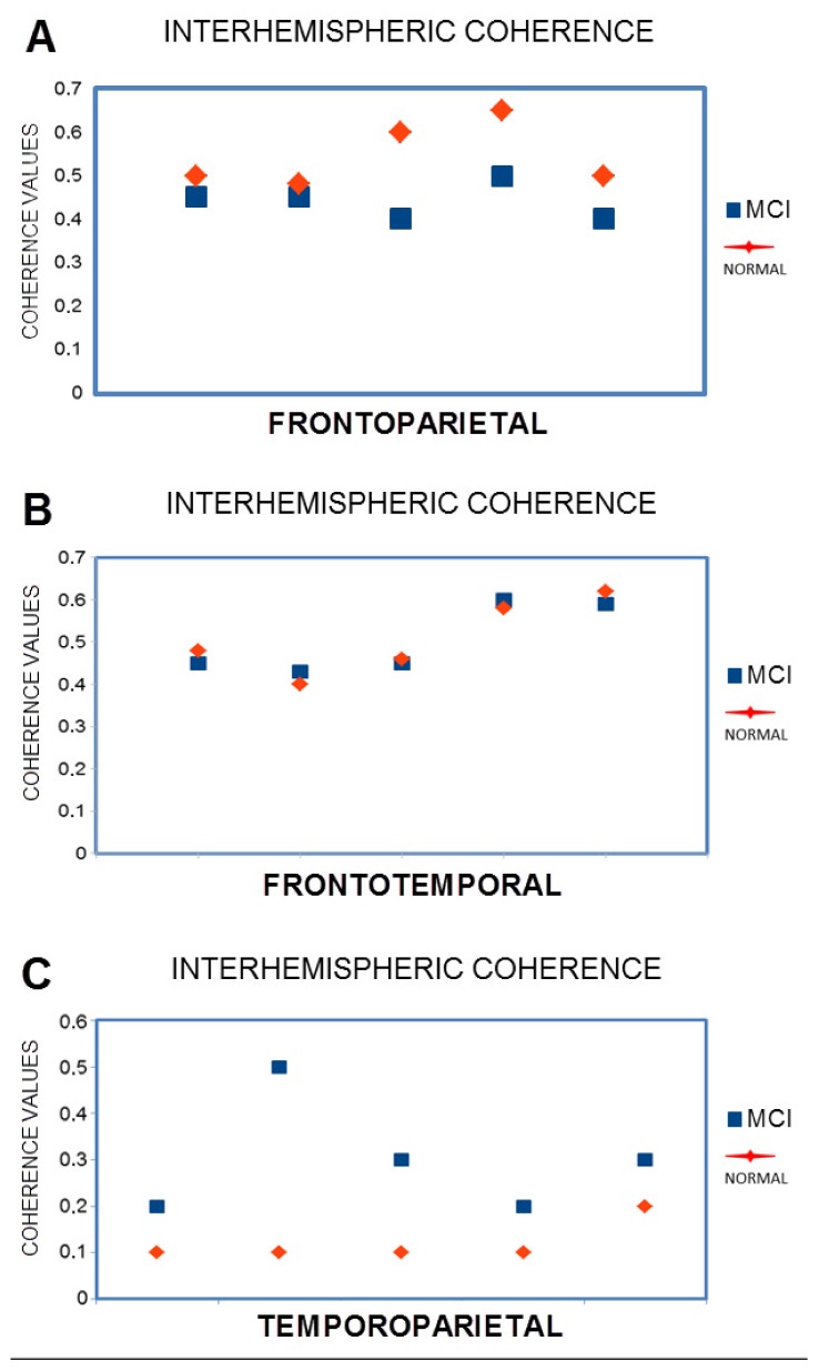
Results: In the higher upper/low α group, memory impairment was more pronounced in both the MRI group and the SPECT MCI groups. An increase in the production of θ oscillations was associated with greater interhemisperic coupling between temporal areas. It also correlated with greater cortical atrophy and lower perfusional rate in the temporoparietal cortex.
Conclusion: High EEG upper/low α power ratio was associated with cortical thinning and lower perfusion in temporoparietal areas. Moreover, both atrophy and lower perfusion rate significantly correlated with memory impairment in MCI subjects. Therefore, the increase in the EEG upper/low α frequency power ratio could be useful in identifying individuals at risk for progression to AD dementia in a clinical context.
Keywords: EEG; MRI; Memory; Prodromal Alzheimer’s disease; SPECT.
Figures
References
-
- Dubois B, Feldman HH, Jacova C, DeKosky ST, Barberger-Gateau P, Cummings J, et al. Research criteria for the diagnosis of Alzheimer’s disease: revising the NINCDS-ADRDA criteria. Lancet Neurol. 2007;6:734–746. - PubMed
-
- Albert MS, DeKosky ST, Dickson D, Dubois B, Feldman HH, Fox NC, et al. The diagnosis of mild cognitive impairment due to Alzheimer’s disease: recommendations from the National Institute on Aging - Alzheimer’s Association workgroups on diagnostic guidelines for Alzheimer’s disease. Alzheimers Dement. 2011;7:270–279. - PMC - PubMed
-
- Hampel H, Bürger K, Teipel SJ, Bokde AL, Zetterberg H, Blennow K. Core candidate neurochemical and imaging biomarkers of Alzheimer’s disease. Alzheimers Dement. 2008;4:38–48. - PubMed
-
- Galluzzi S, Geroldi C, Amicucci G, Bocchio-Chiavetto L, Bonetti M, Bonvicini C, et al. Supporting evidence for using biomarkers in the diagnosis of MCI due to AD. J Neurol. 2013;260:640–650. - PubMed
-
- Frisoni GB, Sabattoli F, Lee AD, Dutton RA, Toga AW, Thompson PM. In vivo neuropathology of the hippocampal formation in AD: a radial mapping MR-based study. Neuroimage. 2006;32:104–110. - PubMed
LinkOut - more resources
Full Text Sources
Other Literature Sources