. 2016 Oct 28;5(12):e1250991.
doi: 10.1080/2162402X.2016.1250991. eCollection 2016.

CD40L confers helper functions to human intra-melanoma class-I-restricted CD4+CD8+ double positive T cells

Affiliations

CD40L confers helper functions to human intra-melanoma class-I-restricted CD4+CD8+ double positive T cells

Tiphaine Parrot et al. Oncoimmunology. .

Abstract

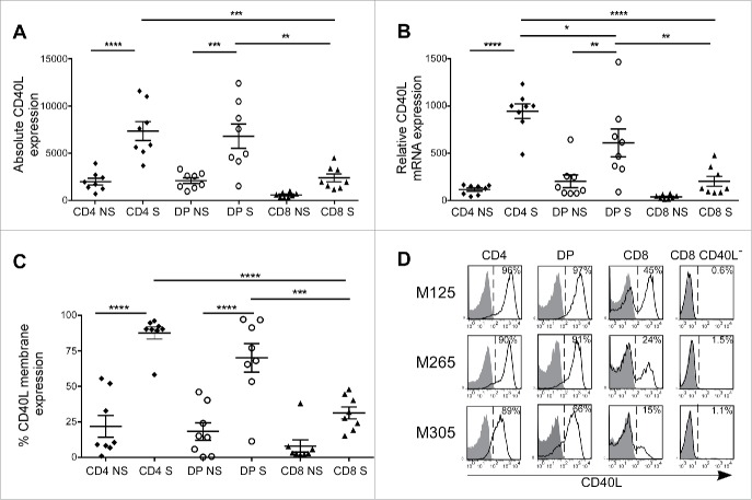
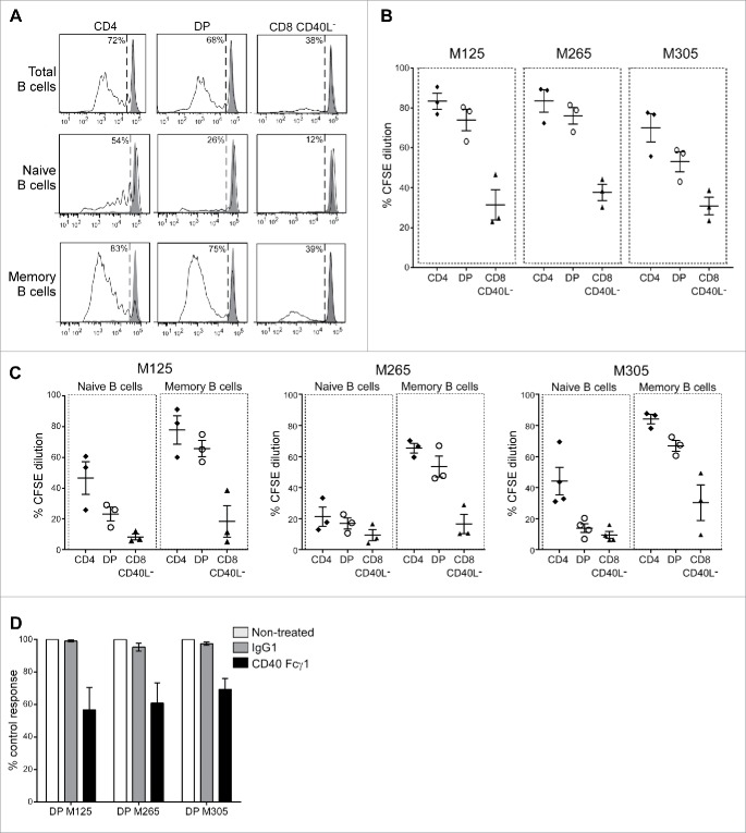
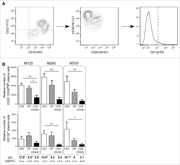
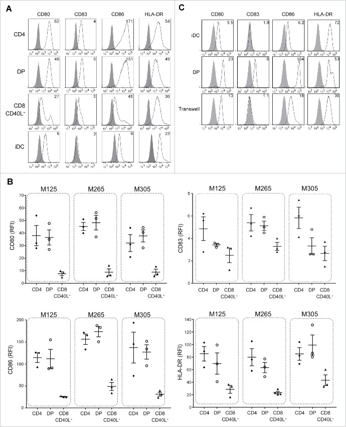
Although CD4+CD8+ double positive (DP) T cells represent a small fraction of peripheral T lymphocytes in healthy human donors, their frequency is often increased under pathological conditions (in blood and targeted tissues). In solid cancers such as melanoma, we previously demonstrated an enrichment of tumor reactive CD4lowCD8highαβ DP T cells among tumor-infiltrating lymphocytes of unknown function. Similarly to their single positive (SP) CD8+ counterparts, intra-melanoma DP T cells recognized melanoma cell lines in an HLA-class-I restricted context. However, they presented a poor cytotoxic activity but a strong production of diverse Th1 and Th2 cytokines. The aim of this study was to clearly define the role of intra-melanoma CD4lowCD8highαβ DP T cells in the antitumor immune response. Based on a comparative transcriptome analysis between intra-melanoma SP CD4+, SP CD8+ and DP autologous melanoma-infiltrating T-cell compartments, we evidenced an overexpression of the CD40L co-stimulatory molecule on activated DP T cells. We showed that, like SP CD4+ T cells, and through CD40L involvement, DP T cells are able to induce both proliferation and differentiation of B lymphocytes and maturation of functional DCs able to efficiently prime cytotoxic melanoma-specific CD8 T-cell responses. Taken together, these results highlight the helper potential of atypical DP T cells and their role in potentiating antitumor response.

Keywords: CD4+CD8+ double positive T cells; CD40L; TIL; helper function; melanoma.


Figures

Figure 1.
Figure 1.
CD40L overexpression is induced on intra-melanoma DP T cells upon activation. CD40L expression of intra-melanoma SP CD4+ (black diamonds), DP (white circles) and SP CD8+ (black triangles) T-cell lines isolated from TILs, stimulated (S) or not (NS) with anti-CD3 mAb for 6 h was determined by (A) microarray analysis, (B) quantitative RT-PCR analysis and (C) flow cytometry (n = 8 melanoma patients). Results are expressed as mean ± SEM. Statistical analysis was performed using the one-way ANOVA analysis, followed by a Tukey multiple comparison test. *p < 0.05, **p < 0.01, ***p < 0.001, ****p < 0.0001. (D) Representative flow cytometry staining of CD40L following anti-CD3 activation on the 3 autologous intra-melanoma SP CD4+, DP, SP CD8+ and SP CD8+ CD40L TIL sub-populations (M125, M265 and M305) selected for further experiments. Cells were co-stained with CD4+, CD8α mAbs and with either the isotype control (filled histogram) or CD40L mAb (open histogram). Numbers indicated represent the percentages of CD40L+ cells.
Figure 2.
Figure 2.
Intra-melanoma DP T cells mediate memory B-cell proliferation through CD40L engagement. Intra-melanoma SP CD4+, DP and SP CD8+ CD40L T-cell lines were co-cultured with CFSE-labeled B cells for 4 d at a 1:1 ratio on anti-CD3-coated microwells, and B-cell proliferation was evaluated by flow cytometry gated on CD19+ cells. (A) Representative CFSE dilution profiles of total (CD19+), naive (CD19+CD27) and memory (CD19+CD27+) B-cell fractions after co-cultured with CD3-activated SP CD4+, DP and SP CD8+ CD40L T cells. B cell proliferation was expressed as the percentage of CFSE dilution. Results of total (B), naive or memory (C) B-cell proliferation stimulated with each T-cell subpopulations derived from the three patients M125, M265 and M305 are gathered on a single graph (n = 3 independent experiments with three different B-cell donors). (D) Memory B-cell proliferation stimulated with DP T cells derived from M125, M265 and M305 patients in the presence of either recombinant human CD40 Fcγ1 (black bars), control IgG1 Fc (gray bars) or without treatment (white bars). Results are normalized to the non-treated control condition (n = 3 independent experiments with three different B-cell donors).
Figure 3.
Figure 3.
Intra-melanoma DP T cells mediate B-cell differentiation. The differentiation status of B cells was determined by flow cytometry after 7 d of co-culture of sorted memory B cells at a 1:1 ratio with anti-CD3 activated DP, SP CD4+ or SP CD8+ CD40L. (A) Gating strategy performed on viable cells to discriminate plasma cells (CD19+CD27+CD38highCD20lowCD138) from highly differentiated plasma cells (CD19+CD27+CD38highCD20lowCD138+). (B) The relative number of plasma cells and highly differentiated plasma cells was determined using counting beads at the end of the 7 d of memory B cells co-cultured with SP CD4+ (white bars), DP (gray bars) or SP CD8+ CD40L (black bars) T-cell subpopulations derived from M125, M265 and M305 melanoma patients (n = 7 independent experiments with seven B-cell donors). Supernatants from the same co-cultures were analyzed at day 7 by ELISA for secretion of IgG. Results are expressed as mean ± SEM. Statistical analysis was performed using the non-parametric Friedman test, followed by Dunn's multiple comparison test. *p < 0.05, **p < 0.01, ***p < 0.001.
Figure 4.
Figure 4.
Intra-melanoma DP T cells induce DC maturation in a contact-dependent manner. iDCs were co-cultured for 36 h with intra-melanoma SP CD4+, DP and SP CD8+ CD40L T-cell subpopulations pre-stimulated for 6 h with OKT3 at ratio of 1:2, and the expression of maturation markers (CD80, CD83, CD86 and HLA-DR) was determined by flow cytometry. (A) Representative flow cytometry analysis of cell surface expression of different maturation markers. DCs were stained with either the isotype control mAb (filled histrogram) or with the indicated maturation marker mAb (open histograms). Results are expressed as the relative fluorescence intensity (RFI). (B) RFI of CD83, CD80, CD86 and HLA-DR expression on DCs after maturation with either SP CD4+ (black diamonds), DP (white circles) and SP CD8+ CD40L (black triangles) T-cell subpopulations derived from TIL of M125, M265 and M305 melanoma patients (n = 3 independent experiments with three different DC donors). Results are expressed as mean ± SEM. (C) Representative flow cytometry analysis of DCs maturation profile after co-culture of intra-melanoma DP T cells with iDCs respectively seeded in the upper chamber and in the lower chamber of a Transwell plate at a 1:2 ratio for 36 h. DCs were stained with either the isotype control (filled histograms) or with CD80, CD83, HLA-DR or CD86 mAbs (open histograms). Data are expressed as the RFI. Control experiments included the standard co-culture of Mo-DCs with or without DP T cells.
Figure 5.
Figure 5.
DCs matured with intra-melanoma DP T cells prime antitumor CD8+ T-cell responses as efficiently as conventional SP CD4+ T cells. CD3 T cells from healthy donors PBMCs were stimulated with Melan-A16-40 A27L peptide loaded HLA-A2+ DCs matured with intra-melanoma SP CD4+ (A, C) or DP (B, D) T cells derived from M125, M265 and M305 melanoma patients. After two weeks, Melan-A-specific CD8+ T cells were sorted and evaluated for their tumor reactivity. (A, B) Production of TNF-α, IFN-γ and IL-2 was determined by flow cytometry on Melan-A-specific CD8+ T cells at rest (filled histograms) or stimulated against a Melan-A-negative HLA-A2-positive melanoma cell line (dashed histogram) or a Melan-A/HLA-A2-positive melanoma cell line (solid line histogram). Results are expressed as the percentages of cytokine-positive T cells. (C, D) Cytolytic activity of Melan-A specific CD8+ T cells was measured by a 51Cr release assay at various effector/target ratios against a Melan-A/HLA-A2-positive melanoma-cell line. Results are expressed as the mean percentage of specific lysis ± SEM in triplicate cultures.

References

    1. Blue ML, Daley JF, Levine H, Branton KR Jr., Schlossman SF. Regulation of CD4 and CD8 surface expression on human thymocyte subpopulations by triggering through CD2 and the CD3-T cell receptor. J Immunol 1989; 142:374-80; PMID:2783435 8461016 - PubMed
    1. Ortolani C, Forti E, Radin E, Cibin R, Cossarizza A. Cytofluorimetric identification of two populations of double positive (CD4+,CD8+) T lymphocytes in human peripheral blood. Biochem Biophys Res Commun 1993; 191:601-9; PMID:8461016; http://dx.doi.org/10.1006/bbrc.1993.1260 - DOI - PubMed
    1. Blue ML, Daley JF, Levine H, Schlossman SF. Coexpression of T4 and T8 on peripheral blood T cells demonstrated by two-color fluorescence flow cytometry. J Immunol 1985; 134:2281-6; PMID:2982943 15044252 - PubMed
    1. Nascimbeni M, Shin EC, Chiriboga L, Kleiner DE, Rehermann B. Peripheral CD4(+)CD8(+) T cells are differentiated effector memory cells with antiviral functions. Blood 2004; 104:478-86; PMID:15044252; http://dx.doi.org/10.1182/blood-2003-12-4395 - DOI - PubMed
    1. Parel Y, Chizzolini C. CD4+ CD8+ double positive (DP) T cells in health and disease. Autoimmun Rev 2004; 3:215-20; PMID:15110234; http://dx.doi.org/10.1016/j.autrev.2003.09.001 - DOI - PubMed

Publication types

LinkOut - more resources