. 2016 Nov 8;5(12):e1253653.
doi: 10.1080/2162402X.2016.1253653. eCollection 2016.

The non-inflammatory role of C1q during Her2/neu-driven mammary carcinogenesis

Affiliations

The non-inflammatory role of C1q during Her2/neu-driven mammary carcinogenesis

Silvio Bandini et al. Oncoimmunology. .

Abstract

There is an ever increasing amount of evidence to support the hypothesis that complement C1q, the first component of the classical complement pathway, is involved in the regulation of cancer growth, in addition to its role in fighting infections. It has been demonstrated that C1q is expressed in the microenvironment of various types of human tumors, including breast adenocarcinomas. This study compares carcinogenesis progression in C1q deficient (neuT-C1KO) and C1q competent neuT mice in order to investigate the role of C1q in mammary carcinogenesis. Significantly accelerated autochthonous neu+ carcinoma progression was paralleled by accelerated spontaneous lung metastases occurrence in C1q deficient mice. Surprisingly, this effect was not caused by differences in the tumor-infiltrating cells or in the activation of the complement classical pathway, since neuT-C1KO mice did not display a reduction in C3 fragment deposition at the tumor site. By contrast, a significant higher number of intratumor blood vessels and a decrease in the activation of the tumor suppressor WW domain containing oxidoreductase (WWOX) were observed in tumors from neuT-C1KO as compare with neuT mice. In parallel, an increase in Her2/neu expression was observed on the membrane of tumor cells. Taken together, our findings suggest that C1q plays a direct role both on halting tumor angiogenesis and on inducing apoptosis in mammary cancer cells by coordinating the signal transduction pathways linked to WWOX and, furthermore, highlight the role of C1q in mammary tumor immune surveillance regardless of complement system activation.

Keywords: C1q; Complement; ErbB2; Her2/neu; genetically engineered mice; immunosurveillance; mammary cancer.


Figures

Figure 1.
Figure 1.
C1q deficiency is responsible for accelerated tumor growth in neuT mice. Tumor incidence (A) and multiplicity (B) of mammary carcinomas in neuT (n = 30, gray line) and neuT-C1KO (n = 18, black line) mice. Earlier incidence (***p < 0.0001, Log-rank Mantel-Cox test) and higher tumor multiplicity (starting from the 17th week of age, p values ranging from 0.04 to <0.0001, Student's t-test) were found in neuT-C1KO as compared with neuT mice. (C) Time required for a 2 mm mean diameter tumor to reach an 8 mm threshold. Tumors that arose in neuT-C1KO (black bar) mice grew significantly faster than those growing in neuT (gray bar) mice (**p = 0.001, two-tailed Student's t-test). (D–I) Representative whole mount images of the fourth (inguinal) mammary glands of 11- (D, G), 15- (E–H), 17- (F, I) week-old neuT (D–F) and neuT-C1KO (G–I) mice. The central oval black shadows are the intra-mammary lymph nodes. Magnification × 6.3. (J, K) Histological and immunohistochemical staining for the PCNA of mammary tumor lesions in neuT (J) and neuT-C1KO (K) mice. Magnification × 400. PCNA+ tumor cell quantification (L) in neuT (gray bar) and in neuT-C1KO (black bar) mice (**p = 0.001, two-tailed Student's t-test). (M, N) Histological and immunohistochemical staining for the active caspase-3 in mammary tumor lesions of neuT (M) and neuT-C1KO (N) mice. Black arrows indicate apoptotic tumor cells. Magnification × 400. Active caspase-3+ tumor cell quantification (O) in neuT (gray bar) and in neuT-C1KO (black bar) mice (**p = 0.002, two-tailed Student's t-test).
Figure 2.
Figure 2.
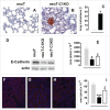
C1q deficiency is associated with anticipated metastatic spread and epithelial-to-mesenchymal transition in neuT tumors. Histological and immunohistochemical staining for Her2/neu of lungs from 17-week-old neuT (A) and neuT-C1KO (B) mice reveal earlier metastatic infiltration in neuT-C1KO mice. Magnification × 400. (C) Percentage of neuT (n = 19, gray bar) and neuT-C1KO (n = 14, black bar) mice (*p = 0.05 Chi-square test) bearing lung metastatic lesions at 17 weeks of age. (D–I) Decreased expression of E-Cadherin in neuT-C1KO and neuT-C3KO vs. neuT tumors. (D) E-Cadherin (upper panel) and actin (lower panel) protein levels as measured using the immunoblotting of whole cell lysates from 6–8 mm mean diameter carcinomas. A representative blot from three independent experiments is shown. (E) Quantification of E-Cadherin protein expression in neuT (gray bar), in neuT-C1KO (black bar) and neuT-C3KO (white bar) tumors (*p < 0.05, two-tailed Student's t-test). (F–H) Representative microscopy images of tumor sections from neuT (F), neuT-C1KO (G), and neuT-C3KO (H) mice (n = 3 per group) labeled with anti-E-Cadherin antibody (red) and DAPI (blue, labeling nuclei). Magnification × 400. (I) E-Cadherin protein was quantified in neuT (gray bar), neuT-C1KO (black bar) (***p < 0.0001, two-tailed Student's t-test) and neuT-C3KO (white bar) tumors (*p = 0.04, two-tailed Student's t-test). Results are represented as means ± SEM.
Figure 3.
Figure 3.
The dispensable role of the classical complement activation pathway in neuT tumor immunosurveillance. C3 fragment deposition at the tumor site is not altered in the absence of C1q or antibodies. (A–C) Confocal microscopy images representative of frozen tumor sections from mammary glands of 17-week-old neuT (A), neuT-C1KO (B), and neuT-BKO (C) mice labeled with anti-C3b/iC3b/C3c antibody (red) and TO-PRO®-3 iodide (blue). Magnification × 400. C3 fragment deposition was quantified (D) in neuT (gray bar), neuT-C1KO (black bar), and neuT-BKO (white bar) mice (n = 10 each group). No differences in C3 fragments deposition were found (p = ns, two-tailed Student's t-test). Results are represented as means ± SEM from 3 × 400 microscopic fields per sample.
Figure 4.
Figure 4.
Decrease of pWWOX and increase of Her2/neu expression in neuT-C1KO tumors. Confocal microscopy images of frozen tumor sections from neuT (A, E, I), neuT-C1KO (B, F, J), and neuT-C3KO (C, G, K) mice (n = 7 per group) labeled with anti-pWWOX (red, A–C), anti-C1q (red, E–G), and anti-Her2/neu (red, I–K) antibodies. Nuclei were stained with TO-PRO®-3 iodide (blue). Magnification ×100. pWWOX (D), C1q (H), and Her2/neu (L) protein quantification was performed in neuT (gray bar), neuT-C1KO (black bar), and neuT-C3KO (white bar) mice (*p = 0.02 for pWWOX; *p = 0.04 and **p = 0.009 for C1q; *p ≤ 0.04 for Her2/neu; two-tailed Student's t-test). Results are represented as means ± SEM from 3 × 400 microscopic fields per sample.
Figure 5.
Figure 5.
C1q deficiency affects intratumoral vessel density but does not modify tumor-infiltrating leukocyte recruitment. (A–C) Representative images of immunohistochemical staining for endothelial cell markers (CD31 and CD105, red) to visualize blood vessels in mouse tumors of equal volume developed in neuT (A) neuT-C1KO (B) and neuT-C3KO (C) mice. Magnification × 200. Quantification of the number (D) of vessels in neuT (gray bar; n = 3), neuT-C1KO (black bar; n = 5), and neuT-C3KO (white bar; n = 5) carcinomas (*p = 0.04; two-tailed Student's t-test). Results are represented as means ± SEM from 5 × 200 microscopic fields per sample. (E–G) Representative confocal microscopy images of tumors from neuT (E) neuT-C1KO (F) and neuT-C3KO (G) mice stained with anti-CD31 antibodies (green). Magnification × 400. Quantification of the vessel area (H) in neuT (gray bar; n = 3), neuT-C1KO (black bar; n = 5), and neuT-C3KO (white bar; n = 5) carcinomas. (***p < 0.0001; two-tailed Student's t-test). Results are represented as means ± SEM from 5 × 200 microscopic fields. (I–M) Flow cytometry analysis of infiltrating leukocytes in 6–8 mm mean diameter tumors from neuT (n = 5; gray bars), neuT-C1KO (n = 7; black bars) and neuT-C3KO (n = 6; white bars) mice. (I) CD3+ leukocytes were gated and CD3+ CD4+ CD25+ FoxP3+ were identified as Tregs (**p = 0.005; two-tailed Student's t-test). (L) CD45+ leukocytes were gated and CD3+ CD4+ cells were identified as CD4+ T, CD3+ CD8+ as CD8 T, CD3+ γδ+ as γδ T and CD3 CD49b+ as NK (**p = 0.005; two-tailed Student's t-test). (M) CD45+ CD11b+ leukocytes were gated and F4/80+ cells were identified as macrophages (MΦ), whereas GR-1+ cells were identified as myeloid-derived suppressor cells (MDSC). Bars represent the percentage of positive cells ± SEM.
Figure 6.
Figure 6.
NeuT-C1KO tumors are less aggressive than those of neuT-C3KO mice. Tumor incidence of mammary carcinomas (A) and overall mice survival (B) in neuT (n = 20, continuous gray line), neuT-C1KO (n = 14, continuous black line) and neuT-C3KO (n = 15, dotted black line) mice. NeuT-C3KO mice displayed earlier tumor incidence (*p = 0.05, Log-rank Mantel-Cox test) and lower overall survival (*p = 0.02, Log-rank Mantel-Cox test) than neuT-C1KO mice.
Figure 7.
Figure 7.
Proposed mechanism of C1q influence on neuT tumor progression. C1q component of C1 complex (C1q, C1s, and C1r) appears to act directly both on tumor vasculature (on the right) and on tumor cells (on the left). Deposition of C1q on vascular endothelium inhibits tumor angiogenesis through a still undefined mechanism. C1q binding with its receptor(s) (C1qR) on tumor cells leads to the phosphorylation of tyrosine 33 (Y33) on WWOX. Activated WWOX in turn inhibits the EMT processes, through directly inducing the expression of E-Cadherin, and induces Caspase-3-mediated apoptosis, probably by engaging p53. We hypothesize that activated WWOX may be also involved in neuT post-translational negative regulation further contributing to tumor inhibition. Green lines: antitumor activities; red lines: pro-tumor activities.

References

    1. Mamidi S, Höne S, Kirschfink M. The complement system in cancer: Ambivalence between tumour destruction and promotion. Immunobiology 2015; 222(1):45-54; PMID:26686908; http://dx.doi.org/10.1016/j.imbio.2015.11.008 - DOI - PubMed
    1. Stover C. Dual role of complement in tumour growth and metastasis (Review). Int J Mol Med 2010; 25:307-13; PMID:20127033; http://dx.doi.org/10.3892/ijmm_00000346 - DOI - PubMed
    1. Pio R, Corrales L, Lambris JD. The role of complement in tumor growth. Adv Exp Med Biol 2014; 772:229-62. Available from: http://www.pubmedcentral.nih.gov/articlerender.fcgi?artid=4379038&to... PMID:24272362; http://dx.doi.org/10.1007/978-1-4614-5915-6_11 - DOI - PMC - PubMed
    1. Markiewski MM, Lambris JD. Is complement good or bad for cancer patients? A new perspective on an old dilemma. Trends Immunol 2009; 30:286-92. Available from: http://www.pubmedcentral.nih.gov/articlerender.fcgi?artid =2704572&tool=pmcentrez&rendertype=abstract; PMID:19428302; http://dx.doi.org/10.1016/j.it.2009.04.002 - DOI - PMC - PubMed
    1. Cooper PD. Complement and cancer: activation of the alternative pathway as a theoretical base for immunotherapy. Adv Immun Cancer Ther 1985; 1:125-66; PMID:3916662; http://dx.doi.org/10.1007/978-1-4612-5068-5_4 - DOI - PubMed

Publication types