. 2016 Nov 21;6(1):111-124.
doi: 10.1016/j.molmet.2016.11.009. eCollection 2017 Jan.

Fetal development of subcutaneous white adipose tissue is dependent on Zfp423

Affiliations

Fetal development of subcutaneous white adipose tissue is dependent on Zfp423

Mengle Shao et al. Mol Metab. .

Abstract

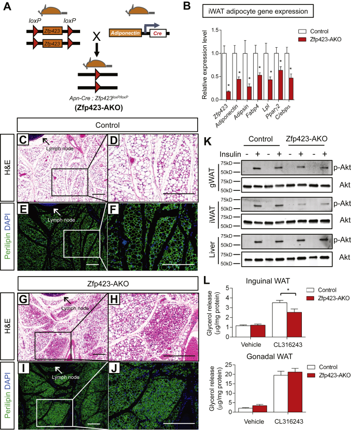
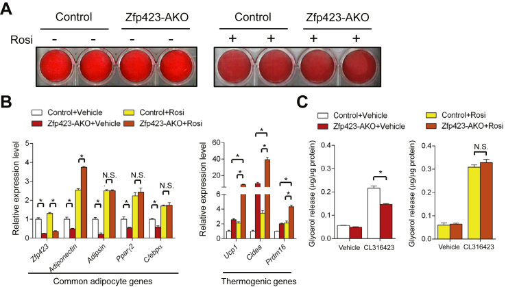
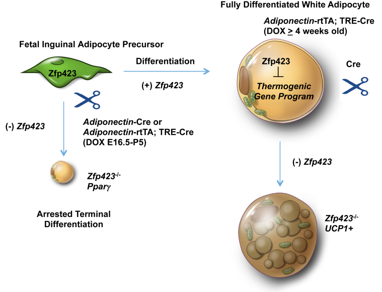
Objective: Zfp423 is a multi zinc-finger transcription factor expressed in preadipocytes and mature adipocytes in vivo. Our recent work has revealed a critical role for Zfp423 in maintaining the fate of white adipocytes in adult mice through suppression of the beige cell thermogenic gene program; loss of Zfp423 in mature adipocytes of adult mice results in a white-to-beige phenotypic switch. However, the exact requirements of Zfp423 in the fetal stages of early adipose development in vivo have not been clarified.

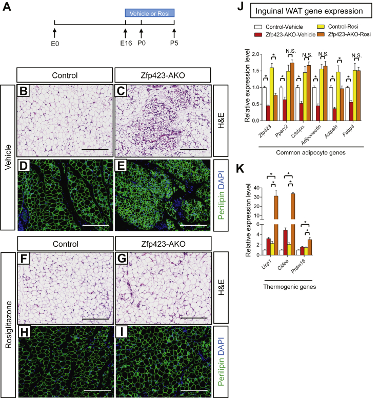
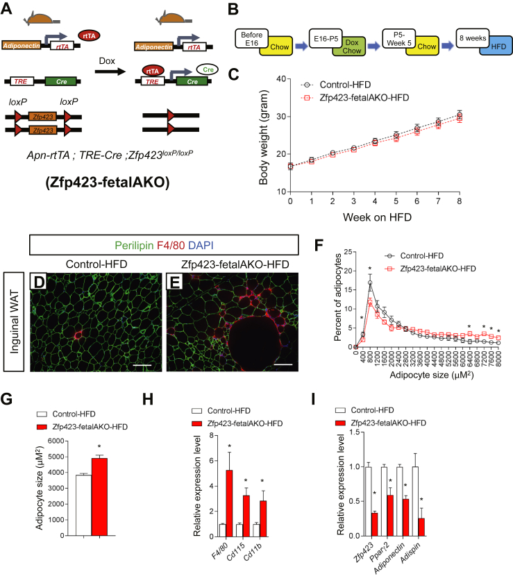
Method: Here, we utilize two models that confer adipose-specific Zfp423 inactivation during fetal adipose development (Adiponectin-Cre; Zfp423loxP/loxP and Adiponectin-rtTA; TRE-Cre; Zfp423loxP/loxP). We assess the impact of fetal adipose Zfp423 deletion on the initial formation of adipose tissue and evaluate the metabolic consequences of challenging these animals with high-fat diet feeding.

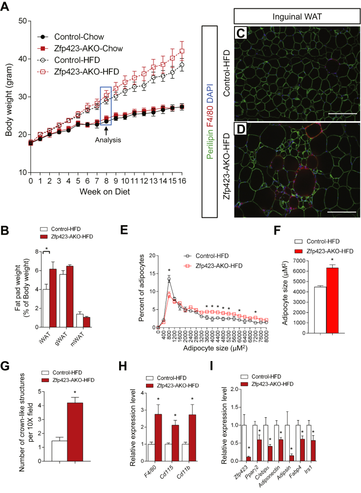
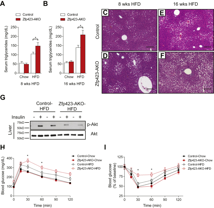
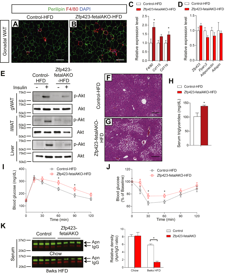
Results: Deletion of Zfp423 during fetal adipose development results in a different phenotype than is observed when deleting Zfp423 in adipocytes of adult mice. Inactivation of Zfp423 during fetal adipose development results in arrested differentiation, specifically of inguinal white adipocytes, rather than a white-to-beige phenotypic switch that occurs when Zfp423 is inactivated in adult mice. This is likely explained by the observation that adiponectin driven Cre expression is active at an earlier stage of the adipocyte life cycle during fetal subcutaneous adipose development than in adult mice. Upon high-fat diet feeding, obese adipose Zfp423-deficient animals undergo a pathological adipose tissue expansion, associated with ectopic lipid deposition and systemic insulin resistance.

Conclusions: Our results reveal that Zfp423 is essential for the terminal differentiation of subcutaneous white adipocytes during fetal adipose tissue development. Moreover, our data highlight the striking adverse effects of pathological subcutaneous adipose tissue remodeling on visceral adipose function and systemic nutrient homeostasis in obesity. Importantly, these data reveal the distinct phenotypes that can occur when adiponectin driven transgenes are activated in fetal vs. adult adipose tissue.

Keywords: Adipogenesis; Insulin resistance; Obesity; Pparg; Preadipocytes; Subcutaneous adipocytes; Zfp423.


Figures

Figure 1
Figure 1
Inguinal adipose tissue development is impaired in Adiponectin-Cre; Zfp423loxP/loxP mice. (A)Adiponectin-Cre; Zfp423loxP/loxP (Zfp423-AKO) mice are generated by breeding Adiponectin-Cre transgenic mice to animals carrying floxed Zfp423 alleles (Zfp423loxP/loxP). (B) Relative mRNA levels of Zfp423 and indicated adipocyte genes in fractionated inguinal white adipose tissue (iWAT) adipocytes from 8 weeks-old control and Zfp423-AKO mice. * denotes p < 0.05 from student's t-test. n = 5 mice. (CF) Representative H&E staining (c, d) and perilipin immunofluorescence staining (e, f) of iWAT sections obtained from 8 weeks-old control mice. Scale bar, 200 μm. (GJ) Representative (g, h) H&E staining and (i, j) perilipin immunofluorescence staining of iWAT sections obtained from 8 weeks-old Zfp423-AKO mice. Scale bar, 200 μm. (K) Western blot analysis of phosphorylated-Akt (p-AKt) and total Akt protein levels in tissue extracts of gonadal WAT (gWAT), iWAT, and liver of 8 weeks-old old control and Zfp423-AKO mice administrated with insulin (1 U/kg). (L) β3-adrenergic receptor induced lipolysis (measured as glycerol release) in ex vivo diced iWAT and gWAT isolated from 8 weeks-old control and Zfp423-AKO mice. *p < 0.05 from two-way ANOVA. n = 6 mice.
Figure 2
Figure 2
Developmental defects caused by Zfp423-deficiency are cell autonomous and can be rescued by Pparγ agonism. (A) Oil Red O staining of in vitro derived adipocytes differentiated from cultures of the inguinal stromal vascular fraction of control and Zfp423-AKO mice. Cultures were treated with vehicle (−) or rosiglitazone (+) throughout differentiation. (B) Relative mRNA levels of common adipocyte genes and thermogenic genes in adipocytes cultures differentiated as shown in (a). * denotes p < 0.05 from two-way ANOVA. N.S. denotes not statistically significant. n = 4 for all conditions. (C) β3-adrenergic receptor induced lipolysis in in vitro derived control and Zfp423-AKO white adipocytes treated with vehicle or rosiglitazone during differentiation. * denotes p < 0.05 from two-way ANOVA. N.S. denotes not statistically significant. n = 3 mice.
Figure 3
Figure 3
Fetal Inguinal WAT development in Adiponectin-Cre; Zfp423loxP/loxP mice is corrected by perinatal administration of the Pparγ agonist, Rosiglitazone. (A) Vehicle or the Pparγ agonist, rosiglitazone (Rosi), was delivered to pregnant/lactating mothers (10 mg kg−1/day) from embryonic day (E) 16 (E16) to postnatal (P) day 5 (P5). Offspring were harvested for analysis at P5. (B, E) Representative H&E staining (B, C) and perilipin immunofluorescence staining (D, E) of iWAT sections obtained from 5 days-old control and Zfp423-AKO pups treated with vehicle. Scale bar, 200 μm. (F, I) Representative H&E staining (F, G) and perilipin immunofluorescence staining (H, I) of iWAT sections obtained from 5 days-old control and Zfp423-AKO pups treated with rosiglitazone. Scale bar, 200 μm. (J) Relative mRNA levels of common adipocyte genes in iWAT from 5 days-old control and Zfp423-AKO pups treated with vehicle or rosiglitazone. * denotes p < 0.05 from two-way ANOVA. N.S. denotes not statistically significant. n = 6 mice. (K) Relative mRNA levels of thermogenic genes in iWAT from 5 days-old control and Zfp423-AKO pups treated with vehicle or rosiglitazone. * denotes p < 0.05 from two-way ANOVA. n = 6 mice.
Figure 4
Figure 4
High-fat diet feeding leads to a pathological inguinal WAT expansion in Adiponectin-Cre; Zfp423loxP/loxP mice. (A) Weekly body weight measurements of control and Zfp423-AKO mice fed chow or high-fat diet (HFD) for 16 weeks following weaning. n = 6–8 mice. (B) Adipose depot mass (normalized to body weight) of control and Zfp423-AKO mice after 8 weeks of HFD feeding. *p < 0.05 from student's test. n = 6–8 mice. (C, D) Representative immunofluorescence images of Perilipin (green) and F4/80 (red) expression in iWAT paraffin sections obtained from control and Zfp423-AKO mice after 8 weeks of HFD feeding. Scale bar, 200 μm. (E) Distribution of adipocyte size in control and Zfp423-AKO iWAT after 8 weeks of HFD feeding. * denotes p < 0.05 from two-way ANOVA. n = 6–8 mice. (F) Average adipocyte size of control and Zfp423-AKO iWAT after 8 weeks of HFD feeding. * denotes p < 0.05 from student's test. n = 6–8 mice. (G) Number of crown-like structures (F4/80 positive) in control and Zfp423-AKO iWAT after 8 weeks of HFD feeding. * denotes p < 0.05 from student's test. n = 6–8 mice. (HI) Relative mRNA levels of macrophage markers (H) and adipocyte-selective genes (I) in control and Zfp423-AKO iWAT after 8 weeks of HFD feeding. * denotes p < 0.05 from student's test. n = 6 mice.
Figure 5
Figure 5
Obese Adiponectin-Cre; Zfp423loxP/loxP mice are insulin resistant and have increased hepatic steatosis. (A, B) Serum triglyceride levels in control and Zfp423-AKO mice after 8 (A) or 16 weeks (B) of chow or HFD feeding. * denotes p < 0.05 from two-way ANOVA. n = 6–8 mice. (C, D) Representative H&E staining of livers from control (C) and Zfp423-AKO (D) mice after 8 weeks of HFD feeding. Scale bar, 200 μm. (E, F) Representative H&E staining of livers from control (E) and Zfp423-AKO (F) mice after 16 weeks of HFD feeding. Scale bar, 200 μm. (G) Western blot analysis of phosphorylated-Akt (p-AKt) and total Akt protein levels in liver extracts from 8 week HFD-fed control and Zfp423-AKO mice administrated with insulin (2 U/kg). (H) Glucose tolerance test of control and Zfp423-AKO mice after 8 weeks HFD feeding. * denotes p < 0.05 from Two-way ANOVA. n = 6–8 mice. (I) Insulin tolerance test of control and Zfp423-AKO mice after 8 weeks HFD feeding. * denotes p < 0.05 from Two-way ANOVA. n = 6–8 mice.
Figure 6
Figure 6
Transient fetal exposure of Adiponectin-rtTA; TRE-Cre; Zfp423loxP/loxP animals leads to inguinal white adipose depot-selective targeting of Zfp423. (A) Inducible inactivation of Zfp423 in adiponectin-expressing cells is achieved by breeding the adiponectin-rtTA transgenic mice to animals expressing Cre recombinase under the control of the Tet-response element (TRE-Cre) and carrying floxed Zfp423 alleles (Zfp423loxP/loxP). Littermates carrying only adiponectin-rtTA and Zfp423loxP/loxP alleles (i.e. Cre–) were used as the control animals. Exposure of animals to doxycycline from E16 to P5 via feeding of mothers results in a model of selective Zfp423 inactivation in the developing inguinal WAT (Zfp423-fetalAKO mice). (B) Control and Zfp423-fetalAKO mice were exposed to doxycycline from E16 to P5 and then kept on standard chow diet until 5 week old before switching to HFD for another 8 weeks. (C) Weekly body weight measurements of control and Zfp423-fetal AKO mice during 8 weeks of HFD feeding. n = 6 mice. (D, E) Representative immunofluorescence images of perilipin (green) and F4/80 (red) expression in iWAT paraffin sections from control (D) and Zfp423-fetalAKO (E) mice after 8 weeks of HFD feeding. Scale bar, 200 μm. (F) Distribution of adipocyte size in control and Zfp423-fetalAKO iWAT after 8 weeks of HFD feeding. * denotes p < 0.05 from two-way ANOVA. n = 6 mice. (G) Average adipocyte size in control and Zfp423-fetalAKO iWAT after 8 weeks of HFD feeding. * denotes p < 0.05 from student's t-test. n = 6 mice. (H, I) Relative mRNA levels of macrophage markers (H) and adipocyte-selective genes (I) in control and Zfp423-fetalAKO iWAT after 8 weeks of HFD feeding. * denotes p < 0.05 from student's t-test. n = 6 mice.
Figure 7
Figure 7
Pathological expansion of subcutaneous WAT is sufficient to trigger impaired systemic nutrient homeostasis. (A, B) Representative immunofluorescence images of perilipin (green) and F4/80 (red) expression in sections of gWAT from control and Zfp423-fetalAKO mice after 8 weeks of HFD feeding. Scale bar, 200 μm. (C, D) Relative mRNA levels of macrophage markers (C) and adipocyte-selective genes (D) in gWAT of control and Zfp423-fetalAKO mice after 8 weeks of HFD feeding. * denotes p < 0.05 from student's t-test. n = 6 mice. (E) Western blot analysis of phosphorylated-Akt (p-AKt) and total Akt protein levels in tissue extracts of gWAT, iWAT, and liver of HFD-fed control and Zfp423-fetalAKO mice administrated with insulin (2 U/kg). (F, G) Representative H&E staining of livers from control and Zfp423-fetalAKO mice after 8 weeks of HFD feeding. Scale bar, 200 μm. (H) Serum triglyceride levels in control and Zfp423-fetalAKO mice after 8 weeks of HFD feeding. * denotes p < 0.05 from student's t-test. n = 6 mice. (I) Glucose tolerance test of control and Zfp423-fetalAKO mice after 8 weeks HFD feeding. * denotes p < 0.05 from student's t-test. n = 6 mice. (J) Insulin tolerance test of control and Zfp423-fetalAKO mice after 8 weeks HFD feeding. * denotes p < 0.05 from student's t-test. n = 6 mice. (K) Western blot analysis and quantification of serum adiponectin levels in control and Zfp423-fetalAKO mice fed with chow or HFD for 8 weeks starting at 5 weeks of age. For quantification, intensity of adiponectin band is normalized to that of IgG band. * denotes p < 0.05 from two-way ANOVA. n = 4 mice.
Figure 8
Figure 8
Establishment and Maintenance of the White Adipocyte Lineage by Zfp423. Zfp423 expression identifies committed preadipocytes and drives adipocyte differentiation through activation of Pparγ, , . Cre-mediated inactivation of Zfp423 in developing fetal inguinal WAT precursors results in arrested terminal adipocyte differentiation. In fully differentiated white adipocytes, Zfp423 functions to suppress the thermogenic gene program, thereby maintaining the white adipocyte phenotype. Inducible, Cre-mediated inactivation of Zfp423 in fully differentiated white adipocytes of adult mice leads to a lineage reprogramming in which mature white adipocytes are converted to thermogenic UCP1-expressing beige-like adipocytes .

References

    1. Stern J.H., Rutkowski J.M., Scherer P.E. Adiponectin, leptin, and fatty acids in the maintenance of metabolic homeostasis through adipose tissue crosstalk. Cell Metabolism. 2016;23(5):770–784. - PMC - PubMed
    1. Harms M., Seale P. Brown and beige fat: development, function and therapeutic potential. Nature Medicine. 2013;19(10):1252–1263. - PubMed
    1. Kissig M., Shapira S.N., Seale P. SnapShot: brown and beige adipose thermogenesis. Cell. 2016;166(1) pp. 258–258 e1. - PMC - PubMed
    1. Cannon B., Nedergaard J. Brown adipose tissue: function and physiological significance. Physiological Review. 2004;84(1):277–359. - PubMed
    1. Unger R.H., Clark G.O., Scherer P.E., Orci L. Lipid homeostasis, lipotoxicity and the metabolic syndrome. Biochimica et Biophysica Acta. 2010;1801(3):209–214. - PubMed

Publication types

MeSH terms

LinkOut - more resources