Review
. 2016 Dec;6(6):632-641.
doi: 10.21037/cdt.2016.09.08.

Complications of inferior vena cava filters

Affiliations
Review

Complications of inferior vena cava filters

Simer Grewal et al. Cardiovasc Diagn Ther. 2016 Dec.

Abstract

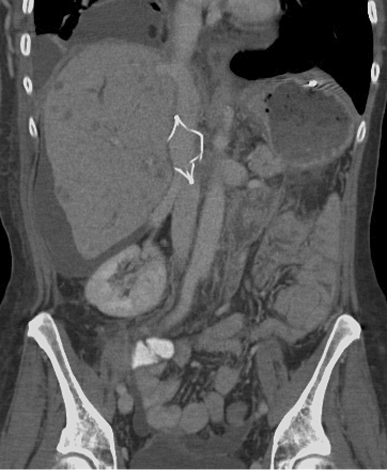
Inferior vena cava (IVC) filter placement is a relatively low risk alternative for prophylaxis against pulmonary embolism in patients with pelvic or lower extremity deep venous thrombosis who are not suitable for anticoagulation. There is an increasing trend in the number of IVC filter implantation procedures performed every year. There are many device types in the market and in the early 2000s, the introduction of retrievable filters brought an additional subset of complications to consider. Modern filter designs have led to decreased morbidity and mortality, however, a thorough understanding of the limitations and complications of IVC filters is necessary to weight the risks and benefits of placing IVC filters. In this review, the complications associated with IVC filters are divided into procedure related, post-procedure, and retrieval complications. Differences amongst the device types and retrievable filters are described, though this is limited by a significant lack of prospective studies. Additionally, the clinical presentation as well as prevention and treatment strategies are outlined with each complication type.

Keywords: Inferior vena cava (IVC) filter; deep vein thrombosis (DVT); fracture; migration; perforation; pulmonary embolism; retrieval; venous thromboembolism (VTE).


Conflict of interest statement

The authors have no conflicts of interest to declare.

Figures

Figure 1
Figure 1
Filter Tilt. Coronal maximum intensity projection from CT data demonstrates right lateral tilt of a Tulip (Cook Medical) IVC filter. A tilt of >15 degrees is reported to be less effective in preventing PE. In addition, tilted filters pose problems during retrieval. IVC, inferior vena cava.
Figure 2
Figure 2
Migration of filter: Plain radiograph of the chest demonstrates a Greenfield filter (Boston Scientific) migrated to right atrium. The filter was surgically removed.
Figure 3
Figure 3
Plain radiograph of the abdomen demonstrates a partially opened Greenfield filter.
Figure 4
Figure 4
Malpositioned filter: Plain radiograph obtained after placement of an IVC Filter (Option, Argon Medical) with intravascular ultrasound guidance demonstrates filter located in the right iliac vein. Filter located in the iliac vein is ineffective in preventing PE that originate in the contralateral leg. The filter was subsequently removed and a new filter was placed in the infrarenal IVC. IVC, inferior vena cava.
Figure 5
Figure 5
Caval thrombosis. Angiogram of the IVC and iliac veins demonstrates a Simon Nitinol filter in the infrarenal IVC with associated caval thrombosis. Also note tilted filter and retroperitoneal collaterals. IVC, inferior vena cava.
Figure 6
Figure 6
Thrombus in the filter. Coronal reconstruction of contrast enhanced CT of the abdomen demonstrates thrombus within a TrapEase (Cordis) IVC filter. Thrombus within a filter may be secondary to captured emboli or in situ thrombosis. IVC, inferior vena cava.
Figure 7
Figure 7
Fractured supra-renal IVC filter. Coronal reconstruction of contrast enhanced CT of the abdomen in a patient with metastatic cancer demonstrates fracture of a suprarenal TrapEase (Cordis) IVC filter. Suprarenal location for IVC filter was chosen due to a pelvic mass compressing the infrarenal IVC and iliac veins. IVC, inferior vena cava.
Figure 8
Figure 8
Plain radiograph of the abdomen demonstrates a tilted, and fractured Titanium Greenfield filter.
Figure 9
Figure 9
Filter perforation. Axial contrast enhanced CT of the abdomen demonstrates filter (Recovery Filter, Bard) perforating the IVC wall with one of its leg in the aortic wall. The patient was asymptomatic. IVC, inferior vena cava.

References

    1. 1. Moore PS, Andrews JS, Craven TE, et al. Trends in vena caval interruption. J Vasc Surg 2010;52:118-125.e3; discussion 125-6. - PubMed
    1. Stein PD, Matta F, Hull RD. Increasing use of vena cava filters for prevention of pulmonary embolism. Am J Med 2011;124:655-61. 10.1016/j.amjmed.2011.02.021 - DOI - PubMed
    1. Alkhouli M, Bashir R. Inferior vena cava filters in the United States: less is more. Int J Cardiol 2014;177:742-3. 10.1016/j.ijcard.2014.08.010 - DOI - PubMed
    1. Andreoli JM, Lewandowski RJ, Vogelzang RL, et al. Comparison of complication rates associated with permanent and retrievable inferior vena cava filters: a review of the MAUDE database. J Vasc Interv Radiol 2014;25:1181-5. 10.1016/j.jvir.2014.04.016 - DOI - PubMed
    1. Kinney TB. Update on inferior vena cava filters. J Vasc Interv Radiol 2003;14:425-40. 10.1097/01.RVI.0000064860.87207.77 - DOI - PubMed

LinkOut - more resources