. 2017 Apr 3;117(4):671-681.
doi: 10.1160/TH16-09-0691. Epub 2017 Jan 26.

Nucleic acids as cofactors for factor XI and prekallikrein activation: Different roles for high-molecular-weight kininogen

Affiliations

Nucleic acids as cofactors for factor XI and prekallikrein activation: Different roles for high-molecular-weight kininogen

Ivan Ivanov et al. Thromb Haemost. .

Abstract

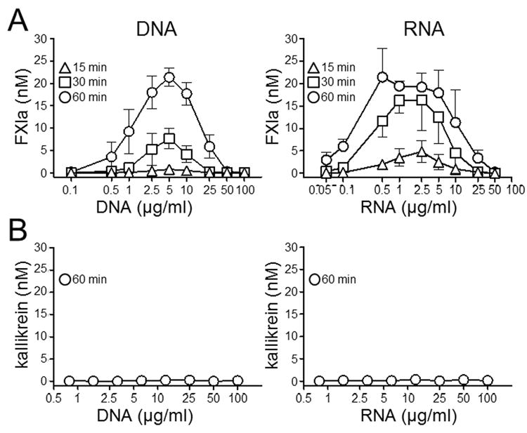
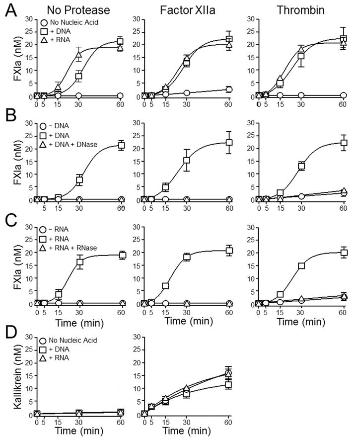
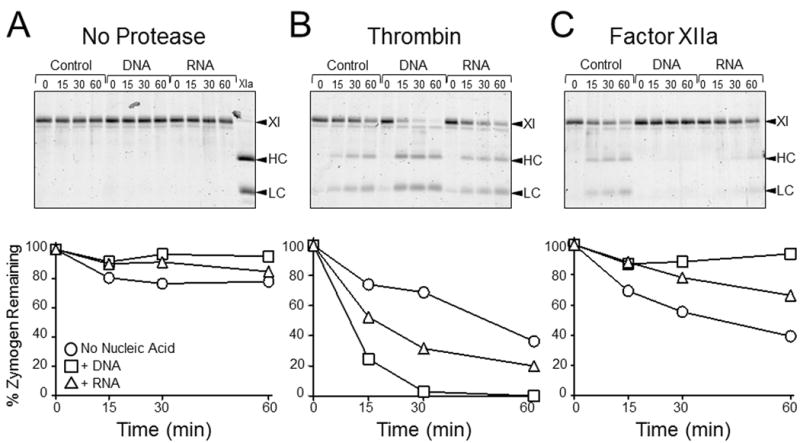
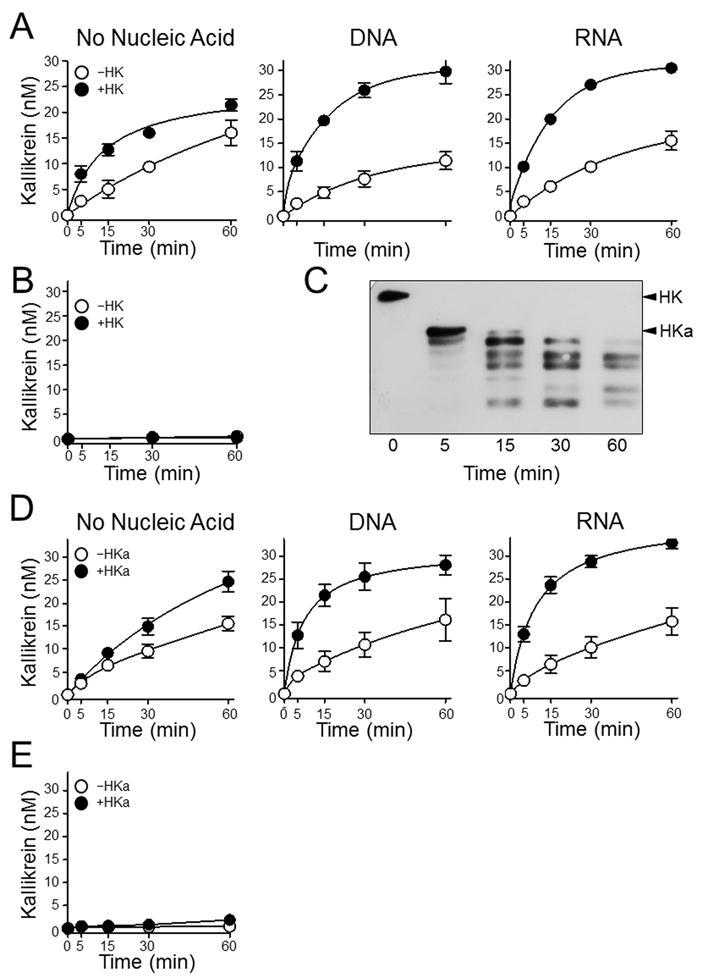
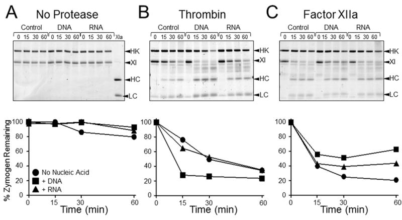
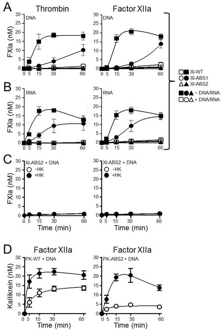
The plasma zymogens factor XI (fXI) and prekallikrein (PK) are activated by factor XIIa (fXIIa) during contact activation. Polyanions such as DNA and RNA may contribute to thrombosis and inflammation partly by enhancing PK and fXI activation. We examined PK and fXI activation in the presence of nucleic acids, and determine the effects of the cofactor high molecular weight kininogen (HK) on the reactions. In the absence of HK, DNA and RNA induced fXI autoactivation. Proteases known to activate fXI (fXIIa and thrombin) did not enhance this process appreciably. Nucleic acids had little effect on PK activation by fXIIa in the absence of HK. HK had significant but opposite effects on PK and fXI activation. HK enhanced fXIIa activation of PK in the presence of nucleic acids, but blocked fXI autoactivation. Thrombin and fXIIa could overcome the HK inhibitory effect on autoactivation, indicating these proteases are necessary for nucleic acid-induced fXI activation in an HK-rich environment such as plasma. In contrast to PK, which requires HK for optimal activation, fXI activation in the presence of nucleic acids depends on anion binding sites on the fXI molecule. The corresponding sites on PK are not necessary for PK activation. Our results indicate that HK functions as a cofactor for PK activation in the presence of nucleic acids in a manner consistent with classic models of contact activation. However, HK has, on balance, an inhibitory effect on nucleic acid-supported fXI activation and may function as a negative regulator of fXI activation.

Keywords: Contact phase; coagulation factors; proteases.


Figures

Figure 1
Figure 1. Nucleic acids induce autoactivation of fXI but not PK
(A) Plasma fXI (30 nM) or (B) PK (60 nM) was incubated with varying concentrations of genomic DNA or total cellular RNA for 15 (∆), 30 (□) or 60 (○) minutes. At designated times fXIa or kallikrein activity where measured by chromogenic assay. Error bars are +/− one SD.
Figure 2
Figure 2. FXI and PK activation by fXIIa or thrombin in the presence of nucleic acids
(A) Plasma fXI (30 nM) was incubated without an activating protease (No Protease) or with 2.5 nM fXIIa, or 2.5 nM thrombin, in the absence of nucleic acid (○), in the presence of 5 μg/ml DNA (□), or in the presence of 1 μg/ml RNA (∆). (B and C) Plasma fXI (30 nM) was incubated without activating protease (No Protease) or with 2.5 nM fXIIa, or 2.5 nM thrombin. Reactions in panel B were run in the absence of nucleic acid (○), in the presence of 5 μg/ml DNA (□), or in the presence of 5 μg/ml DNA treated with DNAse (∆). Reactions in panel C were run in the absence of nucleic acid (○), in the presence of 1 μg/ml RNA (□), or in the presence of 1 μg/ml RNA treated with RNAse (∆). For panels A-C, at indicated time points, aliquots were tested for fXIa activity by chromogenic assay. (D) Plasma PK (60 nM) was incubated without an activating protease (No Protease) or with 50 pM fXIIa, in the absence of nucleic acid (○), in the presence of 5 μg/ml DNA (□), or in the presence of 1 μg/ml RNA (∆). At indicated time points, aliquots were tested for kallikrein activity by chromogenic assay. For all panels error bars are +/−one SD.
Figure 3
Figure 3. Cleavage of fXI-Ala557 by fXIIa or thrombin in the presence of nucleic acids
(Top Row) SDS-PAGE of fXI-Ala557 (200 nM) incubated with vehicle (control), 30 μg/ml DNA, or 5 μg/ml RNA (A) without a protease, (B) with 15 nM thrombin, or (C) with 15 nM fXIIa. Incubation times in minutes are shown at the tops of gels. Abbreviations: XI – zymogen fXI, HC – fXIa heavy chain, LC – fXIa light chain. (Bottom Row) Reduction of the 80 kDa fXI-Ala557 zymogen band on the gels immediately above each graph, as determined by densitometry. Results are for reactions without nucleic acid (○), with DNA (□), or with RNA (∆).
Figure 4
Figure 4. Effects of HK and HKa on PK activation in the presence of nucleic acids
(A) Plasma PK (60 nM) was incubated with 50 pM fXIIa in the absence (○) or presence (●) of 60 nM HK; in the absence of nucleic acids (No Nucleic Acids), in the presence of 5 μg/ml DNA, or in the presence of 1 μg/ml RNA. (B) Plasma PK (60 nM) incubated in the absence (○) or presence (●) of 60 nM HK, without fXIIa or nucleic acids. For panels A and B, at the indicated time points, aliquots were tested for kallikrein activity by chromogenic assay. (C) Western blot for HK using samples from the reaction containing HK in the left-hand image in panel A. Positions of standard for HK and HKa are indicated at the right of the image. (D and E) Same for panels A and B, respectively except that HK was replaced with 60 nM HKa. For panels A, B, D and E, error bars are +/− one SD.
Figure 5
Figure 5. Effects of HK and HKa on fXI activation in the presence of nucleic acids
(A) Plasma fXI (30 nM) was incubated with vehicle (○,●), 5 μg/ml DNA (□,■), or 1 μg/ml RNA (∆,▲) in the absence (○,□,∆) or presence (●,■,▲) of 30 nM HK. Reactions included no activating protease (No Protease), 2.5 nM fXIIa or 2.5 nM thrombin. At indicated time points, aliquots were tested for fXIa activity by chromogenic assay. (B) Western blot for HK using samples from the reactions containing HK and DNA shown in panel A. Positions of standard for HK and HKa are indicated at the right of the images. (C) Same for panel A except that HK was replaced with 30 nM HKa. For panels A and C error bars are +/−one SD.
Figure 6
Figure 6. Cleavage of fXI-Ala557 by fXIIa or thrombin in the presence of nucleic acids and HK
(Top Row) SDS-PAGE of recombinant fXI-Ala557 (200 nM) and plasma HK (200 nM) incubated with vehicle (control), 30 μg/ml DNA, or 5 μg/ml RNA (A) without a protease, (B) with 15 nM thrombin, or (C) with 15 nM fXIIa. Incubation times in minutes are shown at the tops of gels. Abbreviations: HK − high molecular weigh-kininogen, XI − zymogen fXI, HC − fXIa heavy chain, LC − fXIa light chain. (Bottom Row) Reduction of the 80 kDa fXI-Ala557 zymogen band on the gels immediately above each graph, as determined by densitometry. Results are for reactions without nucleic acid (●), with DNA (■), or with RNA (▲).
Figure 7
Figure 7. Activation of fXI and PK anion-binding site (ABS) variants in the presence of nucleic acids
Recombinant fXI species (30 nM) were incubated without (□,○,∆) or with (■,●,▲) (A) 5 μg/ml DNA or (B) 1 μg/ml RNA, and with 2.5 nM thrombin (left) or 2.5 nM fXIIa (right). Symbols: fXI-WT (□,■), fXI-ABS1 (○,●), or fXI-ABS2 (∆,▲). (C) FXI-ABS2 (30 nM) was incubated with 5 μg/ml DNA and 2.5 nM thrombin (left) or 2.5 nM fXIIa (right) without (○) or with (●) 30 nM HK. (D) 60 nM PK-WT (left) or PK-ABS2 (right) were incubated with 50 pM fXIIa in the presence of 5 μg/ml DNA without (○) or with (●) 60 nM HK. For all reactions, at indicated time points, aliquots were tested for fXIa or kallikrein activity by chromogenic assay. Error bars are +/−one SD.
Figure 8
Figure 8. Effect of DNA on fXI-dependent thrombin generation in plasma
(A) Thrombin generation measured in normal plasma after addition of 25 μg/ml DNA without (left hand panel) or with (right hand panel) 0.5 pM TF. Assays were run in the presence of vehicle (solid line), the anti-fXI IgG O1A6 (dotted line) or the fXIIa inhibitor CTI (dashed line). (B) Images of neutrophils stimulated with vehicle (control) or 100 ng/ml phorbol myristate acetate (PMA) with or without 100 U/mL DNAse. Cell permeable Hoechst stain (top row) indicates DNA within intact cell nuclei. Sytox Green staining (bottom row) demonstrates extracellular DNA or DNA within dying cells that have lost the ability to exclude the dye. PMA treated cells form neutrophil extracellular traps (NETs, middle column), which are not observed in the presence of DNase (right column). Images were made on an Inverted Fluorescence Microscope (89404-464) using a V10MP camera (VWR), and Motic Images Plus 2.0 software. (C) Thrombin generation in normal plasma supplemented with neutrophils treated with vehicle (gray lines, –PMA) or PMA (black lines, + PMA), without (solid lines) or with (dashed lines) DNAse in the absence of an inhibitor (No inhibitor), or in the presence of O1A6 or CTI. For reactions containing O1A6, no thrombin generation was detected with or without DNAse treatment. For reactions with CTI, some activity was detected in samples not treated with DNAse.

References

    1. Müller F, Mutch NJ, Schenk WA, Smith SA, Esterl L, Spronk HM, Schmidbauer S, Gahl WA, Morrissey JH, Renné T. Platelet polyphosphates are proinflammatory and procoagulant mediators in vivo. Cell. 2009;139:1143–1156. - PMC - PubMed
    1. Morrissey JH, Choi SH, Smith SA. Polyphosphate: an ancient molecule that links platelets, coagulation, and inflammation. Blood. 2012;119:5972–5979. - PMC - PubMed
    1. Kannemeier C, Shibamiya A, Nakazawa F, et al. Extracellular RNA constitutes a natural procoagulant cofactor in blood coagulation. Proc Natl Acad Sci USA. 2007;104:6388–6393. - PMC - PubMed
    1. Fuchs TA, Brill A, Duerschmied D, Schatzberg D, Monestier M, Myers DD, Wrobleski SK, Wakefield TW, Hartwig JH, Wagner DD. Extracellular DNA traps promote thrombosis. Proc Natl Acad Sci USA. 2010;107:15880–15885. - PMC - PubMed
    1. Fischer S, Preissner KT. Extracellular nucleic acids as novel alarm signals in the vascular system. Mediators of defense and disease Hamostaseologie. 2013;33:37–42. - PubMed

Publication types